Archive for the ‘Chemical Biology’ Category

Simple aptasensor for detecting protein

Graphical abstract: Nicking enzyme based homogeneous aptasensors for amplification detection of proteinChinese scientists have made a simple and sensitive sensor for detecting proteins, which could lead to improved disease detection.

Huang-Hao Yang and colleagues at Fuzhou University used single-stranded nucleic acids known as aptamers to detect thrombin, an important protein involved in blood clotting. 

Although other aptasensors are known, they are more complex than this new sensor, says Yang. And the sensitivity here is three orders of magnitude higher than traditional homogeneous aptasensors. 

The improvement is thanks to a nicking enzyme, which Yang used instead of the more usual polymerase. A nicking enzyme recognises a specific sequence in double-stranded DNA. It then cleaves only one strand, leaving a nick in the DNA.   

The aptasensor is capable of detecting thrombin in real samples and could be expanded to other proteins simply by changing the aptamer sequence. 

To find out more about how it works, download Yang’s ChemComm communication.

Digg This
Reddit This
Stumble Now!
Share on Facebook
Bookmark this on Delicious
Share on LinkedIn
Bookmark this on Technorati
Post on Twitter
Google Buzz (aka. Google Reader)

A model for the single chirality of life

Hot spring
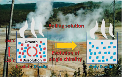
The boiling solutions in prebiotic hot springs could shed light on the emergence of a single chiral form of biomolecules in nature, say Spanish scientists.

Amino acids and sugars exist in living organisms exclusively in one of their two molecular chiral forms, which are mirror images of one another. The reason for this specificity has long puzzled scientists. Conditions on Earth when life first evolved in prebiotic times presumably favoured one form over the other, but the mechanism is so far unresolved.

‘We will most likely be unable to solve this fascinating conundrum, but experiments carried out under environmentally credible conditions may be important signposts to this end,’ says Cristóbal Viedma at the Universidad Complutense, Madrid. With this in mind, he and colleague Pedro Cintas have investigated crystallisation processes that lead to single chiral forms.

Read the full news story in Chemistry World and download Viedma’s ChemComm communication to find out the exact details.

Digg This
Reddit This
Stumble Now!
Share on Facebook
Bookmark this on Delicious
Share on LinkedIn
Bookmark this on Technorati
Post on Twitter
Google Buzz (aka. Google Reader)

Once, twice, three times a potential life saver – electrochemical immunoassay of cancer biomarkers

Early detection and diagnosis of cancer is essential to give sufferers an increased chance of overcoming the disease. The symptoms of liver cancer, in particular, are fairly innocuous – tumours are difficult to detect by physical examination and therefore the disease is not usually discovered until the later stages of development.

Dong Wang and co-workers have developed an electrochemical immunoassay which has the potential to improve the early detection rates of liver cancer by simultaneous detection of not one, not two, but three tumour markers. This simultaneous multianalyte immunoassay (SMIA) has a number of advantages over single-analyte immunoassay methods such as reduced overall cost per assay; improved efficiency; the potential to quantitatively measure the concentrations of proteins detected; as well as having a panel of biomarkers to confirm the diagnosis, lowering the likelihood of false-positive or false-negative results.

The team selected three electrochemical redox species with distinct voltammetric peaks to label three different antibodies as signal tags. These were then loaded onto carbon nanotubes coated with gold nanoparticles to improve the signal response.

Binding of the redox species labelled nanotube-antibody conjugates to the cancer biomarkers enables their electrochemical detection

By deciphering the voltammetric read-out, the team were able to establish successful binding events between the probes and the protein biomarkers, allowing simultaneous identification of the three analytes. Wang and his team then increased the sophistication of the immunoassay further by proving quantitative detection.

Dong Wang and co-workers appeared to have produced a robust SMIA with low detection limits that has the potential to save many lives. This efficient, gold nanoparticle-based technology could also be applied to other types of cancer or diseases – an immunoassay with a Midas touch.

To find out more, download the ChemComm article.

Posted on behalf of Sarah Brown, ChemComm web science writer.

Digg This
Reddit This
Stumble Now!
Share on Facebook
Bookmark this on Delicious
Share on LinkedIn
Bookmark this on Technorati
Post on Twitter
Google Buzz (aka. Google Reader)

Overcoming obstacles in labelling RNA

If you can’t beat them, join them… or rather incorporate, and then join them together. This is the approach adopted by Srivatsan and colleagues for conjugating labels to RNA, which they achieve by incorporating azide functionality into RNA nucleotides.

DNA oligonucleotides are routinely labelled by incorporating modified nucleosides into the desired DNA sequence and introducing the label post-synthesis; however, these standard methods offer low yields of the desired conjugate. Furthermore, modification of RNA is more challenging than DNA modification due to its inherent instability.

Copper catalysed alkyne–azide cycloadditions (CuAAC) and Staudinger ligations are the conjugation reactions du jour due to their high chemoselectivity and reported high yield. So, with this in mind, were Srivatsan and his team able to incorporate the required moieties into RNA oligonucleotides?

Yes! Using some extremely nice chemistry, they synthesised an azide-modified nucleotide and incorporated it into an oligoribonucleotide using in vivo transcription reactions in the presence of a series of promoter-template oligonucleotides. After rigorous testing and analysis of their modified nucleic acid sequence, click reactions were performed to yield biotinylated and fluorescent-labelled click products. They also showed that amine functionality can be introduced through Staudinger ligation.

Although azides are not usually found in nature, they are becoming more useful and versatile in the design of diagnostic and therapeutic biological probes as has been very elegantly demonstrated here.

Read more in Srivatsan’s ChemComm article

Also of interest: Synthetic DNA synthesises RNA by transcription: ‘click’ here for more

Posted on behalf of Sarah Brown, web science writer for ChemComm.

Digg This
Reddit This
Stumble Now!
Share on Facebook
Bookmark this on Delicious
Share on LinkedIn
Bookmark this on Technorati
Post on Twitter
Google Buzz (aka. Google Reader)

Improved DNA chips to detect pathogens

A new probe selection method leads to better DNA chips with fewer false positives and paves the way to improved technology development.

DNA chips are now ubiquitous tools in both genomics and diagnostics, for example pathogen detection. But selecting the correct DNA molecules for immobilising onto the chips’ surface is necessary to improve the chips’ accuracy.

The improved method involves loading the DNA probes onto the surface according to length, melting temperature and specificity. The approach was used to correctly identify 19 different types of human papilloma virus and was used to analyse 1000 clinical samples, giving identical results to sequencing data obtained from the same samples.

Reference:
A generalized probe selection method for DNA chips
S B Nimse, K-S Song, J Kim, V-T Ta, V-T Nguyen and T Kim, Chem. Commun., 2011
DOI:
10.1039/c1cc15137a

Digg This
Reddit This
Stumble Now!
Share on Facebook
Bookmark this on Delicious
Share on LinkedIn
Bookmark this on Technorati
Post on Twitter
Google Buzz (aka. Google Reader)

Synthetic DNA synthesises RNA by transcription: ‘click’ here for more

What’s better than a short synthetic DNA sequence? That’s right: a long synthetic DNA sequence, able to take part in biological processes.

DNA and RNA are routinely synthesised using solid phase synthesis, connecting the bases through phosphodiester linkages.  This method, however, imposes limitations on the length of the strand produced. Current methods employ enzymes to achieve larger oligonucleotides, which have disadvantages such as poor quality and stability and can be laborious.

Tom Brown and Afaf El-Sagheer at the University of Southampton, UK, have now demonstrated that these limitations can be overcome by the flick of a ‘click’ – they chemically ligated oligonucleotides functionalised with chemoselective azide and alkyne moieties using click chemistry to produce long nucleic acid sequences. After personally struggling to conjugate biological moieties using click chemistry for my Ph.D., I can assure you this is no mean feat!

Previous work by the group has shown that oligonucleotides with triazole-modified backbones do not inhibit replication and so they were interested to see if the same would be true for transcription – a fundamentally different process. The primary concern was how modification of the phosphodiester backbone would affect the template strand’s ability to participate in the transcription process.

By synthesising two triazole-modified DNA template strands, one with the triazole unit in the coding sequence and the other with the modification in the T7 RNA promoter region, El-Sagheer and Brown were able to evaluate the impact of changing the template strand backbone in comparison with the equivalent non-modified templates. Both modified strands successfully took part in transcription to produce RNA in good yield.  The result: fully-synthetic, biologically-active DNA templates successfully synthesising RNA. This is not only an elegant demonstration of the abilities of click chemistry but could also be fundamental in changing approaches to the synthesis of biological constructs.

To read more about Brown’s research, download the ChemComm article today.

Posted on behalf of Sarah Brown, web science writer for ChemComm.

Digg This
Reddit This
Stumble Now!
Share on Facebook
Bookmark this on Delicious
Share on LinkedIn
Bookmark this on Technorati
Post on Twitter
Google Buzz (aka. Google Reader)

Fullerene ‘hide and seek’ in lipid membranes

Japanese researchers have experimentally proven that fullerenes reside in the hydrophobic core of liposome membranes.

Liposomes are vesicular structures made up of a phospholipid bilayer. Due to their ability to encapsulate a range of different substances and target numerous cell types, they have great potential as drug delivery agents. Recently, liposomes have been modified with fullerene for a number of uses such as DNA photocleavage, anti-bacterial agents, and photodynamic therapy. Although theoretical simulations have been performed to characterise these fullerene–liposomes, up until now there has been no experimental proof of where the fullerene units end up in the lipid membranes.

Using differential scanning calorimetry and 13C NMR spectroscopy, Atsushi Ikeda and colleagues have determined that the fullerenes self-aggregate in the core of the bilayer, separating themselves from the alkyl chains. The fullerene units are expected to be located in a similar fashion in a cell membrane, say the team. This new insight means efforts can commence on bringing the fullerenes up to the surface of the lipid which may help improve the photoactivity of the fullerenes whilst reducing any deleterious effects they may have.

Download Ikeda’s ChemComm article to find out more…

Digg This
Reddit This
Stumble Now!
Share on Facebook
Bookmark this on Delicious
Share on LinkedIn
Bookmark this on Technorati
Post on Twitter
Google Buzz (aka. Google Reader)

Carbonic anhydrase inhibitors

New drugs need to be found that are capable of targeting carbonic anhydrases – a class of enzyme that catalyses the hydration of carbon dixoide to bicarbonate and H+. By inhibiting or activating these enzymes, a number of pathological disorders can be treated such as glaucoma, osteoporosis and cancer. Unfortunately, many of the drugs developed so far are not selective for the different isoforms of the enzyme.

Representation of the binding mode of an inhibitor compound in the active site cavity of the enzyme

Researchers from Italy have embarked upon investigating the inhibition of mammalian isoforms of carbonic anhydrase using N-substituted benzenesulfonamides. By employing X-ray crystallographic studies, they discovered a completely new binding mode with the enzyme. The team say that by substituting the moieties on the phenyl ring, unexplored regions of the enzyme active site could be targeted, allowing new lead compounds to be identified.

Read the ChemComm article to learn more about their findings.

Digg This
Reddit This
Stumble Now!
Share on Facebook
Bookmark this on Delicious
Share on LinkedIn
Bookmark this on Technorati
Post on Twitter
Google Buzz (aka. Google Reader)

Revolutionising gene studies

A simple method for detecting a natural nucleobase in DNA could revolutionise epigenetic studies, say Japanese scientists.

5-Hydroxymethylcytosine is abundant in neuron cells and embryonic stem cells and plays a critical role in epigenetic regulation. Scientists are eager for a way to detect it, to help them understand how gene function is initialised.

The team discovered that peroxotungstate can detect 5-hydroxymethylcytosine by oxidising it to a thymine derivative, which can be visualised using gel electrophoresis.

Download the ChemComm article today to find out more.

Digg This
Reddit This
Stumble Now!
Share on Facebook
Bookmark this on Delicious
Share on LinkedIn
Bookmark this on Technorati
Post on Twitter
Google Buzz (aka. Google Reader)

Chemosensor could lead to fewer deaths from bacterial infections

Millions of people die each year from bacterial infections. Scientists have been searching for a low-cost way to quickly identify bacteria so disease can be diagnosed and treated at an early stage. 

Graphical abstract: Fluorescent DNA chemosensors: identification of bacterial species by their volatile metabolitesEric Kool and colleagues at Stanford University, USA, have developed fluorescent DNA chemosensors which they claim can sense and distinguish bacteria by the volatile metabolites they release. They tested the sensor on bacteria responsible for tuberculosis, food poisoning, pneumonia and sepsis and showed that it could accurately differentiate the bacterial strains. 

The chemosensors could be developed into quick, cheap and reliable reporters for early identification of bacteria in both patient samples and contaminated food, say the authors.

Want to find out more? Download Kool’s ChemComm communication to read more about how the chemosensors work. You might also be interested in the group’s recent Chemical Science Edge article, where they use fluorescent DNA to sense toxic gases.

Digg This
Reddit This
Stumble Now!
Share on Facebook
Bookmark this on Delicious
Share on LinkedIn
Bookmark this on Technorati
Post on Twitter
Google Buzz (aka. Google Reader)