Archive for the ‘Chemical Biology’ Category

Studying Anticancer Agents with XFM

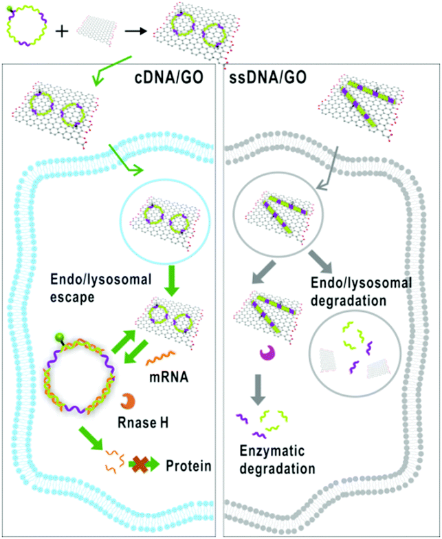
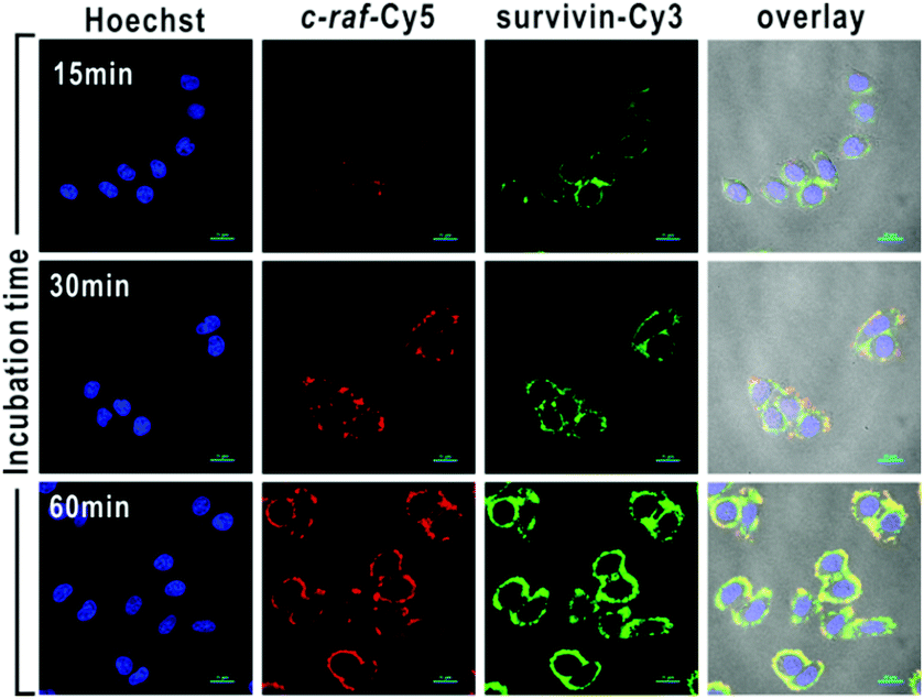
It turns out that rhenium-based compounds have been showing some promising anticancer activity as they’re stable, allow for real time imaging, structurally diverse, and have low off-site toxicity. The most commonly studied complexes are based around a Re(I) tricarbonyl core with the other three binding sites occupied by ligands of varying complexity. Researchers in the US and Australia developed a tricarbonyl Re isonitrile polypyridyl complex fac-[Re(CO)3(dmphen)(para-tolyl-isonitrile)]+, where dmphen = 2,9-dimethyl-1,10-phenanthro-line, called TRIP for short. TRIP showed promising cytotoxicity and can be imaged using confocal fluorescence microscopy, taking advantage of the emissive metal to ligand charge transfer (MLCT) state. The persistence of the emission indicates that the ligands remain bound to the Re even within cells. The complex’s cytotoxicity stems from its inducement of cells to accumulate misfolded proteins, resulting in apoptosis from the unfolded protein response (UPR). UPR induced cell death is relatively uncommon and led the researchers to find a method to characterize the speciation of TRIP in vitro. They used synchrotron X-ray fluorescence microscopy (XFM) to probe the cellular uptake and distribution of TRIP and an iodo-derivative I-TRIP by looking at elemental signals.

Figure 1. Chemical structures of TRIP and I-TRIP

I-TRIP is particularly well-suited to this type of study, as the iodine provides an additional spectroscopic handle on the isonitrile ligand absent in TRIP. Of course, the researchers had to confirm that I-TRIP possessed similar cytotoxicity and working mechanism to TRIP. Various assays and biological studies showed evidence of comparable cytotoxicity and mechanism, demonstrating that altering the substitution of the isonitrile ligand doesn’t significantly impact the bioactivity of the complex. With that settled, the experiments could move to the synchrotron to probe elemental distributions.

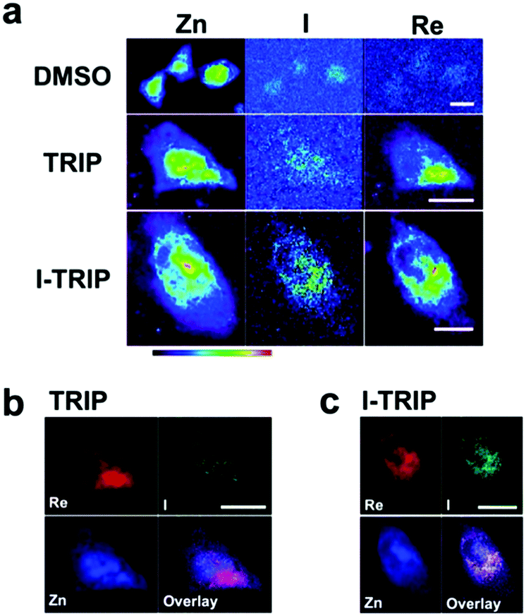
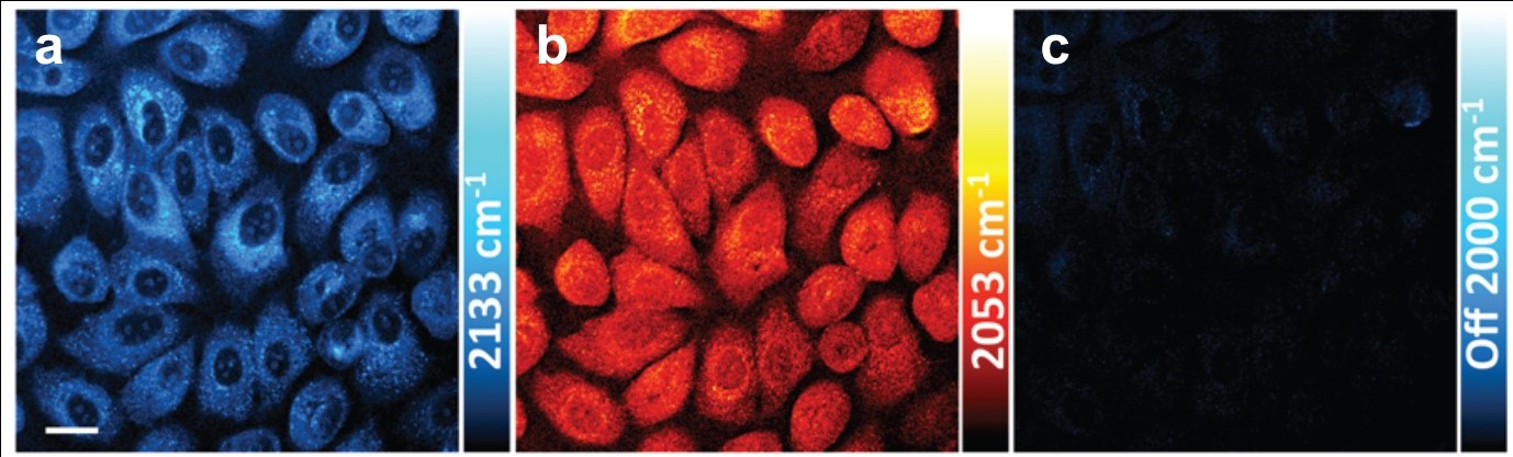
Figure 2. XFM elemental distribution maps of HeLa cervical cancer cells treated with either DMSO (control), TRIP, or I-TRIP.

Cells treated with both TRIP and I-TRIP show a clear Re signal, confirming that they can enter and persist in cells. Critically, the colocalization of the Re and I maps for I-TRIP samples indicate that the isonitrile ligand remains bound as a part of the Re complex inside the cells. This strongly suggests that the Re complex is intact while it induces cell death, adding to the developing mechanistic understanding of their activity. This work shows the utility of XRM as a technique to study the distribution of organometallic complexes in living cells. Additionally, the tunability and stable bioactivity of the Re complexes shows that they’re amenable to study by a wide range of techniques that will allow for further mechanistic probing.

To find out more, please read:

X-Ray fluorescence microscopy reveals that rhenium(I) tricarbonyl isonitrile complexes remain intact in vitro

Chilaluck C. Konkankit, James Lovett, Hugh H. Harris and Justin J. Wilson

Chem. Commun., 2020, 56, 6515-6518

About the blogger:

Dr. Beth Mundy is a recent PhD in chemistry from the Cossairt lab at the University of Washington in Seattle, Washington. Her research focused on developing new and better ways to synthesize nanomaterials for energy applications. She is often spotted knitting in seminars or with her nose in a good book. You can find her on Twitter at @BethMundySci.

Digg This
Reddit This
Stumble Now!
Share on Facebook
Bookmark this on Delicious
Share on LinkedIn
Bookmark this on Technorati
Post on Twitter
Google Buzz (aka. Google Reader)

Targeting the Powerhouse of the Cell to Fight Cancer

Everyone knows that cancer as a disease is awful, but the side effects of currently utilized chemotherapies have their own horrors. Research into natural products as therapies have found some promising compounds, but they face barriers to practical use in patients. One particular molecule, artesunate (ART), recently showed high potential for anticancer activity when in the presence of iron. Unfortunately, ART has major problems that limit its current applicability, including low solubility in water and high instability in biologically relevant conditions.

One approach to get around these issues is to encapsulate the drug (pun intended) in a nanoparticle-based carrier. A carrier with a hydrophobic interior and hydrophilic exterior can bring higher concentrations of drugs with low solubility into a cell and protect them from deleterious conditions in the body. An additional benefit is the relative ease of incorporating targeting ligands into the particles during synthesis. This allows the drugs to only interact with specific cells or, in this specific case, the mitochondria within cells.

Figure 1. Schematic of the nanoparticle synthesis process complete with targeting ligand molecules. The anticancer agent is activated in the presence of iron.

Researchers in China have prepared approximately 200 nm nanoparticle carriers for ART (Figure 1) using triphenyl phosphonium (TPP) as a mitochondrial targeting ligand. These nanoparticles remained stable in biologically relevant conditions for a week, sufficient for in-vitro studies. The studies showed significant decreases in cancer cell growth when the nanoparticles were used compared to the ART alone. The nanoparticles with TPP on the surface showed the highest efficacy, particularly when coupled with iron treatment to activate the ART.

Figure 2. Images of cells exposed to nanoparticles with (bottom) and without (top) a targeting ligand filled with different fluorescent dyes. The increased brightness corresponds to higher uptake of the nanoparticles by the cells.

To further investigate the cell uptake pathway of the nanoparticles, the researchers added fluorescent dye molecules to the inside of the particles. Once the cells took up and ruptured the nanoparticles, the dyes were released and became visible to the researchers (Figure 2). The fluorescence was twice as great in cells exposed to the nanoparticles treated with the TPP targeting ligand, showing its value for cell uptake. The researchers also used fluorescent dyes that react with reactive oxygen species (ROSs), as their generation is how ART kills cancer cells. The in-vitro studies showed an over three-fold increase in fluorescence from reactions with ROSs which, combined with data showing higher rates of cell death, supports the increased activity of ART when combined with this nanoparticle architecture.

To find out more please read:

A mitochondria targeting artesunate prodrug-loaded nanoparticle exerting anticancer activity via iron-mediated generation of the reactive oxygen species

Zhigang Chen, Xiaoxu Kang, Yixin Wu, Haihua Xiao, Xuzi Cai, Shihou Seng, Xuefeng Wang and Shiguo Chen

Chem. Commun., 2019, 55, 4781 – 4784.

About the blogger:


Beth Mundy is a PhD candidate in chemistry in the Cossairt lab at the University of Washington in Seattle, Washington. Her research focuses on developing new and better ways to synthesize nanomaterials for energy applications. She is often spotted knitting in seminars or with her nose in a good book. You can find her on Twitter at @BethMundySci.

Digg This
Reddit This
Stumble Now!
Share on Facebook
Bookmark this on Delicious
Share on LinkedIn
Bookmark this on Technorati
Post on Twitter
Google Buzz (aka. Google Reader)

An enzymatic Rube Goldberg machine: a bioluminescent switch for the detection of uracil DNA-glycosylase

A team of researchers from Shandong Normal University in Jinan, China, have developed a highly sensitive and label-free assay for the detection of uracil-DNA glycosylase, a DNA repair enzyme that removes uracil from DNA molecules. Uracil is an RNA base, and when uracil appears in DNA through deamination of cytosine or misincorporation during DNA synthesis, the error can have mutagenic consequences.

Diminished activity of uracil-DNA glycosylase has been linked to a number of disease states including human immunodeficiency and Bloom syndrome, an inherited disorder associated with an increased risk of cancer (among other symptoms). Developing sensitive methods to quantify uracil-DNA glycosylase would enable early diagnosis of such conditions and improve understanding of the DNA-repair machinery. As a proof-of-concept, the researchers showed that this method could quantify the enzyme in the cell lysate of HeLa cancer cells.

Their method reminds me of Rube Goldberg machines, which achieve a task via a series of connected, mechanical steps. Completion of one step triggers the start of another: such as a line of falling dominos hitting a marble that, in turn, rolls down a track. In this work the action of one enzyme returns a product that is the preferred substrate of another enzyme. At the risk of deviating slightly, one of the more spectacular examples of a Rube Goldberg machine is seen in the music video for OK GO’s ‘this too shall pass’, a single-take shoot of a warehouse sized machine, featuring rolling cars, swinging pianos, flowing water and rolling billiard balls, all to perform the task of (spoiler alert) blasting the band members in the face with coloured paint.

The label-free strategy for detecting uracil-DNA glycosylase results in a bioluminescent signal via tricyclic signal amplification

The strategy starts with the action of uracil-DNA glycosylase and ends with a bioluminescent signal via a cascade of enzymatic reactions

The authors’ strategy involves a series of sequential steps employing seven different enzymes and three nucleic acid probes. It begins with a double stranded DNA probe containing one rogue uracil base: the perfect bait for uracil-DNA glycosylase. The action of this enzyme and two others, in a process involving base excision, DNA backbone cleavage and the addition of thymine-rich sequences, produces a large quantity of single-stranded DNA molecules with long thymine-rich tails. These molecules hybridise with adenine-rich RNA probes to generate RNA-DNA duplexes. An enzyme digests the RNA portion, releasing adenosine monophosphate monomers, which are converted to adenosine triphosphate (ATP), a required energy input to activate firefly luciferase. Luciferase catalyses the oxidation of luciferin to form oxyluciferin, accompanied by a large bioluminescent signal. Thus, uracil-DNA glycosylase is detected with 1-2 orders of magnitude more sensitivity than state-of-the-art fluorescent and luminescent assays.

Unlike conventional Rube Goldberg machines, which are characterised by unnecessary complexity, in this ‘enzymatic Rube Goldberg machine’ each step has a specific purpose and serves to amplify the signal of the last. This is dubbed ‘tricyclic cascade signal amplification’ and it enables highly sensitive detection of the enzyme.

To find out more please read:

Label-free and high-throughput bioluminescence detection of uracil-DNA glycosylase in cancer cells through tricyclic cascade signal amplification

Yan Zhang, Qing-nan Li, Chen-chen Li, Chen-yang Zhang.
Chem. Commun., 2018, 54, 6991-6994
DOI: 10.1039/c8cc03769h

About the author

Zoë Hearne is a PhD candidate in chemistry at McGill University in Montréal, Canada, under the supervision of Professor Chao-Jun Li. She hails from Canberra, Australia, where she completed her undergraduate degree. Her current research focuses on transition metal catalysis to effect novel transformations, and out of the lab she is an enthusiastic chemistry tutor and science communicator.

Digg This
Reddit This
Stumble Now!
Share on Facebook
Bookmark this on Delicious
Share on LinkedIn
Bookmark this on Technorati
Post on Twitter
Google Buzz (aka. Google Reader)

Synthesis of Maleimide Dyes with Colourful Fluorescent Emissions

A group of researchers based at universities spanning the UK, China and Spain have synthesised a diverse library of fluorescent maleimide dyes with the aim of developing a structure-function relationship, relating substituent effects to the optical properties of such molecules. This work is not only important to build upon fundamental understanding of the fluorescence mechanism, but to develop knowledge that may be used to guide the synthesis of organic fluorophores which demand particular optical properties.

Organic fluorescent molecules are used as tools in many areas such as forensics, genetic analysis, DNA sequencing and biotechnology. Maleimides are commonly used as fluorescent labels for proteins, as they can couple with the thiol groups of cysteine residues. They are suited to this purpose as they are stable, easily functionalised, give strong emissions and do not perturb the protein structure to a large extent.

Molecules fluoresce upon absorption of UV or visible light, elevating an electron from a ground state orbital to a higher-energy orbital and resulting in a singlet excited state. Relaxation to the ground state occurs rapidly (~ 10 ns) with concomitant emission of a photon – this is what we observe as ‘fluorescence’. The emitted photon almost always has a longer wavelength than the absorbed light, a phenomenon known as the ‘Stokes shift’.


Structures of selected aminohalomaleimides and alkoxyhalomaleimides

Structures of selected amino-halo-maleimides and alkoxy-halo-maleimides synthesised for the study

With three dihalomaleimide precursers in hand (Cl, Br and I) the researchers assembled a library of amino-halo-maleimides, amino-alkoxy-maleimides, and amino-thio-maleimides. They varied the R groups bound to the N, O and S heteroatoms to include aliphatic, phenyl and benzyl examples.

The optical properties of the amino-halo-maleimides in diethyl ether were examined and the emission wavelengths were measured to be 461-487 nm, giving green-blue fluorescence. The fluorescence quantum yields, a measure of the quantity of emitted photons compared to absorbed photons and an indication of emission brightness, decreased with the electronegativity of the halide (Cl: 37%, Br: 30%, I: 8%). Like many fluorescent molecules in solution the compounds exhibited solvafluorochromism: when the polarity of the solvent alters the optical properties. In protic solvents (methanol and water) the fluorescence quantum yields decreased to below 1% and the emission wavelengths increased by 73-109 nm. On the other hand, in non-polar solvents (cyclohexane) the fluorescence quantum yield increased, up to 56% for the chloro analogue.

a) The UV and emission spectra of fluorescent maleimides bearing amino (2a-c) and alkoxy (3a, 3b) substituents. b) The quantum yields of selected amino and alkoxymaleimides. c) The solvafluorochromism effect for three aminomaleimides (2a-c) in increasingly non-polar solvents.

a) The UV and emission spectra of fluorescent maleimides bearing amino (2a-c) and alkoxy (3a, 3b) substituents. b) The quantum yields of selected amino and alkoxymaleimides. c) The solvafluorochromism effect for three maleimides (2a-c) in various solvents.

Compared to their amino-substituted counterparts, alkoxy-halo-maleimides have lower quantum yields (reduction of 20-25%), indicating the increased electron-donating capacity of the amine substituent is important for fluorescence intensity. Furthermore, the slight decrease in the emission wavelengths of alkoxy-halo-maleimides (458-465 nm) gives them blue fluorescent emissions. Amino-thio-maleimides, with greater electron-donating capacity than both the amino and alkoxy analogues, have increased emission wavelengths (526-564 nm), thus yellow fluorescent emissions.

This study is a worthwhile read for anyone who uses fluorescent molecules in their work, those wishing to understand a little more about the practical principles of fluorescence and all those curious minds who like to form their own hypotheses.

To find out more please read:

Rational design of substituted maleimide dyes with tunable fluorescence and solvafluorochromism

Yujie Xie, Jonathan T. Husband, Miquel Torrent-Sucarrat, Huan Yang, Weisheng Liu, Rachel K. O’Reilly.
Chem. Commun., 2018, 54, 3339 – 3342
DOI: 10.1039/C8CC00772A

About the author:

Zoë Hearne is a PhD candidate in chemistry at McGill University in Montréal, Canada, under the supervision of Professor Chao-Jun Li. She hails from Canberra, Australia, where she completed her undergraduate degree. Her current research focuses on transition metal catalysis to effect novel transformations, and out of the lab she is an enthusiastic chemistry tutor and science communicator.

Digg This
Reddit This
Stumble Now!
Share on Facebook
Bookmark this on Delicious
Share on LinkedIn
Bookmark this on Technorati
Post on Twitter
Google Buzz (aka. Google Reader)

Gold Rocks the Casbah

Researchers at the University of Texas have developed an inventive method to deliver molecules into the cell’s nucleus. Advances in gene therapy and the development of drugs that target DNA, the transcription machinery and other regulatory systems all rely on effective transport of molecules into the nucleus. Furthermore, achieving selective delivery of drugs reduces toxicity to non-target organs while maintaining the therapeutic effect.

Towards this aim, the authors delivered liposomes coated with clusters of gold nanoparticles into the cytoplasm. Laser irradiation of the cells heats the nanoparticles to high temperatures resulting in vapourisation of the water-based cytosol, and the transient formation of nanobubbles. The effect of this is an increase in the porosity of the nuclear envelope, enabling the transfer of various macromolecules from the cytoplasm into the nucleus. The authors describe this technique as ‘nanomechanical transduction’ because it is hypothesised that the mechanical effects brought on by the rapid growth and collapse (20 – 50 ns lifetimes) of the bubbles is responsible for the observed increase in porosity.

Local heating of gold nanoparticles and the subsequent formation of nanobubbles occurs due to ‘plasmon resonance’, whereby an electromagnetic field interacts with gold on the surface of the liposome and drives free-electron oscillation in resonance with the incident laser.

A diagram showing nanomechanical transduction. A gold-coated nanoparticle liposome enters the cell and, upon activation by a laser pulse, creates nanobubbles which causes mechanical disruptions in the cell and increased permeability of the nuclear membrane so molecules such as plasmids can enter.

A diagram showing nanomechanical transduction. A gold-coated liposome enters the cell and, upon activation by a laser pulse, creates nanobubbles and mechanical disruption within the cell, resulting in increased permeability of the nuclear membrane.

As a proof-of-concept the authors investigated whether nanomechanical transduction can improve the nuclear localisation of two different types of macromolecule: a dextran polymer labelled with a fluorescent dye, and a plasmid encoding the green fluorescent protein. In the first experiment, cells containing the dextran polymer were incubated with plasmonic liposomes and subjected to a near-infrared laser pulse. Up to 70% fluorescence intensity was observed in the nucleus compared to the cytoplasm, far exceeding the result from control experiments using electroporation to increase cell membrane permeability. In a similar way, nanomechanical transduction resulted in a 2.7 fold increase in the expression of the green-fluorescent protein compared to using electroporation, demonstrating efficient delivery of the plasmid into the nucleus.

The authors entitle their paper ‘rock the nucleus’ and, unintentional reference or not, I think a Casbah (one meaning is the central keep, or citadel, of a walled city) is a rather fitting analogy for the nucleus: the command post of the cell, and safeguard of genetic information. The authors of this work offer a sophisticated yet general method for molecules to breach the walls.

To find out more please read:

Rock the nucleus: significantly enhanced nuclear membrane permeability and gene transfection by plasmonic nanobubble induced nanomechanical transduction

Xiuying Li, Peiyuan Kang, Zhuo Chen, Sneha Lal, Li Zhang, Jeremiah J. Gassensmith and Zhenpeng Qin.
Chem. Commun., 2008, Advance Article
DOI: 10.1039/c7cc09613e

About the author:

Zoë Hearne is a PhD candidate in chemistry at McGill University in Montréal, Canada, under the supervision of Professor Chao-Jun Li. She hails from Canberra, Australia, where she completed her undergraduate degree. Her current research focuses on transition metal catalysis to effect novel transformations, and out of the lab she is an enthusiastic chemistry tutor and science communicator.

Digg This
Reddit This
Stumble Now!
Share on Facebook
Bookmark this on Delicious
Share on LinkedIn
Bookmark this on Technorati
Post on Twitter
Google Buzz (aka. Google Reader)

Shoot the Messenger: Circular DNA-Graphene Oxide Material Targets mRNA in Living Cells

Schematic of the circular DNA cDNA/GO graphene oxide platform fabrication for intracellular mRNA messenger RNA imaging and gene therapy.

Scheme showing how cDNA/GO enters the cell and interacts with mRNA

Did you know that the combined length of DNA in your body’s cells is a number so large that the only references I could find use cosmic distances as a reference? Try twice the diameter of the solar system, or the distance to the moon and back 1500 times. Despite the complexity and infinite detail encountered when studying science, it is often something so simple as size that gives us pause. How can DNA be both uncomprehendingly huge and tiny at the same time?

The major function of DNA is to encode proteins, a process which begins with the transcription of genes into single-stranded messenger RNA (mRNA) molecules. It is mRNA that is directly translated into the strands of amino acids which fold to form proteins.

A team of researchers at Fuzhou University in China have developed a graphene oxide and circularised single-stranded DNA (cDNA/GO) hybrid material capable of penetrating living cells and binding mRNA. The material’s utility is shown in two practical applications: mRNA imaging and nucleotide therapeutics. The authors chose the mRNA of survivin and c-raf kinase as targets, because the enzymes are involved in carcinogenesis, and the mRNA are overexpressed in cancer cells and can be used as biomarkers.

cDNA was chosen for its increased stability over linear single-stranded DNA, which is rapidly degraded in vivo by exonucleases. For mRNA imaging the material is designed with a fluorescent dye coupled to the cDNA. GO was chosen as a hydrophilic delivery scaffold capable of adsorbing cDNA and quenching the dye. When cDNA/GO was incubated with HeLa cells (a cancer cell strain) a time-dependent increase in fluorescence was observed in the cytoplasm. Fluorescence is restored when cDNA encounters the target and desorbs from the GO to form a duplex with the mRNA.

CLSM images acquired for HeLa cells treated with both survivin and c-raf targeted cDNA/GO for duplexed intracellular mRNA imaging

The mRNA of both survivin and c-raf kinase can be imaged in living cells with cDNA/GO.

The researchers also probed whether the material might serve as a therapeutic agent: if formation of the cDNA-mRNA duplex blocks translation it may reduce the load of c-raf kinase and survivin in the cell and influence cancer cell growth. Accordingly, the researchers found that when the HeLa cells were incubated with cDNA/GO, cell proliferation was inhibited in a dose-dependent manner.

This research contributes a robust design which can be applied to diverse mRNA targets because optimisable properties such as stability, bioavailability and selectivity are largely independent of the sequence of nucleotides.

To find out more please read:

Circular DNA: a stable probe for highly efficient mRNA imaging and gene therapy in living cells

Jingying Li, Jie Zhou, Tong Liu, Shan Chen, Juan Li and Huanghao Yang
Chem. Commun., 2018, Advance Article
DOI: 10.1039/C7CC08906F

About the author

Zoë Hearne is a PhD candidate in chemistry at McGill University in Montréal, Canada, under the supervision of Professor Chao-Jun Li. She hails from Canberra, Australia, where she completed her undergraduate degree. Her current research focuses on transition metal catalysis to effect novel transformations, and out of the lab she is an enthusiastic chemistry tutor and science communicator.

Digg This
Reddit This
Stumble Now!
Share on Facebook
Bookmark this on Delicious
Share on LinkedIn
Bookmark this on Technorati
Post on Twitter
Google Buzz (aka. Google Reader)

In-Vivo Visualization of Glucose Metabolism with a Two-Color Imaging Technique

A group of scientists from Columbia University in United States have developed a state-of-the-art probing technique that can simultaneously map glucose uptake and incorporation activities in living cells.

Glucose is a ubiquitous “fuel” for most living organisms. Its metabolism, including uptake and incorporation, is vital to sustain the energy consumption of living organisms. Visualization of glucose metabolism is of critical importance for clinical diagnostics and fundamental biological researches. However, current imaging techniques are destructive to living cells, poorly resolved or incapable of probing uptake and incorporation at the same time.

Now in ChemComm, Prof. Min Wei’s research team demonstrates a breakthrough based on a vibrational imaging technique coupled with stimulated Raman scattering microscopy. This technique utilizes two glucose analogues to present the glucose metabolism, the 13C-labelled 3-O-propargyl-D-glucose (3-OPG-13C3) for glucose uptake and the D7-glucose for glucose incorporation. Conventional Raman spectroscopy is unable to distinguish the aforementioned two species due to their overlapping Raman peaks. The authors addressed this challenge by labelling 3-OPG with 13C that exhibits a blue shifted Raman peak, thus separating it from the peak of D7-glucose. Decoupling of the two peaks allows in-vivo imaging and simultaneous observation of glucose uptake and incorporation in cells with sub-cellular resolution.

Figure 1 shows the two-color mapping images collected for human cancer cells, PC-3. The blue (panel a) and red (panel b) areas display the regions where glucose incorporation and uptake are taking place, respectively. The two images can be easily obtained by tuning the wavenumber of the incident light to match with corresponding Raman peak positions. Use of light with other wavenumbers results in the black image (panel c) containing virtually no colored regions, showing the excellent selectivity of the technique. Additionally, this approach differentiates between cancer cells and healthy cells by comparing the blue to red color intensity ratio.

This novel and versatile imaging technique is expected to serve as a useful tool in advanced bio-imaging and future cancer diagnostics.

Figure 1. Two-color mapping images of PC-3 cells highlighting the (a) glucose-incorporation regions (Raman peak: 2133 cm-1) and (b) glucose-uptake regions (Raman peak: 2053 cm-1). (c) An image collected with a wavenumber (2000 cm-1) that does not match with either of the Raman peaks. Scale bar: 20 µm.


To find out more please read:

Two-color Vibrational Imaging of Glucose Metabolism Using Stimulated Raman Scattering

Rong Long, Luyuan Zhang, Lingyan Shi, Yihui Shen, Fanghao Hu, Chen Zeng and Wei Min

Chem. Commun. 2018, DOI: 10.1039/C7CC08217G

About the blogger:

Tianyu Liu obtained his Ph.D. in Physical Chemistry from University of California, Santa Cruz in United States. He is passionate about scientific communication to introduce cutting-edge research to both the general public and scientists with diverse research expertise. He is a web blog writer for Chem. Commun. and Chem. Sci. More information about him can be found at http://liutianyuresearch.weebly.com/.

Digg This
Reddit This
Stumble Now!
Share on Facebook
Bookmark this on Delicious
Share on LinkedIn
Bookmark this on Technorati
Post on Twitter
Google Buzz (aka. Google Reader)

A warm welcome to Sandeep Verma, our new ChemComm Associate Editor

We are excited to welcome new Associate Editor Sandeep Verma (Indian Institute of Technology Kanpur) to the ChemComm Editorial Board

Professor Sandeep Verma

Sandeep Verma holds the positions of Professor of Chemistry and Shri Deva Raj Endowed Chair Professor at the Department of Chemistry, Indian Institute of Technology Kanpur, which he joined in 1997. His work has been recognized by numerous awards such as Swarnajayanti Fellowship (2005), Shanti Swarup Bhatnagar Prize in Chemical Sciences (2010), Department of Atomic Energy-Science Research Council Outstanding Investigator Award (2012), Ranbaxy Research Award in Pharmaceutical Sciences (2013), J C Bose National Fellowship (2013), Silver Medal, Chemical Research Society of India (2017), and National Prize for Research on Interfaces between Chemistry and Biology (2017).

His main research interests include peptide/protein assemblies for disease modeling, soft biomaterials, bioimaging, and surface chemistry of metal complexes. In particular, his group focuses on heterogeneous catalysts designed by developing polymeric templates based on nucleobase frameworks for application to interesting chemical and biochemical reactions. His work also focuses on the construction of architectures mimicking biological assemblies and metal-organic frameworks.

As a ChemComm, Sandeep will be handling submissions to the journal in the above areas. Why not submit your next paper to his Editorial Office?

Read Professor Verma’s recent articles published in ChemComm and its sister journals:

Chemical sensing in two dimensional porous covalent organic nanosheets
Gobinda Das, Bishnu P. Biswal, Sharath Kandambeth, V. Venkatesh, Gagandeep Kaur, Matthew Addicoat, Thomas Heine, Sandeep Verma and Rahul Banerjee
Chem. Sci., 2015, 6, 3931-3939

Organostannoxane-supported nucleobase arrays: synthesis and supramolecular structures of polymeric and molecular organotin complexes containing guanine, uracil and 2-aminopurine
Subrata Kundu, N. Nagapradeep, Balaram Mohapatra, Sourav Biswas, Sandeep Verma and Vadapalli Chandrasekharn
CrystEngComm, 2016, 18, 4807-4817

Assembly, postsynthetic modification and hepatocyte targeting by multiantennary, galactosylated soft structures
Anisha Thomas, Akansha Shukla, Sri Sivakumarb and Sandeep Verma
Chem. Commun., 2014, 50, 15752-15755

Digg This
Reddit This
Stumble Now!
Share on Facebook
Bookmark this on Delicious
Share on LinkedIn
Bookmark this on Technorati
Post on Twitter
Google Buzz (aka. Google Reader)

Elizabeth New: Winner of the 2017 ChemComm Emerging Investigator Lectureship

On behalf of the ChemComm Editorial Board, we are delighted to announce Elizabeth New from the University of Sydney, Australia, as the winner of the 2017 ChemComm Emerging Investigator Lectureship – congratulations, Liz!

Elizabeth New

Liz finished her BSc (Advanced, Hons 1 and Medal) and MSc in Chemistry at the University of Sydney before embarking on a PhD programme at Durham University, UK, working with Professor David Parker. After being awarded her PhD in Chemistry in January, 2010, she was a Royal Commission for the Exhibition of 1851 Postdoctoral Fellow at the University of California at Berkeley within the group of Professor Christopher Chang. She then returned to the University of Sydney as an ARC DECRA Fellow to start her independent research career in 2012, establishing herself at the cutting-edge of molecular imaging and developing novel chemical imaging tools to supplement existing imaging platforms.

She developed the first set of reversible sensors for cellular redox environment containing flavins as the sensing group, including the first examples of ratiometric reversible cytoplasmic sensing, reversible mitochondrial sensing, and ratiometric mitochondrial sensing. She has also developed the first fluorescent sensor for a platinum metabolite, enabling the unprecedented visualisation of cisplatin metabolism, and a subsequent sensor to study the metabolism of transplatin analogues. Her research group is one of the very few in the world to be investigating cobalt complexes as responsive magnetic resonance contrast agents, and she has developed new methods for ratiometric fluorescent sensing, as well as new strategies to control subcellular targeting. Her research excellence has been recognised by a number of awards, among them the NSW Early Career Researcher of the Year (2016) and the Asian Biological Inorganic Chemistry Early Career Researcher Award (2014).

Passionate about communicating science, she has spoken about her research to high school students (as the Royal Australian Chemical Institute (RACI) Nyholm Youth Lecturer, 2014-5, and the RACI Tasmanian Youth Lecturer, 2017), to the general public (as a NSW Young Tall Poppy Awardee, 2015), and to politicians and policy-makers (as elected executive member of the Australian Academy of Science’s Early-Mid Career Researcher Forum). She is currently a Senior Lecturer and Westpac Research Fellow in the School of Chemistry at the University of Sydney, where her group continues to focus on the development of molecular probes for the study of biological systems.

As part of the Lectureship, Elizabeth will present a lecture at three locations over the coming year, with at least one of these events taking place at an international conference, where she will be formally presented with her Emerging Investigator Lectureship certificate. Details of her lectures will be announced in due course – keep an eye on the blog for details.

Read these articles by Elizabeth New:

A cobalt(II) complex with unique paraSHIFT responses to anion
E. S. O’Neill, J. L. Kolanowski, P. D. Bonnitcha and E. J. New
Chem. Commun., 2017, 53, 3571-3574
DOI: 10.1039/C7CC00619E, Communication

On the outside looking in: redefining the role of analytical chemistry in the biosciences
Dominic J. Hare and Elizabeth J. New
Chem. Commun., 2016, 52, 8918-8934
DOI: 10.1039/C6CC00128A, Feature Article
From themed collection 2016 Emerging Investigators

Fluorescent sensing of monofunctional platinum species
Clara Shen, Benjamin D. W. Harris, Lucy J. Dawson, Kellie A. Charles, Trevor W. Hambley and Elizabeth J. New
Chem. Commun., 2015, 51, 6312-6314
DOI: 10.1039/C4CC08077G, Communication,  Open Access

Imaging metals in biology: balancing sensitivity, selectivity and spatial resolution
Dominic J. Hare, Elizabeth J. New, Martin D. de Jonge and Gawain McColl
Chem. Soc. Rev., 2015, 44, 5941-5958
DOI: 10.1039/C5CS00055F, Tutorial Review,  Open Access

A FRET-based ratiometric redox probe for detecting oxidative stress by confocal microscopy, FLIM and flow cytometry
Amandeep Kaur, Mohammad A. Haghighatbin, Conor F. Hogan and Elizabeth J. New
Chem. Commun., 2015, 51, 10510-10513
DOI: 10.1039/C5CC03394B, Communication

The annual ChemComm Emerging Investigator Lectureship recognises emerging scientists in the early stages of their independent academic career. Nominations for the 2018 Emerging Investigator Lectureship will open later in the year – keep an eye on the blog for details, and read more about our previous winners.

2016:    Ang Li from the Shanghai Institute of Organic Chemistry, China

2015:    Deanne D’Alessandro from the University of Sydney, Australia

    Yong Sheng Zhao from the Beijing National Laboratory for Molecular Sciences, China

2014:    Xinliang Feng from the Max Planck Institute for Polymer Research, Germany

2014:    Tomislav Friščić from McGill University, Canada

2014:    Simon M. Humphrey from the University of Texas at Austin, USA

2013:    Louise A. Berben from the University of California at Davis, USA

2013:    Marina Kuimova from Imperial College London, UK

2012:    Hiromitsu Maeda from Ritsumeikan University, Japan

2011:    Scott Dalgarno from Heriot-Watt University, Edinburgh, UK

Also of interest: You can read the 2016 ChemComm Emerging Investigators Issue which highlights research from outstanding up-and-coming scientists and watch out for our 2017 Emerging Investigators issue – coming very soon. You can also take a look at our previous Emerging Investigator issues in 2011, 2012, 2013, 2014 and 2015.

Digg This
Reddit This
Stumble Now!
Share on Facebook
Bookmark this on Delicious
Share on LinkedIn
Bookmark this on Technorati
Post on Twitter
Google Buzz (aka. Google Reader)

Professor Itaru Hamachi joins as Associate Editor

We are very pleased to welcome Professor Itaru Hamachi from Kyoto University as a new Associate Editor to the ChemComm team and look forward to working with him over the coming years.

Itaru is a chemical biologist with expertise in live-cell organic chemistry, chemical biology, bioorganic and bioinorganic chemistry, and supramolecular biomaterials. He is now accepting submissions to ChemComm in the area of chemical biology.

Itaru is looking froward to his new role:

I would like to encourage that new chemistry and chemical approaches between the chemistry and biology interfaces will appear in ChemComm, in order to decipher a lot of chemical-biology problems and also to create novel bio-inspired materials.

About Itaru:

Professor Itaru Hamachi was born in Fukuoka Prefecture, Japan in 1960 and received his Ph.D. in 1988 from Kyoto University under the guidance of the late Professor Iwao Tabushi. Immediately thereafter he joined Kyushu University, where he worked as an Assistant Professor for three years in the Kunitake laboratory before he became an Associate Professor in the Shinkai laboratory in 1992. In 2001, he became a Full Professor at IFOC, Kyushu University and moved to Kyoto University in 2005 where he currently heads the bioorganic chemistry wing.

Professor Hamachi has been a PRESTO investigator for 7 years (from 2000 to 2006) and a team leader of two CREST projects (from 2008 to 2013 and then from 2013 to 2018), which all are supported by the Japan Science and Technology (JST) Agency.

Submit your next top-notch, high-impact research now to Itaru Hamachi’s Editorial Office.


Itaru’s recent articles in ChemComm and other Royal Society of Chemistry journals include:*

Protein recognition using synthetic small-molecular binders toward optical protein sensing in vitro and in live cells
Ryou Kubota and Itaru Hamachi
Chem. Soc. Rev., 2015, 44, 4454-4471
DOI: 10.1039/C4CS00381K, Review Article

Ligand-directed dibromophenyl benzoate chemistry for rapid and selective acylation of intracellular natural proteins
Yousuke Takaoka, Yuki Nishikawa, Yuki Hashimoto, Kenta Sasaki and Itaru Hamachi
Chem. Sci., 2015, 6, 3217-3224
DOI: 10.1039/C5SC00190K, Edge Article
OA iconOpen Access

Hoechst tagging: a modular strategy to design synthetic fluorescent probes for live-cell nucleus imaging
Akinobu Nakamura, Kazumasa Takigawa, Yasutaka Kurishita, Keiko Kuwata, Manabu Ishida, Yasushi Shimoda, Itaru Hamachi and Shinya Tsukiji
Chem. Commun., 2014, 50, 6149-6152
DOI: 10.1039/C4CC01753F, Communication

*Access is free until 30/09/2016 through a registered RSC account.

Digg This
Reddit This
Stumble Now!
Share on Facebook
Bookmark this on Delicious
Share on LinkedIn
Bookmark this on Technorati
Post on Twitter
Google Buzz (aka. Google Reader)