Archive for the ‘Chemical Biology’ Category

Personal glucose sensors can be adapted to detect cancer too

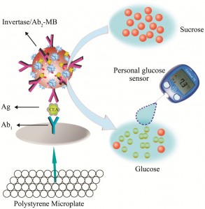
Personal glucose sensors (PGS) can be used to detect cancer, say Chinese scientists.

The team loaded magnetic beads with invertase (an enzyme that catalyses the hydrolysis of sucrose to glucose) and an antibody. The beads acted as a label for a lung cancer biomarker that had been captured on an antibody-coated ELISA plate. By monitoring the production of glucose from sucrose with a PGS, they could indirectly measure the amount of the biomarker down to the sub-nanogram per millilitre level.

Graphical Abstract


Link to journal article
Personal glucose sensor for point-of-care early cancer diagnosis
Jiao Su, Jin Xu, Ying Chen, Yun Xiang, Ruo Yuan and Yaqin Cha
Chem. Commun., 2012, Accepted Manuscript, DOI: 10.1039/C2CC32729E

Digg This
Reddit This
Stumble Now!
Share on Facebook
Bookmark this on Delicious
Share on LinkedIn
Bookmark this on Technorati
Post on Twitter
Google Buzz (aka. Google Reader)

Tomayto, tomato? Enantiospecific kinking of DNA

A few years ago, when I discovered what an intercalator was, I thought it would be a great name for a burger bar (probably best situated near a chemistry department). In scientific terms (and not catering as sadly the idea didn’t take off), intercalators have attracted a great deal of attention and are best known for their use in anticancer treatments.

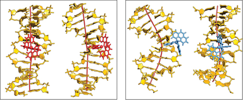
Chelate compounds of polycyclic heteroaromatics with transition metals can bind to DNA. The polycyclic moeities intercalate between the base pairs of the DNA, a little like the burger in a bun.  This can have a profound effect on the DNA’s structure, separating the base pairs and causing the helix to kink. However, the extent of this effect is dependent on parameters such as the ligand and which enantiomer of the instrinsically chiral compounds is involved.

A study by Anna Reymer and Bengt Nordén into the ruthenium compound, [Ru(phenanthroline)3]2+,  investigates its two enantiomers (Δ and Λ) and the effect each one has on binding specificity with DNA. Using molecular dynamics simulations, they demonstrated that the Δ-form induced a kink of 53° whilst the Λ-form produced a more typical bend of only 16°. They also reveal information about binding affinities of the compounds and how ‘deeply’ they can insert themselves into the base stack.

This interesting simulation is significant in the context of chiral recognition and evolutionary selection. It also gives further insight into the behaviour of DNA-protein interactions; an analogous kink as produced by Δ- [Ru(phenanthroline)3]2+ have been observed for several classes of operatory proteins.

To find out more download Reymer and Nordén’s communication.

Digg This
Reddit This
Stumble Now!
Share on Facebook
Bookmark this on Delicious
Share on LinkedIn
Bookmark this on Technorati
Post on Twitter
Google Buzz (aka. Google Reader)

Stopping bugs in their tracks

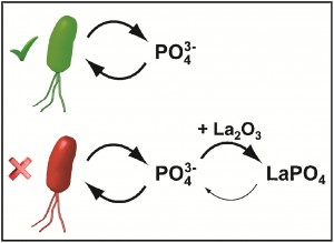
To prevent the spread of bugs, scientists in Switzerland have starved microbes of phosphate by using lanthanum oxide nanoparticles. The nanoparticles compete against the microbes for available phosphate and so the microbes can’t grow. The team says that the strategy is of particular technical interest as it can bypass toxic material release and provides an antimicrobial solution with small environmental footprint.

Phosphate starvation as an antimicrobial strategy

Link to journal article
Phosphate starvation as an antimicrobial strategy: the controllable toxicity of lanthanum oxide nanoparticles
L C Gerber et al
Chem. Commun., 2012, DOI: 10.1039/c2cc30903c

Digg This
Reddit This
Stumble Now!
Share on Facebook
Bookmark this on Delicious
Share on LinkedIn
Bookmark this on Technorati
Post on Twitter
Google Buzz (aka. Google Reader)

Cheap and non-toxic porphyrins for detecting cells deep within the body

Near-infrared fluorescent imaging can be used for rapid and sensitive detection of cells deep within the body. However, common NIR dyes and quantum dots are expensive and/or toxic.

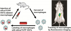
Porphyrins are economical fluorescent dyes, but their emission and excitation wavelengths are shorter than the NIR region and they can be toxic and poorly water-soluble. Scientists in Japan have made silica–porphyrin hybrid nanotubes that have no acute toxicity and higher water solubility compared to porphyrin. They used them to label macrophages, injected them into mice and tracked their distribution by fluorescence imaging with good results.

c2cc17444h

Link to journal article
Silica/Porphyrin Hybrid Nanotubes for In Vivo Cell Tracking by Near-Infrared Fluorescence Imaging
K Hayashi, M Nakamura and K Ishimura
Chem. Commun., 2012, DOI: 10.1039/c2cc17444h

Digg This
Reddit This
Stumble Now!
Share on Facebook
Bookmark this on Delicious
Share on LinkedIn
Bookmark this on Technorati
Post on Twitter
Google Buzz (aka. Google Reader)

Proteins perform (useful) tricks via DNA-based self assembly

Proteins are very useful molecules and when they work together, or assemble, they can display biocatalytic cascades, performing sequential multistep transformations of substrates. Scientists have tried to mimic nature for years, by creating artificial multi-enzyme complexes to replicate these biomolecules’ ability to catalyse reactions for use in biofuels, bioelectronics, bioproduction etc.

The arrangement of the proteins’ active sites relative to one another is intrinsic to the success of these reactions. One method of synthetically engineering these arrangements is through the use of DNA nanostructures.  DNA aptamers can be used as scaffolds to encourage the proteins’ assembly and even introduce other functional properties – imagine this as the bottom layer of a human pyramid in Cirque du Soleil.

However, the DNA scaffolds are reported to degrade and the protein assemblies decompose. (Now, imagine someone telling a really good joke to the bottom layer of the human pyramid and it all falling apart.)  The scaffolds and proteins are difficult to separate and this has limited the application of this strategy. Until now….

Masahiro Goto and co-workers have managed to arrange protein molecules (in this case, thrombin) on a DNA scaffold with the use of a DNA aptamers. With the addition of a chemical cross-linker, the neighbouring protein molecules were covalently cross-linked and retained their activity.

Programmable protein-protein conjugation via DNA-based self-assembly

Using a DNA template for thrombin binding aptamers, and hybridising that with three thrombin binding aptamers with sticky ends, they formed a comb-like structure with branched arms. The thrombin molecules bind with these arms and a chemical cross-linker encourages the neighbouring thrombins to cross-link. This has been intonated on the diagram with ‘holding hands’. (Told you they were inspired by Cirque du Soleil).

Using polyacrylamide gel electrophoresis (PAGE), the group elegantly illustrated their results, successfully demonstrating that DNA scaffolds can produce successful protein-protein conjugation. The group continue to develop and improve their work to overcome limitations in the size of conjugate proteins, efficiency and applications.

Find out more – download the ChemComm communication, free for 4 weeks.

Digg This
Reddit This
Stumble Now!
Share on Facebook
Bookmark this on Delicious
Share on LinkedIn
Bookmark this on Technorati
Post on Twitter
Google Buzz (aka. Google Reader)

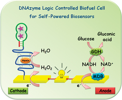
DNAzyme logic-controlled biofuel cell for self-powered biosensors

US scientists have made the first DNAzyme-controlled biofuel cell, an important step on the road to self-powered medical diagnostics, they claim. 

Biofuel cells (BFCs) use enzymes or microbes to oxidise fuels. Integrating them with logic-based biosensing systems provides a way to correlate the relationship between multiple target analytes in complex samples according to Boolean logic (which uses AND, OR and NOT operators) without the need for external power. 

Joseph Wang and colleagues at the University of California, San Diego, used DNAzyme-based biochemical signals to control the power output of a BFC. DNAzymes are biocatalytic nucleic acids that are widely used for biosensing but have never before been used to control a BFC. Wang demonstrated that the biosensor can determine the presence of a specific target in the absence of another related target in a single test.

DNAzyme logic controlled biofuel cell

While this is a proof-of-concept study, Wang says that self-powered diagnostics may be realised if pathologically relevant targets were applied to the BFC.

Read Wang’s ChemComm communication, free for a limited period.

Also of interest:
DNAzymes for sensing, nanobiotechnology and logic gate applications

Digg This
Reddit This
Stumble Now!
Share on Facebook
Bookmark this on Delicious
Share on LinkedIn
Bookmark this on Technorati
Post on Twitter
Google Buzz (aka. Google Reader)

Monooxygenase blends

When deciding which material to use for a particular application, it’s often necessary to weigh up the pros and cons of each candidate. Wouldn’t it be great if you could combine the best bits from each one to produce the ideal material?

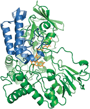
This is exactly what Marco Fraaije and his team from the University of Groningen did to create a new monooxygenase enzyme capable of performing Baeyer–Villiger oxidations with ultimate catalytic properties. For biocatalytic applications, enzymes need to be robust and should ideally be able to catalyse a broad range of substrates. Unfortunately, the only monooxygenase shown to be thermally stable (phenylacetone monooxygenase, PAMO) has narrow substrate specificity. On the other hand, there is cyclohexanone monooxygenase, CHMO, which can oxidise hundreds of substrates yet cannot be used at elevated temperatures.

The monooxygenase. Original PAMO structure is shown in green; the replaced sub-domain is shown in blue.

By replacing the substrate-binding domain of PAMO with that of CHMO or steroid monooxygenase (STMO), Fraaije was able to engineer an enzyme that was thermally robust and able to accept a wide range of substrates. Not only were the team able to combine the best of both worlds but in some cases, supersede them as they found when evaluating the conversions and enantiomeric excesses. It seems that the enzyme blend is not necessarily an average of the parent enzymes but can exhibit new properties.

Read the ChemComm article to find out more on how the team were able to improve the properties of Baeyer–Villiger monooxygenases.

Also of interest… ChemComm‘s Enzymes and Proteins web theme issue guest edited by Professors Nicholas Turner, Wilfred van der Donk and Herbert Waldmann.

Digg This
Reddit This
Stumble Now!
Share on Facebook
Bookmark this on Delicious
Share on LinkedIn
Bookmark this on Technorati
Post on Twitter
Google Buzz (aka. Google Reader)

Look into my MRI: Non-invasive detection of melanin formation

Magnetic Resonance Imaging (MRI) is used as a medical tool to image soft tissue. It can give insights into the physical state of brain, identify sports injuries and diagnose cancer without ionising radiation. Its main drawback is the low sensitivity of the contrast enhancing probes it uses. These are the paramagnetic metal complexes, often containing gadolinium or manganese, that distinguish between healthy and damaged tissue by altering the relaxation times of the water protons in the body. The challenge is to increase the concentration of the contrast reagents in the tissues.

One solution is to use nanocarriers to deliver a high concentration of the contrast agent to the specific site of interest. Silvio Aime and his team recently used the nanocarrier, apoferritin, to transport solid MnOOH, which was subsequently reduced to paramagnetic Mn2+. Their recent ChemComm details how they have now taken this one step further to find a way of generating the Mn2+ contrast agent using a naturally occurring reductant. The result: a probe for melanin.

Melanin is produced from the successive oxidation of tyrosine – a process that is up-regulated in malignant melanoma. Aime exploited this oxidation process by introducing MnOOH-loaded apoferritin into melanoma cells – the MnOOH was reduced, generating the contrast agent and enabling the cells to be successfully imaged by MRI.

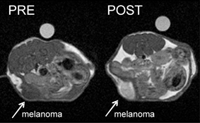
in vivo MRI images of tumour-bearing mice before and after administration of the contrast agent loaded nanocarrier

The team tested the sensor on melanogenic cells (melanin-forming cells) against non-melanogenic cells. They demonstrated that the signal generated belonged only to the melanin-producing cells which had internalised the Mn(III)OOH–apoferriton payloads. Animal studies revealed enhanced signal intensity in melanogenic tumours.

This interesting research has the potential to provide non-invasive, early diagnoses of skin cancers and evaluate the development of tumours, critical for saving people’s lives. The in vivo sensor may also be used to monitor other processes involving massive oxidative processes. The work of Silvio Aime is one to watch.

To find out more, download the ChemComm article today…

Posted on behalf of Sarah Brown, web science writer for ChemComm.

Digg This
Reddit This
Stumble Now!
Share on Facebook
Bookmark this on Delicious
Share on LinkedIn
Bookmark this on Technorati
Post on Twitter
Google Buzz (aka. Google Reader)

Simple detection of RNA depurination by RIPs

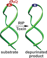
A class of protein toxins exists with an extremely apt acronym, RIP. This stands for Ribosome Inactivating Proteins but may as well stand for “Rest in Peace” when applied to cellular RNA. These proteins are known to attack the link between a purine base (adenine or guanosine) and its sugar causing depurination. This results in the formation of abasic sites (bases that are neither pyrimidines or purines) which in turn, causes the ribosome containing the sequence to have a lower affinity to elongation factors that are crucial for protein synthesis, ultimately leading to cell death.

Methods exist to detect RIPs such as ELISAs and antibody-based immunoassays or by monitoring the specific depurination activity through fluorescence, radiolabelling and immunoaffinity chromatography, amongst others. These techniques require sophisticated or elaborate set-ups, limiting the potential for high throughput (HTP) screening in a bid to discover potential inhibitors of these destructive toxins.

Seergazhi Srivatsan and co-workers have produced a label-free fluorescence hybridisation assay to detect the depurination activity of saporin, a RIP toxin, using a fluorescence ligand that specifically binds to the cytosine opposite an abasic site. If depurination takes place, then the ligand can bind and its fluorescence intensity will be quenched. This technique allows for a plethora of information to be obtained about the depurination activity of saporin as well as many other RIP toxins. This opens the door to using high throughput screening to find inhibitors of such toxins.

Download the article to read more…

Also of interest: Overcoming obstacles in labelling RNA

Posted on behalf of Sarah Brown, web science writer for ChemComm.

Digg This
Reddit This
Stumble Now!
Share on Facebook
Bookmark this on Delicious
Share on LinkedIn
Bookmark this on Technorati
Post on Twitter
Google Buzz (aka. Google Reader)

Hybrid biofuel cell

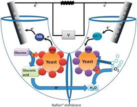
Scientists from Israel have designed a biofuel cell that combines the advantages of both enzymatic and microbial fuel cells.

Biofuel cells use redox enzymes to convert chemical energy into electricity. These cells can be divided into two categories: enzymatic fuel cells which require the enzymes to be purified and microbial fuel cells which make use of an entire microorganism. There are pros and cons to both strategies – enzymatic fuel cells tend to have increased power output whilst microbial fuel cells enable full oxidation of a wider range of fuels.

Yet now, Lital Alfonta and co-workers demonstrate that by designing a hybrid cell, one can have the best of both worlds. The team have modified yeast to display redox enzymes on their surface and then introduced this into both the anode and cathode compartments. This approach removes the need to purify the enzymes and enables regeneration of both fuel compartments.

To find out more about Alfonta’s biofuel cell device, read the ChemComm article today.

Digg This
Reddit This
Stumble Now!
Share on Facebook
Bookmark this on Delicious
Share on LinkedIn
Bookmark this on Technorati
Post on Twitter
Google Buzz (aka. Google Reader)