Archive for the ‘Hot Articles’ Category

Looking Inside Nanocomposites with Tomography

As a nanocrystal chemist, it pains me to say that sometimes nanoparticles aren’t enough. One strategy for engineering materials with complex functionalities is to embed nanoparticles into a larger host matrix structure. This has been widely studied for polymer-nanoparticle assemblies, but challenges abound for inorganic host matrices. Difficulties stem from problems controlling the microstructure of the host, as nanoparticles tend to accumulate at the borders of individual crystals in polycrystalline materials. Even when higher quality inorganic materials are made in the presence of nanocrystals loadings are below 1 wt%. A possible route to address these issues is by combining inorganic matrices with polymer-functionalized nanoparticles. The polymers can also form into vesicle structures that contain the nanoparticles which are larger and more compatible with characterization techniques.

One of those techniques is cryo-ptychographic X-ray computed tomography (cryo-PXCT), a fascinating and literally cool characterization method to image the internal structure of crystals. This is a variation on imaging techniques used heavily in medicine archeology to non-destructively visualize the interior of humans or artifacts. Cryo-PXCT cools the sample to -180 oC and has spatial resolution on the order of 50-70 nm. The researchers synthesized polymer vesicles and worms of approximately 232 nm in diameter and over 1 micron in length, respectively. The nanocomposites were made via an ammonia diffusion method with a solution of calcium chloride containing the polymer nano-structure exposed to gaseous ammonia and carbon dioxide to form CaCO3 crystals with nano-structure occlusions. The morphology of the nanocomposite crystal altered based on the type of occlusion – the vesicle/calcite combination retained a traditional calcite rhombohedral structure, while the worm/calcite composite crystals featured several rounded sides, an elongated shape, and only three flat faces. These composites possessed 15 – 25 wt% occlusions, significantly higher than prior work with pure nanoparticle incorporation.

Figure 1. SEM images of vessicle/calcite (left) and worm/calcite (right) nanocomposite single crystals.

Once prepared, the researchers examined the crystals by cryo-PXCT to determine the locations of the occlusions within the composites. In the vesicle/calcite composite the vesicles are non-uniformly distributed, with several layers of vesicle density, starting with a vesicle poor core, followed by a vesicle rich region, surrounded by another vesicle poor layer, with a slight vesicle enrichment near the surface. On average the vesicles are 300 nm apart and they maintain their shape, with the larger vesicles preferentially occluding in regions of higher occlusion densities.

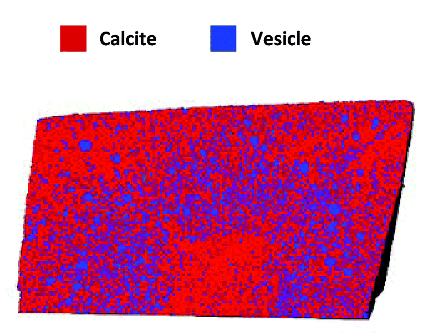
Figure 2. Rendering of slice through the vesicle/calcite nanocomposite colored to show both components.

The worm/calcite composite crystals show a very different distribution of occlusions, with an hourglass of low density in the center of the crystal, surrounded by a worm rich zone, and an exterior worm poor layer. These zoning effects are likely determined by the interactions between the polymers and the growing crystal surfaces or the calcium cations in the solution. Cryo-PXCT offers a fascinating way to probe the internal structure of novel multicomponent crystals in three dimensions with nanoscale resolution, providing valuable information to eventually help determine structure-function relationships.

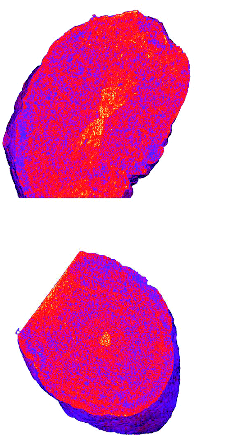
Figure 3. Tomographs of worm/calcite nanocomposites showing the localization of worms in an hourglass shape in the center of the crystal.

To find out more, please read:

Ptychographic X-ray tomography reveals additive zoning in nanocomposite single crystals

Johannes Ihli, Mark A. Levenstein, Yi-Yeoun Kim, Klaus Wakonig, Yin Ning, Aikaterini Tatani, Alexander N. Kulak, David C. Green, Mirko Holler, Steven P. Armes and Fiona C. Meldrum

Chem. Sci., 2020, 11, 355-363.

About the blogger:

Beth Mundy is a PhD candidate in chemistry in the Cossairt lab at the University of Washington in Seattle, Washington. Her research focuses on developing new and better ways to synthesize nanomaterials for energy applications. She is often spotted knitting in seminars or with her nose in a good book. You can find her on Twitter at @BethMundySci.

Digg This
Reddit This
Stumble Now!
Share on Facebook
Bookmark this on Delicious
Share on LinkedIn
Bookmark this on Technorati
Post on Twitter
Google Buzz (aka. Google Reader)

Molecular Switches from DNA

The idea of using DNA-based devices to create highly specific sensors, diagnostic tools, and therapeutics has inspired widespread research. DNA molecular switches, a basic class of DNA nanostructures, turn some process on and off depending on whether a substrate binds to the switch. They are typically coupled with some form of signal amplification process to increase sensitivity and should theoretically provide enhanced signal-to-noise. Unfortunately, current amplification procedures have a significant amount of background reactions, called leakage, that limit their current utility. One approach to design better switches is to find ways to observe single molecule dynamics. Combining transient binding of complimentary oligonucleotides with high resolution fluorescence microscopy allows for the development of highly sensitive molecular switches without leakage problems.

To do this, researchers in China developed a series of three-way junction DNA-based molecular switches. These TWJs possess a recognition domain, which interacts with the target of interest and induces a structural change in the TWJ, and a transduction domain, which then becomes accessible and binds to a fluorescent molecule (Figure 1).

Figure 1. General scheme of three-way junction molecular switch with two domains noted.

Experimentally, the TWJs were captured on the imaging surface and only fluorophores bound to a TWJ would remain in place long enough for signal to be acquired by the camera with a 500 ms integration time. The researchers found that shorter transduction domains with 5 or fewer base pairs were not stable enough to allow the fluorescent probes access in the absence of a bound target. The next generation of TWJs feature a hybridization probe to allow the switch to recognize specific DNA inputs. In the presence of inputs, the researchers observed transient binding behavior of the fluorophore, whereas they observed only nonspecific binding in the absence of inputs. The ability to differentiate between non-specific and transient binding in the single-molecule system gives a detection limit of 10 fM without concerns about leakage.

Building on this work, the researchers utilized the same general framework and substituted aptamer sequences for the hybridization probe in the recognition domain. They utilized split aptamer fragments that only draw together when bound to a target molecule. This motif was tested on ATP, a small molecule, and thrombin, a protein. These aptamer-coupled TWJs exhibited sensitivity to concentrations as low as 20 – 50 pm with high sensitivity (Figure 2). In the presence of molecular analogs to ATP or thrombin, the signal level showed no significant difference from that of a blank.

Figure 2. A) Split aptamer-based molecular switch schematic. B) Single-molecule fluorescence-time trajectory data in the presence (top) or absence (bottom) of targets. C) and D) Linear relationship between thrombin concentration and signal and specificity when compared to analogs. E) and F) Linear relationship between ATP concentration and signal and specificity when compared to analogs.

Another advantage of this system is its ability to provide information on the binding affinity of substrates, as it should impact the kinetics of the fluorescent probes. The dwell times of the fluorescence on and off states demonstrated exponential trends with changing input concentrations and could be fit to extract time constants. These time constants can then be used to derive the kinetics parameters and binding affinities of the target species. The general stability of the molecular switch framework allows for studying these types of interactions in a range of pH and salinity conditions, useful for mimicking different environments relevant to future applications. This provides a platform for studying the fundamental interactions that will allow DNA-based nanotechnology to move forward.

To find out more, please read:

Single-molecule dynamic DNA junctions for engineering robust molecular switches

Shuang Cai, Yingnan Deng, Shengnan Fu, Junjie Li, Changyuan Yu and Xin Su

Chem. Sci., 2019, 10, 9922-9927.

About the blogger:

Beth Mundy is a PhD candidate in chemistry in the Cossairt lab at the University of Washington in Seattle, Washington. Her research focuses on developing new and better ways to synthesize nanomaterials for energy applications. She is often spotted knitting in seminars or with her nose in a good book. You can find her on Twitter at @BethMundySci.

Digg This
Reddit This
Stumble Now!
Share on Facebook
Bookmark this on Delicious
Share on LinkedIn
Bookmark this on Technorati
Post on Twitter
Google Buzz (aka. Google Reader)

Metallohelices to kill microbes

I feel pretty safe saying that the development of effective antimicrobial drugs (looking at you penicillin!) was one of the most significant pharmacological events of recent history. Unfortunately, the widespread and often indiscriminate use of antibiotics has created an environment where bacteria with evolved drug resistances, colloquially known as “superbugs,” pose a serious threat to global health. While development of new small-molecule antimicrobial drugs is still ongoing, scientists are exploring alternative approaches as well. Of interest are antimicrobial peptides, found in plants and animals as a part of native immune systems. The most common are cationic antimicrobial peptides (CAMPs) comprised of 10-50 amino acids that possess both cationic and hydrophobic subunits and an overall positive charge. This allows them to strongly electrostatically interact with negatively charged bacterial cell membranes without disrupting neutral animal cells. Unfortunately, CAMPs have proved challenging to commercialize with generally unfavorable activities and issues with large-scale manufacture.

Figure 1. General synthetic scheme, structure, and key for components of metallohelices.

Researchers in the UK and Czech Republic recently developed a range of cationic metallohelices (Figure 1) that demonstrate structure-dependent activity against both Gram-positive and -negative bacteria. The diamine ligands form cleanly in the presence of [15]-crown-[5] and upon combination with 2-pyridinecarboxaldehyde and a metal salt self-assemble into the iron or zinc metallohelices. The metallohelices consist of a single enantiomer, as the ligands are optically pure, as determined by NMR and single-crystal X-ray diffraction when possible. Altering the aryl linker unit in the ligand the overall size and shape of the metallohelix in both zinc and iron derivatives. The iron metallohelices are water compatible, with lifetimes exceeding 10 days even in highly acidic conditions, and thus suitable for antimicrobial activity screening.

The researchers used ­in vitro studies to find minimum inhibitory concentrations (MICs), the lowest concentration necessary to see bacterial growth inhibition. While the metallohelices demonstrate the highest activity towards Gram-positive bacteria, some showed lethal effects on Gram-negative E. coli in 20-40 minutes. The 5b helices with a para-benzene bridging group acted most selectively on E. coli with the Λ-5b enantiomer acting as the champion compound and selected for further mechanistic study. The researchers exposed a notorious E. coli strain to inhibitory levels of Λ-5b in an attempt to isolate resistant mutants. The 17 isolates showed only slight tolerance increases rather than true resistance and could be classified into 4 genetic sub-types. Two of the sub-types developed mutations that altered the biophysical properties of their outer membrane, a third lost the ability to produce the vitamin B12 transporter protein, and the fourth lost the pO157 virulence plasmid (which makes this particularly E. coli strain particularly unpleasant). Taken in concert, these 4 sub-types show that tolerance can be derived from disrupting the ability of Λ-5b to interact with and cross the cellular membrane.

Figure 2. Fluorescence images of cells treated with Λ-5b coupled to a fluorescent probe, with arrows pointing to the localization of Λ-5b.

Given data suggesting the ability of Λ-5b to cross the cellular membrane, despite the relatively large of the metallohelices, the researchers used fluorescence microscopy to probe Λ-5b localization by coupling Λ-5b with a fluorescent label. The labeled metallohelix preferentially localized to regions in growing cells that contain anionic phospholipids (Figure 2). This indicates that Λ-5b can cross the cellular membrane and acts internally to the cell rather than simply acting via electrostatic interaction that disrupts the membrane. Overall, this work provides an exciting approach to developing novel anti-microbial drugs that mimic CAMPs with higher stability, activity, and easier synthesis.

To find out more please read:

Metallohelices that kill Gram-negative pathogens using intracellular antimicrobial peptide pathways

Daniel H. Simpson, Alexia Hapeshi, Nicola J. Rogers, Viktor Brabec, Guy J. Clarkson, David J. Fox, Ondrej Hrabina, Gemma L. Kay, Andrew K. King, Jaroslav Malina, Andrew D. Millard, John Moat, David I. Roper, Hualong Song, Nicholas R. Waterfield and Peter Scott

Chem. Sci., 2019,10, 8547-8557

About the blogger:

Beth Mundy is a PhD candidate in chemistry in the Cossairt lab at the University of Washington in Seattle, Washington. Her research focuses on developing new and better ways to synthesize nanomaterials for energy applications. She is often spotted knitting in seminars or with her nose in a good book. You can find her on Twitter at @BethMundySci.

Digg This
Reddit This
Stumble Now!
Share on Facebook
Bookmark this on Delicious
Share on LinkedIn
Bookmark this on Technorati
Post on Twitter
Google Buzz (aka. Google Reader)

Driving Molecular Motors with Visible Light

While nanobots are still a thing of science fiction, the development of the fundamental scientific concepts that could lead to highly complex molecular-scale machines has been an active area of research for over 2 decades. Molecular motors rely on isomerization through metastable intermediate states that leads to unidirectional rotation via a rotor portion of the molecule. Initial work utilized molecules that convert high-energy UV-light into this directional motion. However, these systems are limited in their future utility, particularly in biological applications, if they need UV-light for activation. Recent work focuses on driving molecular motors with visible light via a range of approaches, including altering the absorbance profile of the motor molecule itself. Unfortunately, changing the electronics of the molecule has previously substantially decreased the quantum yield while resulting in only slight red shifts.

Researchers in The Netherlands recently developed 2nd generation molecular motors featuring a mixture of electron-donating and electron-withdrawing groups that exhibit substantial red-shifts. Density functional theory (DFT) calculations predicted that adding cyano- and methoxy- groups to opposite halves of the motor would shift the absorbance past 410 nm. The new molecules were synthesized via a general procedure shown in Figure 1, with yields ranging from 3 – 10%.

Figure 1. General synthetic route to cyano- and methoxy- substituted molecular motors and photoisomerization reaction that results in unidirectional motion.

The addition of the cyano- and methoxy- groups shifted the absorbance maxima corresponding to the HOMO-LUMO transitions to from 422 – 453 nm, an increase of over 60 nm from the parent molecule. When irradiated, the molecules transitioned from their stable ground state to the metastable isomers. These were characterized by UV-Vis spectroscopy, with the emergence of further red-shifted features (Figure 2), and 1H NMR.  The 1H NMR spectra were obtained with in-situ irradiation (which sounds like a sweet experimental setup) at various wavelengths and the ratios of several specific protons on the rotor. The derivative 2 with a methoxy- group on the rotor and cyano- groups on the stator demonstrated activity with irradiation at wavelengths up to 530 nm.

Figure 2. UV-Vis absorption spectra of stable (solid) and metastable (dashed) isomers of the molecular motors.

The energy of activation of the rotation for all three derivatives was determined by Eyring analysis and corroborated by DFT calculations. All were around 90 kJ/mol, with 2 requiring the most energy to elongate the central alkene bond and isomerize. The quantum yields of the motors for the forward reaction range from 5.8 – 11.5%, comparable to state-of-the-art UV absorbing motors. The quantum yields for the back reactions were calculated to be significantly lower than those for the forward reaction, which corresponds to the excess of metastable isomers observed under active irradiation. These motors also exhibit high photostability, with no significant change in the ground state absorbance after irradiation and cycling. This is promising for smart materials applications where stability is crucial. This work pushes forward the design of molecular motor systems that utilize visible rather than UV light.

To find out more please read:

Photoefficient 2nd generation molecular motors responsive to visible light

Lukas Pfeifer, Maximilian Scherübl, Maximilian Fellert, Wojciech Danowski, Jinling Cheng, Jasper Pol and Ben L. Feringa

Chem. Sci., 2019, 10, 8768-8773.

About the blogger:

Beth Mundy is a PhD candidate in chemistry in the Cossairt lab at the University of Washington in Seattle, Washington. Her research focuses on developing new and better ways to synthesize nanomaterials for energy applications. She is often spotted knitting in seminars or with her nose in a good book. You can find her on Twitter at @BethMundySci.

Digg This
Reddit This
Stumble Now!
Share on Facebook
Bookmark this on Delicious
Share on LinkedIn
Bookmark this on Technorati
Post on Twitter
Google Buzz (aka. Google Reader)

Can Al3+ Indeed Intercalate Layered Metal Oxides in Aqueous Electrolytes?

Intercalation of multivalent ions, e.g., Al3+, has received increasing attention as an energy-boosting strategy for rechargeable batteries. The charge storage process of the wide-spread Li-ion batteries relies on Li+ intercalating electrodes with layered structures. Multivalent-ion batteries could accommodate more charges than Li-ion batteries because their ions carry more charge than Li+. This concept, however, has now been challenged by a research team led by BenoÎt Limoges and Véronique Balland of Université de Paris, France. Their mechanistic investigation, published in Chemical Science, revealed that Al3+ ions were unable to intercalate electrodes in aqueous electrolytes.

The authors selected TiO2 arrays as the object of their study. These ~1 µm-high arrays were grown using the glancing angle deposition technique. By applying negative potentials to the TiO2 arrays, cations such as Al3+ could diffuse through the inter-array slits and interact with TiO2 (Figure 1).

Figure 1. Structure of the TiO2 arrays. (left) Scanning electron micrographs and (right) a cartoon illustrating ion diffusion pathways.

Electrochemistry tests elucidated that the charge-storage process of TiO2 in an Al3+-containing aqueous electrolyte correlated to proton intercalation. This conclusion was mainly based on the nearly identical cyclic voltammograms (Figure 2, top) and capacity vs. potential curves (Figure 2, bottom) of the TiO2 arrays in both AlCl3 and acetic acid aqueous electrolytes. Since acetic acid solution had no Al3+, the observed charge-storage activity could not be attributed to Al3+ intercalation. Instead, the authors argued that protons dissociated either from hydrated Al3+ cations, [Al(H2O)6]3+ or acetic acid must intercalate TiO2 and result in the observed charge-storage capacities.

Figure 2. (top) Cyclic voltammograms and (bottom) capacity vs. potential curves of TiO2 in (left) Al3+-containing and (right) acetic acid aqueous electrolytes. The electrolytes are 0.3 M KCl with different concentrations of AlCl3 or acetic acid: 0 M (black), 25 mM (blue), 50 mM (purple), 100 mM (magenta), and 250 mM (red).

The authors believe that the misconception of Al3+ intercalation is due to the overlooking of Al3+ hydration, which is inevitable when Al3+ is present in aqueous electrolytes. Removing the water shell (a prerequisite for ion intercalation) is energy costly for Al3+ because of the strong binding force between water molecules and Al3+. Additionally, even if Al3+ ions intercalate TiO2, their movement is strongly hindered by Coulombic interactions within the TiO2 lattice. The immobilized intercalated ions would then block other ions from entering the TiO2 lattice. Together, both factors prevent Al3+ from intercalating into TiO2.

In summary, this work demonstrates that the charge-storage capacity of TiO2 in Al3+-containing aqueous electrolytes is most probably due to proton intercalation. This conclusion also applies to other multivalent cations, including Zn2+ and Mn2+, as shown in this work.


To find out more, please read:

On the Unsuspected Role of Multivalent Metal Ions on the Charge Storage of A Metal Oxide Electrode in Mild Aqueous Electrolytes

Yee-Seul Kim, Kenneth D. Harris, BenoÎt Limoges, and Véronique Balland

Chem. Sci., 2019, doi: 10.1039/c9sc02397f

Tianyu Liu acknowledges Zachary L. Croft of Virginia Tech, the U.S., for his constructive comments on this post.

About the blogger:

Tianyu Liu obtained his Ph.D. (2017) in Chemistry from University of California, Santa Cruz in the United States. He is passionate about the communication of scientific endeavors and cutting-edge research to both the general public and other scientists with diverse research expertise. He is a blog writer for Chem. Commun. and Chem. Sci. More information about him can be found at http://liutianyuresearch.weebly.com/.

Digg This
Reddit This
Stumble Now!
Share on Facebook
Bookmark this on Delicious
Share on LinkedIn
Bookmark this on Technorati
Post on Twitter
Google Buzz (aka. Google Reader)

Breaking C-H bonds with water, iron, and electricity

As we transition to an energy future composed primarily of intermittent, renewable technologies, finding ways to store the excess generated charges will be critical. While the general public typically thinks of batteries, another area of massive interest is storing the electrons in a chemical bond. This can be accomplished by creating electrocatalysts that can couple an electric current with chemicals like water and oxygen to generate stable new bonds in molecules for later use as fuels. These renewably generated fuels would then be available whenever needed, like at night. However, finding efficient electrocatalysts composed of earth-abundant materials has proven challenging. Many researchers have turned to nature for inspiration by designing molecules that mimic the active sites of enzymes.

Researchers in the United States used this approach, focusing on creating high-valent iron-oxo species, which others previously identified as the key catalytic intermediates in multiple enzymatic reactions. These types of species have traditionally been synthesized by reacting a reduced iron complex with an oxygen transfer reagent, but the researchers developed a system to generate highly reactive species using electricity as the reaction driving force and water as the oxygen source. The studied catalyst is a commercially available iron(III)-aquo complex with a tetraamido macrocyclic ligand (TAML) as the ancillary ligand (Figure 1B).

Figure 1. A. Cyclic voltammagram of (TAML)Fe in acetonitrile. B. Structure of (TAML)Fe and cyclic voltammagram showing increased current upon the addition of ethylbenzene.

When analyzed by cyclic voltammetry, an electrochemical technique where you cycle the voltage between set points and measure the current output, the (TAML)Fe shows two redox events at around 650 and 1250 mV that the researchers attributed to generating the FeIV-OH and FeV(O) species (Figure 1). Addition of ethylbenzene, which should react with the FeV species, increased the current at voltages of 1250 mV and higher, indicating (TAML)Fe turnover. However, isolating the Fev(O) species proved challenging as it reacts rapidly with the (TAML)Fe to form an FeIV dimer. This also limits the efficiency of the overall system by decreasing the amount of the most reactive species in solution.

Figure 2. A, B. Products generated by oxidation of various substrates screened with (TAML)Fe for electrocatalysis with isolated yield and calculated conversion in parenthesis. C. Substrates that did not react with the (TAML)Fe complex.

Both the FeIV dimer and FeV(O) species proved capable of oxidizing C-H bonds in ethylbenzene, but the FeV(O) is much more reactive and increases the oxidation rate at high electrochemical potentials. The researchers tested the scope of (TAML)Fe reactivity using a series of compounds with benzylic C-H bonds (Figure 2). They found that the (TAML)Fe performed well with electron-rich and electron-neutral derivatives, with an electron-deficient nitro-substituted derivative showing lower reactivity. Several substrates with non-benzylic C-H bonds showed high selectivity for oxidation at the benzylic C-H bond. (TAML)Fe also showed high electrocatalytic activity for oxidizing alcohols and converted substrates as simple as cyclohexanol and as complex as a steroid to ketones in high yields (up to 97%).

This study of an earth-abundant, stable, and commercially available electrocatalyst acts as a baseline for further studies with other similar metal complexes. Despite the efficiency limits attributed to dimerization, the high stability and selectivity of the (TAML)Fe could lead to its use with a broader range of substrates with varied functional groups.

To find out more please read:

Electrochemical C–H oxygenation and alcohol dehydrogenation involving Fe-oxo species using water as the oxygen source

Amit Das, Jordan E. Nutting and Shannon S. Stahl

Chem. Sci., 2019, 10, 7542-7548

About the blogger:

Beth Mundy is a PhD candidate in chemistry in the Cossairt lab at the University of Washington in Seattle, Washington. Her research focuses on developing new and better ways to synthesize nanomaterials for energy applications. She is often spotted knitting in seminars or with her nose in a good book. You can find her on Twitter at @BethMundySci.

Digg This
Reddit This
Stumble Now!
Share on Facebook
Bookmark this on Delicious
Share on LinkedIn
Bookmark this on Technorati
Post on Twitter
Google Buzz (aka. Google Reader)

Making Gyroid Polymer Films to Speed up Proton Conduction

Proton exchange membranes (PEMs) are essential to the functionality of fuel cells. They conduct protons in electrolytes and drive electricity generation by oxidizing fuels. Following the success of Nafion® –– a family of commercial proton-conductive fluoropolymers –– materials researchers around the globe are developing innovative PEMs with high proton conductivities and affordable prices.

A group of Japanese researchers has recently synthesized self-standing polymer films with a gyroid nanostructure. These films possess two unique characteristics that other PEMs rarely have: a high proton conductivity in the order of 10-1 S/cm and retention of the conductivity across a wide temperature range (20-120 °C). This finding has been published in Chem. Sci. (doi: 10.1039/C9SC00131J).

The authors used a tailor-made macromolecule, Diene-GZI (Figure 1a), as the building block. It had an amphipathic structure, with one end being a hydrophilic zwitterionic group and another end of a hydrophobic alkyl chain. When mixed with bis(trifluoromethanesulfonyl)imide and water, multiple Diene-GZI molecules could assemble together into a gyroid network –– an infinitely periodic minimal surface (Figure 1b). After the self-assembly, ultra-violet-irradiation-induced polymerization solidified the morphology of the gyroid nanostructure.

Figure 1. (a) The molecular structure of Diene-GZI. (b) Solidification of the self-assembled gyroid via polymerization.

The high proton conductivity of the polymer film originated from its three-dimensional gyroid structure. Since the gyroid surface was densely coated with the hydrophilic zwitterionic chains, the film could readily uptake as high as 15.6 wt.% of water at a relative humidity of 90%. The adsorbed water layers formed a three-dimensional continuous pathway along the gyroid surface, serving as proton-conduction expressways and resulting in a high conductivity in the order of 10-1 S/cm. Due to the strong binding force between water and the zwitterionic groups, heating the polymer film to 120 °C did not decrease the water content significantly, and thus, the proton conductivity remained high. Additionally, the control films with no gyroid structures were unable to compete with the gyroid film in terms of proton conductivities within the measured temperature range (Figure 2).

Figure 2. The dependence between proton conductivities and temperature. Legends: red solid circles – gyroid film; others – control samples without the gyroid nanostructure.

This work highlights the critical role of rational design of raw materials to augment the proton conductivities of PEMs. The advantage of the gyroid phase in speeding up ion diffusion could also inspire innovative materials in applications demanding ultrafast ion transport, e.g., supercapacitor electrodes.


To find out more, please read:

Gyroid Structured Aqua-Sheets with Sub-Nanometer Thickness Enabling 3D Fast Proton Relay Conduction

Tsubasa Kobayashi, Ya-xin Li, Ayaka Ono, Xiang-bing Zeng, and Takahiro Ichikawa

Chem. Sci., 2019, 10, 6245-6253

About the blogger:

Tianyu Liu obtained his Ph.D. (2017) in Chemistry from University of California, Santa Cruz in the United States. He is passionate about scientific communication to introduce cutting-edge research to both the general public and scientists with diverse research expertise. He is a blog writer for Chem. Commun. and Chem. Sci. More information about him can be found at http://liutianyuresearch.weebly.com/.

Digg This
Reddit This
Stumble Now!
Share on Facebook
Bookmark this on Delicious
Share on LinkedIn
Bookmark this on Technorati
Post on Twitter
Google Buzz (aka. Google Reader)

Tryptophan, Featuring BN/CC Isosterism

Ever wanted to find a way to replace a carbon-carbon (CC) double bond with another bond that will change the physical and chemical properties of a molecule without significantly altering its sterics? Look no further than a boron-nitrogen bond! BN/CC isosterism involves substituting a CC double bond with a BN bond, which can substantially change the electronic properties of a molecule while keeping it the same size. This isosterism could be a powerful tool in biomedical studies of biologically relevant arene-containing organic molecules, which are plentiful. However, few studies report on the differences in functions cause by substituting a BN bond into an arene. Initial results suggest that the BN compounds can have similar or increased activity and availability when compared to the natural, all carbon molecules.

Figure 1. Image of naturally occuring tryptophan and the BN-tryptophan analogue.

Researchers in the United States synthesized a BN-analogue of tryptophan (Figure 1) for use as an unnatural amino acid (UAA) to study and intentionally alter the properties of proteins. Tryptophan, in addition to making Americans sleepy at Thanksgiving, is relatively rare, but participates in pi system interactions and is the primary source of native protein fluorescence. This makes it an important target for UAA research. The researchers synthesized the sodium salt of BN-tryptophan in a 6-step process, which can be modified to resolve the two enantiomers by chiral HPLC. The BN-tryptophan exhibits noticeably red-shifted absorbance and emission spectra, with the fluorescence maximum shifted by almost 40 nm in the BN compound.

In order to test whether the BN-tryptophan could be incorporated into proteins, researchers incorporated it into media without tryptophan and monitored whether E. coli cells that lacked the ability to produce tryptophan would grow. They found that the cells grew when in the presence of BN-tryptophan, but to a significantly lesser degree than with an equivalent quantity of natural tryptophan. However, cell growth increased when the media contained both BN-tryptophan and natural tryptophan. This suggests that cells will accept BN-tryptophan as a tryptophan analogue, but they don’t tolerate full replacement well.

Figure 2. Representation of the protein sequence, structures of other tryptophan analogues, and fluorescence plot for the studied substrates.

Further studies incorporated BN-tryptophan and three other previously utilized tryptophan analogues into a green fluorescent protein (GFP). For fluorescence to be detected, the analogue must be incorporated into the protein and then accurately read by cells. The BN-tryptophan performs as well or better than the established tryptophan analogues, proving its functionality (Figure 2). The proteins with BN-tryptophan also demonstrate several different properties than those containing natural tryptophan; their fluorescence is red shifted and they are more susceptible to oxidation by hydrogen peroxide. These alterations in activity could prove useful in future studies.

To find out more please read:

Synthesis and characterization of an unnatural boron and nitrogen-containing tryptophan analogue and its incorporation into proteins

Katherine Boknevitz, James S. Italia, Bo Li, Abhishek Chatterjee and Shih-Yuan Liu

Chem. Sci., 2019, 10, 4994–4998.

About the blogger:

Beth Mundy is a PhD candidate in chemistry in the Cossairt lab at the University of Washington in Seattle, Washington. Her research focuses on developing new and better ways to synthesize nanomaterials for energy applications. She is often spotted knitting in seminars or with her nose in a good book. You can find her on Twitter at @BethMundySci.

Digg This
Reddit This
Stumble Now!
Share on Facebook
Bookmark this on Delicious
Share on LinkedIn
Bookmark this on Technorati
Post on Twitter
Google Buzz (aka. Google Reader)

HOT Chemical Science articles for April

We are happy to present a selection of our HOT articles for April. To see all of our HOT referee-recommended articles from 2019, please find the collection here.

As always, Chemical Science articles are free to access.

The full dynamics of energy relaxation in large organic molecules: from photo-excitation to solvent heating

Vytautas Balevičius Jr, Tiejun Wei, Devis Di Tommaso, Darius Abramavicius, Jürgen Hauer, Tomas Polívka and Christopher D. P. Duffy*

Chem. Sci., 2019, 10, 4792-4804

DOI
: 10.1039/C9SC00410F, Edge Article

______________________________________________________


Chiral diversification through the assembly of achiral phenylacetylene macrocycles with a two-fold bridge

Ryo Katoono,* Keiichi Kusaka, Yuki Saito, Kazuki Sakamoto and Takanori Suzuki

Chem. Sci., 2019, 10, 4782-4791

DOI
: 10.1039/C9SC00972H, Edge Article

_________________________________________________


NosL is a dedicated copper chaperone for assembly of the CuZ center of nitrous oxide reductase

Sophie P. Bennett, Manuel J. Soriano-Laguna, Justin M. Bradley, Dimitri A. Svistunenko, David J. Richardson, Andrew J. Gates* and Nick E. Le Brun

Chem. Sci., 2019, 10, 4985-4993

DOI
: 10.1039/C9SC01053J, Edge Article

_________________________________________________


Brønsted acid catalysis – the effect of 3,3′-substituents on the structural space and the stabilization of imine/phosphoric acid complexes

Maxime Melikian, Johannes Gramüller, Johnny Hioe, Julian Greindl and Ruth M. Gschwind*

Chem. Sci., 2019, 10, 5226-5234

DOI
: 10.1039/C9SC01044K, Edge Article

______________________________________________________


Theoretical design of a technetium-like alloy and its catalytic properties

Wei Xie* and Michihisa Koyama*

Chem. Sci., 2019, 10, 5461-5469

DOI
: 10.1039/C9SC00912D, Edge Article


______________________________________________________


Highly regioselective complexation of tungsten with Eu@C82/Eu@C84: interplay between endohedral and exohedral metallic units induced by electron transfer

Lipiao Bao, Pengyuan Yu, Ying Li, Changwang Pan, Wangqiang Shen, Peng Jin,* Shuquan Liang* and Xing Lu*

Chem. Sci., 2019, 10, 4945-4950

DOI
: 10.1039/C9SC01479A, Edge Article


______________________________________________________

Digg This
Reddit This
Stumble Now!
Share on Facebook
Bookmark this on Delicious
Share on LinkedIn
Bookmark this on Technorati
Post on Twitter
Google Buzz (aka. Google Reader)

One-Pot MOFs

Metal-organic frameworks, commonly known as MOFs, are one to three-dimensional structures composed of metal ions coordinated to organic linkers. They’ve drawn substantial research interest given their highly porous nature, the extreme tunability of their properties, and, in the early days, relative ease of synthesis. As the field has matured, the syntheses of the organic linkers have increased in complexity. New linkers require substantial expertise in synthetic organic chemistry and can be time and cost intensive to produce. One strategy to avoid the linker-induced bottleneck in MOF development is to create one-pot procedures, generating both the linker and the MOF in a single vessel. While the idea is straightforward, in practice it involves carefully balancing conditions to crystalize the MOF without producing unwanted side reactions.

Figure 1. Reaction motifs utilized for in-situ ligand generation. a) nitro-compound reduction, b) diazo coupling of nitro compounds, c) condensation of boronic acids, and d) imidization of an anhydride and an amine.

Researchers in China recently examined several classes of organic reactions to test the viability of in-situ ligand and MOF synthesis. The basic procedure involves complex ligand generation, formation of small metal clusters, and finally crystallization of the final MOF structure. They chose reduction and diazo coupling of nitro compounds, condensation of boronic acids, and imidization between anhydrides and amines (Figure 1). A rigid, nitro-containing dicarboxylic acid proved the most robust for the reduction studies.  When combined with a hydrated metal salt (copper, zinc, and indium nitrates and manganese chloride), exposed to a protic solvent, and heated, a MOF formed in a single vessel without the addition of a purposeful reductant. This specific ligand didn’t have the proper geometry to produce MOFs via diazo couplings, but a similar motif was used to create a new ligand with greater distance separating the carboxylic acid groups. The researchers dissolved the ligand and various metal salts in DMF, then added proton source, and heated the mixture. The reactions with zirconium, zinc, cadmium, and indium all produced MOFs. The reaction conditions varied from metal to metal, producing different forms of the ligand in-situ that resulted in MOFs of a range of morphologies (Figure 2).

Figure 2. Structures of zirconium, zinc, cadmium, and indium-based MOFs synthesized by ligands generated via diazo coupling.

While these MOFs formed via strong, irreversible reactions, the covalent organic framework literature utilizes the plethora of reversible reactions to expand the scope of possible MOFs. This inspired the researchers to use boronic acid derivatives as a proof of concept. When a ligand with both boronic and carboxylic acid motifs reacted with zirconium or hafnium and formic acid, the researchers isolated a MOF with tetrahedral cages. This approach also proved successful when combining two ligands in a Schiff base synthesis to form a zirconium-based MOF. The reversible reactions required meticulous tuning of the acid source to effectively crystalize and assemble the MOFs. However, the ease of reaction set up allows more rapid screening of conditions than full-scale ligand synthesis.

This relatively simple and efficient strategy for producing new MOFs likely has broader applications than the few reactions currently explored. This will hopefully increase the speed of new MOF discovery with increasingly complex ligands.

To find out more please read:

Constructing new metal-organic frameworks with complicated ligands from “One-Pot” in situ reactions

Xiang-Jing Kong, Tao He, Yong-Zheng Zhang, Xue-Quian Wu, Si-Nan Wang, Ming-Ming Xu, Guang-Rui Si, and Jian-Rong Li

Chem. Sci., 2019, 10, 3949 – 3955.

About the blogger:

Beth Mundy is a PhD candidate in chemistry in the Cossairt lab at the University of Washington in Seattle, Washington. Her research focuses on developing new and better ways to synthesize nanomaterials for energy applications. She is often spotted knitting in seminars or with her nose in a good book. You can find her on Twitter at @BethMundySci.

Digg This
Reddit This
Stumble Now!
Share on Facebook
Bookmark this on Delicious
Share on LinkedIn
Bookmark this on Technorati
Post on Twitter
Google Buzz (aka. Google Reader)