Author Archive

Surface Charge Determines Success of Encapsulating Proteins into a ZIF-8 Metal Organic Framework

Australian scientists from The University of Adelaide and Graz University of Technology recently made a breakthrough in protein chemistry. They revealed that the key to successfully encasing proteins into metal organic frameworks (MOFs, a family of highly porous organic-metal coordination molecules) was the surface charge of the proteins. Their work was recently published in Chemical Science.

Encapsulating proteins into MOFs, a process termed “biomimetic mineralisation”, is an efficient way to protect and preserve proteins. This process is typically initiated by mixing the proteins and the precursors of a MOF. The MOF starts to grow at the protein surface and eventually fully covers the protein. As the growth of the MOF begins with nucleation on the protein surface, the surface properties play an important role in controlling the rate and quality of the encapsulation. Unfortunately, the interplay between protein surface and MOF growth has not been well understood, leading to inefficient reactions that require excess MOF precursors and long reaction times.

The authors demonstrated for the first time that the surface charge is one of the key factors that affects the possibility of biomimetic mineralisation. Specifically, they discovered that proteins with strongly negative charged surfaces, such as pepsin and bovine serum albumin, were able to be spontaneously incorporated into ZIF-8 (a benchmark MOF). Conversely, those with naturally positively charged or slightly negatively charged surfaces, including haemoglobin, were incapable of forming composites with ZIF-8. The authors further showed that changing the surface charge could allow or prohibit the encapsulation. For example, after reacting the lysine groups of haemoglobin with succinic anhydride, the surface of haemoglobin became more negative and ZIF-8 could now readily wrap around the protein (Figure 1). The surface potential threshold to induce biomimetic mineralisation was determined to be -30 mV.

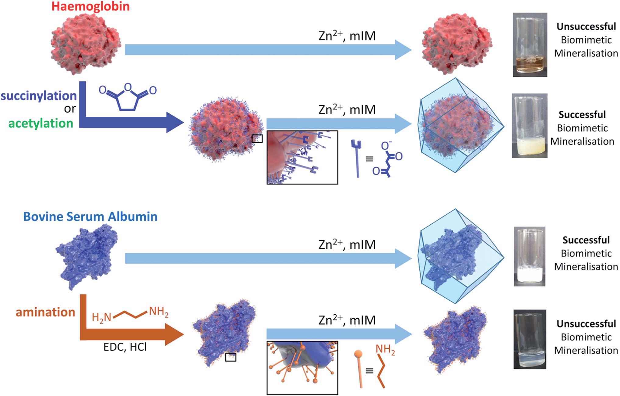
Figure 1. Schematic illustrations of the biomimetic mineralisation of haemoglobin and bovine serum albumin. Haemoglobin with slightly negatively charged surface is unable to form composites with ZIF-8, but becomes active after succinylation or acetylation to make its surface strongly negatively charged. On the contrary, bovine serum albumin with a strongly negatively charged surface readily combines with ZIF-8, but loses its activity when its surface becomes less negatively charged via amination.

The mechanism of the aforementioned observations was attributed to the electrostatic attraction between the protein surface and Zn2+, one of the MOF precursors. The more negatively the surface is charged, the more easily the Zn2+ will attach to and accumulate at the protein surface. The adsorbed Zn2+ ions then serve as the nucleation sites for the MOF to grow around the protein. This hypothesis is proven by a number of control experiments and also validated by computational studies.

This study highlights the surface potential of a protein as a critical factor in its ability to induce biomimetic mineralisation with MOFs. The conclusions could potentially be extended to biomolecules other than proteins (e.g. viruses and cells) to facilitate their integration with various MOFs.


To find out more please read:

Protein Surface Functionalisation as A General Strategy for Facilitating Biomimetic Mineralisation of ZIF-8

Natasha K. Maddigan, Andrew Tarzia, David M. Huang, Christopher J. Sumby, Stephen G. Bell, Paolo Falcaro and Christian J. Doonan

Chem. Sci., 2018 , DOI: 10.1039/c8sc00825f

About the blogger:

Tianyu Liu obtained his Ph.D. (2017) in Physical Chemistry from University of California, Santa Cruz in United States. He is passionate about scientific communication to introduce cutting-edge research to both the general public and scientists with diverse research expertise. He is a blog writer for Chem. Commun. and Chem. Sci. More information about him can be found at http://liutianyuresearch.weebly.com/.

Digg This
Reddit This
Stumble Now!
Share on Facebook
Bookmark this on Delicious
Share on LinkedIn
Bookmark this on Technorati
Post on Twitter
Google Buzz (aka. Google Reader)

Measuring the Strength of Hydrogen Bonds

For the first time, a group of scientists from University of California, San Diego in United States has quantitatively measured the strength of hydrogen bonds between two complex molecules. They also observed an abnormal trend regarding the bond strength in the absence and presence of electron transfer. This work contributes to the understanding of how the hydrogen bond strength changes, an important point that reveals the way biological systems function.

Hydrogen bonds are a type of electrostatic attraction between hydrogen atoms and certain highly electronegative atoms including N, O and F. These bonds help to bind individual water molecules together and keep water as liquid at room temperature, a critical condition for the origin of life.

The researchers, led by Prof. Kubiak, picked two ruthenium-based complexes joined by hydrogen bonds as their studying platform. As shown in Figure 1, the two-molecule system has three states depending on whether the ends are charged or not: the neutral state when both ends are not charged (left), the singly reduced state when only one end is negatively charged (middle), and the doubly reduced state when both ends are negatively charged (right). The group utilized infrared spectroscopy, UV-vis spectroscopy and electrochemical measurements to experimentally determine the strength of hydrogen bonds in these different redox states.

The results from the study found that the hydrogen bond energy of the neutral state and the doubly reduced state was in the range of 2.56-2.88 kcal/mol and 4.50-4.63 kcal/mol, respectively. Surprisingly, the hydrogen bond energy of the singly charged state did not lie between that of the neutral state and the doubly reduced state. It ranged from 7.78 kcal/mol to 8.31 kcal/mol, indicating the hydrogen bond is much stronger than for both the neutral and doubly-reduced states. The authors ascribed such an abnormality to the reinforcement brought by electron transfer i.e., the movement of the negative charge between the two ends.

This work is the first demonstration that hydrogen bond strength can be significantly enhanced by electron delocalization.

Figure 1. A schematic diagram showing the hydrogen bond strength of the singly reduced state (middle), the neutral state (left) and the doubly reduced states (right). [Note: In this figure, the lower the state lies, the stronger its hydrogen bond is. ET: electron transfer.]

To find out more please read:

Effects of Electron Transfer on the Stability of Hydrogen Bonds

Tyler M. Porter, Gavin P. Heim and Clifford P. Kubiak

Chem. Sci. DOI: 10.1039/c7sc03361c

About the blogger:

Tianyu Liu is a Ph.D. in chemistry graduated from University of California, Santa Cruz in United States. He is passionate about scientific communication to introduce cutting-edge researches to both the general public and the scientists with diverse research expertise. He is a web blogger for the Chem. Commun. and Chem. Sci. blog websites. More information about him can be found at http://liutianyuresearch.weebly.com/.

Digg This
Reddit This
Stumble Now!
Share on Facebook
Bookmark this on Delicious
Share on LinkedIn
Bookmark this on Technorati
Post on Twitter
Google Buzz (aka. Google Reader)

Drilling Holes on Metal Organic Framework Crystals with Phosphoric Acid

Synthesis of porous materials with tunable pore size remains a long-standing challenge for materials research. These materials, particularly hierarchical porous materials with copious different sized pores, are attractive candidates as catalysts, battery electrodes, and guest molecule hosts etc. An ideal synthesis protocol of hierarchical porous materials should be easy and capable of fine tuning pore size within a wide size range as well as able to preserve structural integrity. Unfortunately, it is extremely challenging for the currently developed methods to achieve the aforementioned three characteristics simultaneously.

Recently, this challenge has been addressed by Kimoon Kim and co-workers from the Institute of Basic Science and the Pohang University of Science and Technology in Republic of Korea. Their strategy targeted at metal organic frameworks (MOFs), a family of highly porous crystalline materials built by interconnected metal-organic complexes, as the starting materials. Phosphoric acid was chosen to selectively break down the building blocks and create pores with tunable diameters (Figure 1). Detailed synthesis procedures are published in Chemical Science.

Figure 1. Schematic illustration of the major steps of the demonstrated strategy.

The authors specifically chose the octahedral-shaped MIL-100(Fe) MOF crystals as an example, and demonstrated that their method could readily turn the original smooth crystals into highly porous ones merely by phosphoric acid aqueous solutions. Moreover, simply changing the concentration of phosphoric acid was able to tune the diameter of the created pores from 2.4 nm to 18.4 nm (Figure 2).

Figure 2. Scanning electron microscopy images and pore diameter distributions (showing pores larger than 2 nm only) of different MOFs: (a) the untreated MIL-100(Fe) and the acid treated MIL-100(Fe) with phosphoric acid concentration of (b) 20 mM, (c) 40 mM, (d) 60 mM and (e) 80 mM. All scale bars represent 200 nm.

Structural evolution analysis revealed that the etching process initiated preferably by cleaving the coordination bonds between metal cores and organic ligands around the hexagonal windows on (2 2 0) crystal planes. It further propagated to dig out the inner part of MIL-100(Fe), forming pores on surface.

This method is expected to be applicable to other MOFs coupled with properly selected etchants, thus making the hierarchical porous crystals with tailorable porous structures readily available to worldwide materials researchers.

To find out more please read:

Hollowing Out MOFs: Hierarchical Micro- and Mesoporous MOFs with Tailorable Porosity via Selective Acid Etching

Jaehyoung Koo, In-Chul Hwang, Xiujun Yu, Subhadeep Saha, Yonghwi Kim and Kimoon Kim

DOI: 10.1039/c7sc02886e

About the blogger:

Tianyu Liu is a Ph.D. in chemistry graduated from University of California-Santa Cruz. He is passionate about scientific communication to introduce cutting-edge researches to both the general public and the scientists with diverse research expertise. He is a web writer for the Chem. Commun. and Chem. Sci. blog websites. More information about him can be found at http://liutianyuresearch.weebly.com/.

Digg This
Reddit This
Stumble Now!
Share on Facebook
Bookmark this on Delicious
Share on LinkedIn
Bookmark this on Technorati
Post on Twitter
Google Buzz (aka. Google Reader)

Mimicking Biological Systems with Supramolecular Assemblies: A Step Closer

Biological systems, such as circadian rhythms and ion pumps, are generally able to respond autonomously to chemical stimuli. Such ability is at the root of certain amphiphilic molecules (i.e. molecules containing both hydrophilic and hydrophobic parts) that can change their configurations temporally at the presence of the stimulants. In recent decades, building artificial biological systems with active, adaptive and autonomous behaviours similar to natural biological systems has become a hot topic.

Supramolecular assemblies are among the most extensively explored building materials. A supramolecular assembly is a system consisting of complex molecules held together by non-covalent bonds, such as DNA. One of the major technological hurdles for developing artificial biological systems using supramolecular assemblies is to acquire assemblies with an amphiphilic nature and the ability to undergo a transient change of configuration when stimulated. The change should also be highly reversible and durable. Currently, extensive research efforts are committed to finding candidates that address the aforementioned challenge.

A research group from Jawaharlal Nehru Centre for Advanced Scientific Research in India recently made a step forward and published their work in Chemical Science. They developed an amphiphilic supramolecular motif, coined PN-VN foldamer, which could switch its conformation upon contact with oxidizing and reducing agents. As shown in Figure 1a, the skeleton of the supramolecular motif is composed of three sections. The green head is an electron donor pyranine (PN), the red tail is an electron acceptor called viologen (VN) rendering the hydrophobic nature, and the blue body is a flexible hydrophilic hexaethylene glycol that connects the electron donor and acceptor.

Figure 1. (a) The structure of the amphiphilic PN-VN foldamer. (b) Unfolded and folded states of the PN-VN foldamer correspond to sheet and vesicle morphologies, respectively.

The researchers demonstrated the assembly pattern of PN-VN foldamers (Figure 1b) by using two chemical fuels, sodium dithionite and glucose, as the stimulants. The transformation process is depicted in Figure 2a. When undisturbed, the negatively charged PN (PN3-) and the positively charged VN (VN2+) can attract each other via a charge transfer interaction, folding the entire molecular chains to vesicles. When placed in a solution containing sodium dithionite and glucose, the VN2+ terminal can be instantaneously reduced by sodium dithionite to its radical cation form (VN•+). The reduction weakens the charge transfer interaction, and subsequently unfolds PN-VN vesicles to sheets. Meanwhile, catalyzed by glucose oxidase (an enzyme), the excess glucose in the same solution can oxidize VN•+ back to its original state, resulting in the folded state that recovers vesicles. The transition is directly confirmed by transmission electron microscopy as shown in Figure 2b. Since the reduction process proceeds much faster than the oxidation process, it is observed that PN-VN vesicles first extend to sheets momentarily and gradually but automatically fold back to vesicles within minutes. The rate of the transition can be well tuned by varying the concentration of glucose oxidase.

Figure 2. (a) Schematic of the transient configuration change between vesicles and sheets. SDT: sodium dithionite; GOx: glucose oxidase. (b) Transmission electron microscopic images of the vesicles (left) and the sheets (right). Inset in the left panel shows the distribution of the wall thickness of the vesicles. Inset in the right panel is a confocal fluorescence microscopy image of a selected sheet (scale bar: 2 μm).

To find out more please read:

Temporal Switching of an Amphiphilic Self-assembly By a Chemical Fuel-driven Conformational Response

Krishnendu Jalani, Shikha Dhiman, Ankit Jain, and Subi J. George

DOI: 10.1039/c7sc01730h

About the author:

Tianyu Liu is a Ph.D. in chemistry graduated from University of California-Santa Cruz. He is passionate about scientific communication to introduce cutting-edge researches to both the general public and the scientists with diverse research expertise. He is a web writer for the Chem. Commun. and Chem. Sci. blog websites. More information about him can be found at http://liutianyuresearch.weebly.com/.

Digg This
Reddit This
Stumble Now!
Share on Facebook
Bookmark this on Delicious
Share on LinkedIn
Bookmark this on Technorati
Post on Twitter
Google Buzz (aka. Google Reader)