Author Archive

How can cooling rate define the nature of nano-structures formed using block copolymers?

Block copolymers (BCPs) consist of amphiphilic molecules that can self-assemble in selective solvents and generate various types of nano- and micro-dimensional structures. The unique self-assembly process is low-cost and relatively straight forward in solution phase.  The final structures have morphological diversity and complexity.  These self-assembled structures have been widely used in various applications such as drug delivery, catalysis, and water purification. The self-assembly process follows a heating step for dissolution of all the components and a subsequent cooling step. Both the steps and the parameters are vital for final structural characteristics of the assembled structures. A group of scientists from two esteemed universities in Canada recently studied the effects of rate of cooling in the self-assembly process.

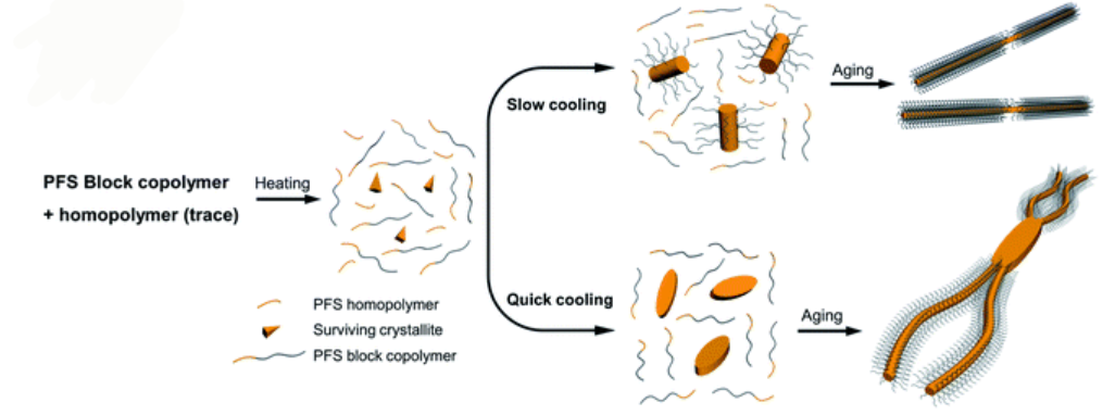
Schematic representation of how cooling rate can change the morphology of formed micellar structures using PFS BCP and homopolymers

The authors used a systematic approach to explain the influence of cooling rate on micelle morphologies for a series of PFS based BCPs. The cooling rate greatly influences the size and the shape of colloidal structures. Rapid cooling increases branching and opens a new avenue to manipulate micelle morphologies. The study finds that rapid cooling reduces crystallinity, as polymer chains do not have enough time to pack in ordered structures.

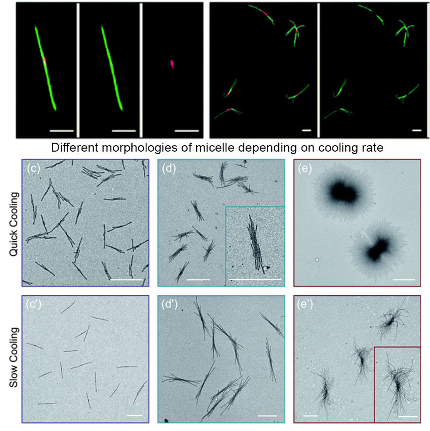
The authors standardized sample preparation protocol and then varied the cooling times, with quick cooling of 2.5 min producing flower like structures and median cooling time of 50 mins leading to the same structural features with larger size. Co-self-assembly of homopolymer BCP mixtures with variable cooling rate also shows that quick cooling generates uniform sized branched micellar structures with elongated central platelets whereas slow cooling led to a long single fiber with a dark circle platelet in the centre.

With several examples and optimization conditions, the effect of cooling in the formation of self-assembled micellar structures has been evaluated. The main outcome of this study is that the cooling rate is another parameter to manipulate crystallization-driven self-assembly and to control micelle morphologies. There exists a lot of possibilities to use the findings and apply them to generate BCPs with a crystallizable block with important optical or electronic properties.

For details, please visit the entire article at https://doi.org/10.1039/D1SC05937H

About the author:

Dr Damayanti Bagchi is a postdoctoral researcher in Irene Chen’s lab at University of California, Los Angeles, United States. She obtained her PhD in Physical Chemistry from Satyendra Nath Bose National Centre for Basic Sciences, India. Her research is focused on spectroscopic studies of nano-biomaterials. She is interested in exploring light enabled therapeutics. She enjoys food and experimenting with various cuisines, which she found resembles products/ side products of chemical reactions!

Digg This
Reddit This
Stumble Now!
Share on Facebook
Bookmark this on Delicious
Share on LinkedIn
Bookmark this on Technorati
Post on Twitter
Google Buzz (aka. Google Reader)

A new class of bioluminescent substrate-enzyme pair for deep tissue multi-colour imaging

Bioluminescent enzymes (luciferases) generate light via the oxidation of small molecule luciferins. The process is highly specific and accurate even at heterogeneous environment. Luciferin-luciferases based imaging technique is highly appreciated for specificity in tracking cell movements, cell proliferation, and numerous other features in living organisms.

Imaging of in-depth organ tissues requires emission at NIR region for effective penetration through tissue layer. There existed a big gap in successful synthesis followed by appropriate multiplexed imaging application of bioluminescent pairs. Researchers from University of California, Irvine recently developed a unique class of orthogonal, NIR emitting luciferins that could promise more accessible, long-wavelength bioluminescent pairs for in-vivo imaging.

Fig. 1 Red-emitting orthogonal bioluminescent probes designed from fluorophores. (a) D-Luciferin is oxidized by firefly luciferase (Fluc) to produce oxyluciferin and a photon of light. (b) Coumarin fluorophores were used as templates for red-shifted luciferins. (c) Retrosynthetic analysis of the CouLuc-1 analogs.

The authors focused on a new class of luciferins (CouLuc-1s) comprising both an elongated pi-system and a 4-tri-fluoromethylcoumarin unit (Fig 1). The synthesis follows two-step route to bridge the fluorescent coumarin heterocycle with the key thiazoline unit necessary for luciferin bioluminescence. The small size of the coumarin core require only minimal enzyme engineering to identify complementary luciferases that were identified via a parallel engineering approach.

Fig. 2 Multi-component imaging with three NIR-emitting probes.

The brightest luciferase-CouLuc-1 pair exhibited higher luminescent signals compared to native bioluminescent probes and can be immediately adopted for biological imaging. Multiplexed NIR imaging could also be attained using three different analogues of the newly prepared luciferins (Fig 2). In a broader sense, synthesis of novel luminophores from simple fluorophores pave a step forward in the bioluminescent imaging field.

For details: please visit https://pubs.rsc.org/en/content/articlelanding/2021/sc/d1sc03114g

About the blogger:

Dr. Damayanti Bagchi is a postdoctoral researcher in Irene Chen’s lab at University of California, Los Angeles, United States. She has obtained her PhD in Physical Chemistry from Satyendra Nath Bose National Centre for Basic Sciences, India. Her research is focused on spectroscopic studies of nano-biomaterials. She is interested in exploring light enabled therapeutics. She enjoys travelling and experimenting with various cuisines.

You can find her on Twitter at @DamayantiBagchi.

Digg This
Reddit This
Stumble Now!
Share on Facebook
Bookmark this on Delicious
Share on LinkedIn
Bookmark this on Technorati
Post on Twitter
Google Buzz (aka. Google Reader)

Watch out for artifacts in your next multi-colour fluorescence imaging experiment

The discovery of super resolution microscopy, followed by the announcement of 2014 Chemistry Nobel prize, facilitated a great expansion in the use of multi colour fluorescence imaging to study cellular or sub-cellular systems. Super resolution localization microscopy requires highly photostable, sufficiently bright fluorophores to achieve the blinking which is necessary to distinguish individual fluorophores within the diffraction limit. To validate all these criteria, organic dyes are a most obvious choice as fluorophores. However, chemical conversion of organic dyes upon prolonged laser exposure exhibit multicolour image artifacts leading to false-positive colocalization. Researchers from Pohang University, South Korea demonstrate a detailed protocol to understand and avoid such artifacts.

The researchers labelled cell membrane using far-IR dye (A647) which shows a red photoluminescence. But surprisingly, upon photobleaching of the A647 dye, which is blue in colour, it turned to red. This photobleached product also emits at red region, coinciding with the original emission of A647. This phenomenon is called blue-conversion. A range of commonly used organic dyes are evaluated for blue-conversion occurrences which indicates cyanine dyes show multiple blue-converted species. Interestingly, among all the dye groups there is not a single group that exhibit no blue-conversion at all.

Blue-conversion of far-red organic dyes upon photobleaching. A647 dissolved in DMSO before (left) and after (right) photobleaching using direct laser illumination. (a) TIRF images of A647-EGFR on COS7 cells in the far-red (upper panels) channel excited at 642 nm and the red (lower panels) channel excited at 561 nm before (left panels) and after (right panels) photobleaching of A647-EGFR.

The researchers also study multicolour fluorescence imaging by colocalization of two well-known dyes. They have observed that the single-molecule brightness of the blue-converted species contributed to the production of the artifact in the reconstructed images. Finally, they concluded sufficient care must be taken in multicolour imaging applications, including colocalization, and other fluorescence-based multi-well plate format assays, to prevent false positives produced by blue-conversion of organic dyes.

Although they primarily discussed the negative effect of the blue-conversion of organic dyes, they are also hopeful to use this new photoconversion pathway of cyanine dyes for advantages of fluorescence imaging applications. They propose that super-resolution techniques require the photoactivation of organic dyes, which might exert some undesirable effects in live cells. However, the photoactivation of the blue-converted species occurs without any external stimuli and can be inferred as an advantage for super resolution techniques.

For details please read: https://doi.org/10.1039/D1SC00612F

About the blogger:

Dr. Damayanti Bagchi is a postdoctoral researcher in Irene Chen’s lab at University of California, Los Angeles, United States. She has obtained her PhD in Physical Chemistry from Satyendra Nath Bose National Centre for Basic Sciences, India. Her research is focused on spectroscopic studies of nano-biomaterials. She is interested in exploring light enabled therapeutics. She enjoys travelling and experimenting with various cuisines.

You can find her on Twitter at @DamayantiBagchi.

Digg This
Reddit This
Stumble Now!
Share on Facebook
Bookmark this on Delicious
Share on LinkedIn
Bookmark this on Technorati
Post on Twitter
Google Buzz (aka. Google Reader)

Chemical domain image recognition using autocatalysis

A reaction in which one of the products speeds up further product formation is called an autocatalytic reaction. Autocatalysis plays an important role in living systems including DNA replication, apoptosis, and even in the origin of life, due to self-sustaining growth and oscillation. Researchers from Brown University employ this nature of autocatalytic click chemistry to generate an artificial neural network that can be used for image classification.

Autocatalytic reaction rate depends on the concentration of product and shows a non-linear dependency of product formation with progress in reaction time. In this view, a network of autocatalytic reactions is analogous to an artificial neural network. An artificial neuron is a basic learning unit, inspired by biological neurons, which multiplies it’s inputs by a set of weights and transforms their sum through a nonlinear operator. Researchers used this resemblance to formulate a winner-take-all neural network.

Fig 1: Kinetics of autocatalysis. (a) Reagent and autocatalytic product evolution over time (b) Rate of product concentration change over time for the reaction simulated in a, showing the accelerated production typical of an autocatalytic process.

Copper-catalyzed azide–alkyne cycloaddition (CuAAC) reaction was chosen for autocatalysis as it is fast, can occur under mild conditions and produce high yield. Also, CuAAC reaction involves colored copper–ligand complexes and can be quantitatively monitored using UV-vis spectroscopy.

In a winner-take-all neural network, winner is determined by it’s achievement to reach to a particular condition. Here, they have used the reaction half-way point (t1/2) as the condition of image classification. Experiment wise, they have used automated liquid handling equipment to remove a certain volume and then added it together into individual pools for potential image class. The pool that reaches the transition time first is determined as the winner.

Fig 2: An overview of the copper (C) catalyzed azide–alkyne cycloaddition reaction, showing the buildup of triazole branches on the amine backbone of (A) after each azide (B) incorporation. The threebranched product (D) catalyzes its own generation by promoting the reduction of Cu(II). Experimental setup for evaluating a chemical WTA network (Right: upper panel). (Right lower panel) Network training and in silico simulation. (a) Example images from each of the considered classes. (b) Trained weights for each class.  

This study shows an interesting adaptation of autocatalysis as a platform for non-linear activation function necessary for artificial neural network classification. The findings are expected to improve future development of chemical-domain computing systems.


For further details, please go through:

Leveraging autocatalytic reactions for chemical domain image classification

Christopher E. Arcadia, Amanda Dombroski, Kady Oakley, Shui Ling Chen, Hokchhay Tann, Christopher Rose, Eunsuk Kim, Sherief Reda, Brenda M. Rubensteinb and Jacob K. Rosenstein*

Chem. Sci., 2021, 12, 5464


About the blogger

Dr Damayanti Bagchi is a postdoctoral researcher in Irene Chen’s lab at University of California, Los Angeles, United States. She has obtained her PhD in Physical Chemistry from Satyendra Nath Bose National Centre for Basic Sciences, India. Her research is focused on spectroscopic studies of nano-biomaterials. She is interested in exploring light enabled therapeutics. She enjoys travelling and experimenting with various cuisines, which she found resembles with products/ side products of chemical reactions!

You can find her on Twitter at @DamayantiBagchi.

Digg This
Reddit This
Stumble Now!
Share on Facebook
Bookmark this on Delicious
Share on LinkedIn
Bookmark this on Technorati
Post on Twitter
Google Buzz (aka. Google Reader)