Magnetic nanoparticles detect low levels of cyanide

Chinese scientists have reported dumbbell-shaped nanoparticles capable of detecting cyanide concentrations as low as 4 x 10⁻⁷ M in drinking water. This low detection limit, which is below the acceptable limit advised by the World Health Organization, is attributed to a combination of fluorescence detection with a filtering process called ‘magnetic concentration-washing’.

The toxicity of cyanide and its potential prevalence in drinking water has made easy detection of cyanide an active area of research. Gold nanoparticles have attracted attention as cyanide sensors due to their selectivity for cyanide over other anions. Now, Shaojun Dong and colleagues at the Changchun Institute of Applied Chemistry, Chinese Academy of Sciences, have adapted a bifunctional nanoparticle made up of Au and magnetic Fe3O4 nanoparticles for sensitive detection of cyanide, even in the presence of interfering species. 

Schematic depiction of the turn-on fluorescent detection of cyanide based on the dual-functional Au–Fe3O4 dumbbell nanoparticles.

The group detected cyanide by relying on what is known as the ‘inner filter effect’, observed when two molecules present in a solution have overlapping absorption and emission wavelengths. The fluorescence emission from a fluorophore is blocked, or reabsorbed, by the second molecule, reducing or even quenching the overall fluorescence of the solution. In the present system, the excitation and emission energy for the fluorophore Rhodamine B is quenched by the Au-Fe3O4 nanoparticles. However, addition of cyanide to the sample reduces the gold component of the nanoparticles by forming an Au(CN)2 complex. This change in the structure alters the nanoparticles’ absorbance spectra and the Rhodamine B emissions are no longer fully absorbed. The team calculated the amount of cyanide present in a sample by monitoring the change in fluorescence.

To extend this detection method for use with environmental samples, which may contain dyes or other contaminants that absorb or emit at a similar wavelength to Rhodamine B, Dong and colleagues implemented a ‘magnetic concentration-washing process’. In it, the Au-Fe3O4 nanoparticles are magnetically separated from the sample solution after they have reacted with any cyanide present in the sample. This magnetic separation retains the bifunctional nanoparticles and leaves any interfering species behind. Pure buffered water containing additional Rhodamine B is added and the fluorescence is monitored. The authors showed that two cycles of this process were enough to remove any interfering species and accurately detect cyanide levels. 

To find out more, read the full article:

Dual-Functonal Au-Fe3O4 Dumbbell Nanoparticles for Sensitive and Selective Turn-on Fluorescent Detection of Cyanide Based on the Inner Filter Effect
Yueming Zhai, Lihua Jin, Ping Wang and Shaojun Dong, Chem. Commun., 2011, DOI: 10.1039/C1CC13149D 

Posted on behalf of Patricia Pantoș, web writer for ChemComm.

Digg This
Reddit This
Stumble Now!
Share on Facebook
Bookmark this on Delicious
Share on LinkedIn
Bookmark this on Technorati
Post on Twitter
Google Buzz (aka. Google Reader)

Lysozymes on silica – seeing is believing

Human lysozyme on mesoporous silica (SBA-15) has been visualised in 3D, say scientists in Italy.

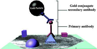
Ultrathin section of embedded mesoporous materials

Maura Monduzzi and her co-workers at Università di Cagliari, used an immunochemical method to investigate the location of human lysozyme molecules loaded onto SBA-15. Using post-embedding immunogold staining and transmission electron microscopy results in a 3D reconstruction and shows that the protein molecules were located on the external and internal surfaces of the mesoporous silica. This result may have important consequences in biocatalysis and applications requiring sustained drug release.

Want to know more? Then download the ChemComm communication, which will be free to access until the 15th July 2011.

Digg This
Reddit This
Stumble Now!
Share on Facebook
Bookmark this on Delicious
Share on LinkedIn
Bookmark this on Technorati
Post on Twitter
Google Buzz (aka. Google Reader)

Improving bulk-heterojunction solar cells

Making new n-type materials for bulk-heterojunction solar cells (BHJ) can be achieved by simply modifying the fullerene framework, say scientists in Japan.

Looking at LUMO and charge-density of modified fullerene

The BHJ solar cells may be an alternative to conventional silicon-based solar cells, if the low power conversion efficiency can be overcome. Yasujiro Murata and colleagues from Kyoto University have shown that structurally manipulating the C60 skeleton is a promising approach for creating better acceptor materials for BHJ solar cells. This new approach means that the team can effectively control the lowest unoccupied molecular orbital (LUMO) level of the  C60 pi-system, resulting in a clear improvement in open-circuit voltage – a promising development.

Fancy finding out more? Then download the ChemComm communication, which will be free to access until the 15th July 2011.

Digg This
Reddit This
Stumble Now!
Share on Facebook
Bookmark this on Delicious
Share on LinkedIn
Bookmark this on Technorati
Post on Twitter
Google Buzz (aka. Google Reader)

New template has potential for bone tissue engineering

British scientists at working at Imperial College, London have synthesised a macroporous hydroxyapatite bioceramic with potential uses in bone regeneration.

Porous hydroxyapatite (HA) bioceramics are widely studied for their potential use in bone regeneration. These materials are biocompatible and have a structure that contains many connected pores. This allows fluid to flow through the material and provides nucleation points for bone growth. The best results are obtained when the the pores are smaller than 1 μm and therefore synthetic methods that give good control over the pore size and material structure are highly desirable.

Prof. Molly Stevens and her team have now demonstrated a way of making an HA bioceramic with a 3D ordered (DOM) macroporous structure. They used an ordered macroporous carbon template (OMC)  in combination with a sol-gel method to form the desired bioceramic. They found that the DOM HA, when compared with HA material made without the OMC template, allowed the nucleation of calcium phosphate on the surface of the pores, thus demonstrating that the template method used affects the crystallisation of material.

hydroxyapatite bioceramics

The team hopes that these materials will find applications not only in the generation of bone but also in the generation of other materials. Want to find out more? Then download the ChemComm article for free today and leave a comment below to let us know what you think.

Digg This
Reddit This
Stumble Now!
Share on Facebook
Bookmark this on Delicious
Share on LinkedIn
Bookmark this on Technorati
Post on Twitter
Google Buzz (aka. Google Reader)

Lanthanide discs learn to chillax

A team of UK scientists have made gadolinium and dysprosium discs that can ‘chill well and relax slowly’ with interesting magnetic properties.

Structure of the gadolinium heptametallic complex

David Collison and his colleagues from the University of Manchester have designed heptametallic discs (i.e. with 7 metal centers) and have studied their structures and magnetic properties. The dysprosium disc shows single molecule magnetism behaviour and the gadolinum analogue shows a large magnetocaloric effect.

 Fancy finding out more? Then download the ChemComm communication, which will be free to access until the 15th July 2011.

Also, are you interested in molecule-based magnetism research? Then take a look at Chem Soc Rev‘s recently published themed issue on Molecule-based Magnets, (Issue 6, 2011) guest edited by Joel Miller and Dante Gatteschi.
Digg This
Reddit This
Stumble Now!
Share on Facebook
Bookmark this on Delicious
Share on LinkedIn
Bookmark this on Technorati
Post on Twitter
Google Buzz (aka. Google Reader)

Probing cells’ power generators

UK scientists have developed a probe to monitor bicarbonate concentrations in mitochondria – components in living cells that generate chemical energy. Monitoring bicarbonate levels will improve researchers’ understanding of its role in cellular reaction mechanisms. 

A challenge when designing cellular probes is ensuring that the probe is not only selective for its target but can also be delivered to the site of interest within the cell. A team of scientists led by David Parker at the University of Durham has made a probe that can overcome this challenge. 

Stained HeLa cellsThe luminescent probe features an azaxanthone moiety, which is linked to a europium complex by an amide bond. The azaxanthone allows the probe’s uptake into cells and localisation within the mitochondria, and the europium complex has an affinity for bicarbonate ions. The ability to probe bicarbonate levels ‘can offer an unprecedented insight into signalling mechanisms’, says Parker.

Read the rest of this story in Chemistry World and download Professor Parker’s ChemComm communication, which is free to access for a limited period.

Also of interest:
Definition of the uptake mechanism and sub-cellular localisation profile of emissive lanthanide complexes as cellular optical probes
Elizabeth J. New, Aileen Congreve and David Parker, Chem. Sci., 2010, 1, 111-118

Digg This
Reddit This
Stumble Now!
Share on Facebook
Bookmark this on Delicious
Share on LinkedIn
Bookmark this on Technorati
Post on Twitter
Google Buzz (aka. Google Reader)

RSC Centenary Prize awarded to…

Many congratulations to Jonathan Sessler, ChemComm‘s very own US Associate Editor for supramolecular chemistry, who has been awarded the Centenary Prize for his groundbreaking research on polypyrrolic systems in the areas of supramolecular and medicinal chemistry.

This year the Centenary Prize was also awarded to two other outstanding chemists: Marius Clore and Graham Cooks. All three winners will present lectures in the British Isles in association with this prize.

Well done Jonathan from the ChemComm Editorial team here in Cambridge!

Digg This
Reddit This
Stumble Now!
Share on Facebook
Bookmark this on Delicious
Share on LinkedIn
Bookmark this on Technorati
Post on Twitter
Google Buzz (aka. Google Reader)

Across the barrier for tumour imaging

Brain and structure of nanoprobeA probe that can cross the blood-brain barrier to allow high sensitivity brain tumour imaging has been made by Chinese scientists. The probe could be used to pinpoint the location and extent of a tumour before an operation and be used for image-guided tumour removal. 

Establishing the position, extent and structure of brain tumours is crucial for their successful removal. But, current tumour imaging agents used in magnetic resonance imaging are limited by short circulation lifetimes, non-targeted specificity and poor blood-brain barrier permeability. The results of these limitations are that low grade tumours and 20-30 per cent of advanced brain tumours with an intact blood-brain barrier go unnoticed.

Cong Li from Fudan University, Shanghai, and his team made the probe starting with a dendrimer – a branched molecule with a long circulation lifetime – and attached functional groups with different tasks. One such group, a lipoprotein ligand angiopep-2, helps to deliver the probe across the blood-brain barrier and targets the lipoprotein’s receptors, which are present in increased amounts on tumour cells. High-resolution images can be generated thanks to imaging reporters, including fluorescence dyes, attached to the dendrimer. 

Read the rest of this story in Chemistry World and download Li’s ChemComm communication, which is free to access for the rest of the month.

Digg This
Reddit This
Stumble Now!
Share on Facebook
Bookmark this on Delicious
Share on LinkedIn
Bookmark this on Technorati
Post on Twitter
Google Buzz (aka. Google Reader)

New acetylene polymerisation catalyst is best yet

A team of chemists working in the UK and Australia have developed a new, highly active catalyst for acetylene polymerisation.

Polyacetylene is an important material as it has a degree of conjugation that leads to good conductive properties. It has found uses in areas such as synthetic metals and plastic electronics. New routes to make this polymer are constantly sought and, to date, the majority of catalysts for this task have low activities.

This problem has now been solved by two teams at the University of Tasmania and Imperial College London led by David McGuinness and George Britovsek, respectively. They took a bis(imino)pyridine iron catalyst, normally used for ethylene polymerisation, along with methylaluminoxane and successfully produced polyacetylene.

Graphical abstract: High activity acetylene polymerisation with a bis(imino)pyridine iron(ii) catalyst

An activity of 62 500 h-1 (turn over frequency; TOF) was obtained, which compares excellently with the 100  h-1 TOF obtained when using a standard existing acetylene polymerisation catalyst under the same conditions. This represents the most active acetylene polymerisation reported to date. Curently, the team are studying the effects of ligand modification and chain transfer reagents on the reaction.

Want to find out more? Then download  the full ChemComm article for free today.

Digg This
Reddit This
Stumble Now!
Share on Facebook
Bookmark this on Delicious
Share on LinkedIn
Bookmark this on Technorati
Post on Twitter
Google Buzz (aka. Google Reader)

New sensor for naked-eye fluoride detection

Fluoride, in its organic and inorganic forms, has been increasingly present in the food and beverage chain over the years. It has been added to toothpastes to prevent cavities; different forms of fluoride have been included in pesticides; several categories of food, including cereals, have been fortified with fluoride and even water for public consumption has been artificially fluorinated for decades.

Graphical abstract: A highly selective colorimetric and ratiometric fluorescent chemodosimeter for imaging fluoride ions in living cellsDespite being a useful supplement to support the healthy growth of hair, nails and teeth and the strengthening of bones, an excessive intake of fluorine can lead to adverse effects on development. It can cause mottling of teeth and skeletal fluorosis (causing joint pain and abnormalities in the skeletal structure). Other effects have only been studied in animal models and at concentrations unlikely to be encountered by humans.

Regardless of the debate about its toxicity, it cannot be denied that as such a ubiquitous chemical in the human food chain, the development of simple methodologies and techniques to accurately detect the concentration of this anion in vivo plays an important role in biochemical research.

One of these techniques employs the use of fluorescent indicators that detect the presence of fluoride anions in solution.

Recent research by the Chinese group of Ma, Du and Zhang has been focused on the realisation of a novel “chemodosimeter” for fluoride that responds to the requisites of ease of synthesis, activity in highly aqueous solutions and buffers, cell permeability, quantitative response and high selectivity.

Their sensor, incorporating benzothiazolium hemicyanine as the fluorophore, was tested in fluorine detection in water:ethanol solutions (7:3) containing phosphate buffered saline at a pH of 7.4. In these conditions, the fluorescence of the sensor was not only quantitatively responsive to changes in fluoride concentration, but showed a change in the fluorescence spectrum, with emission at a different wavelength when in presence of the analyte and even noticeable to the naked eye. The versatility and selectivity of the system was also assessed by performing competition experiments in the presence of other anions, such as CO32-, SO42-, NO3, Cl, I and selected aminoacids and proteins like cysteine and human serum albumin, demonstrating a remarkable preferential response for fluoride.

Tests were also performed on living cells in order to determine the cytotoxicity of the chemodosimeter, using HeLa cells as the test substrate, showing low toxicity under the operational conditions.

The selectivity over different anions and analytes of biological relevance, the ability to operate in strongly aqueous solutions, the reliability and quantitative response and the applicability to living cells may make this new chemodosimeter a beneficial tool for biomedical researchers.

To find out more, read the full article:

A highly selective colorimetric and ratiometric fluorescent chemodosimeter for imaging fluoride ions in living cells
Baocun Zhu, Fang Yuan, Rongxia Li, Yamin Li, Qin Wei, Zhenmin Ma, Bin Du and Xiaoling Zhang
Chem. Commun., 2011, DOI: 10.1039/C1CC11308A

Posted on behalf of Dr. Giorgio De Faveri, Web Writer for Catalysis Science & Technology.

Digg This
Reddit This
Stumble Now!
Share on Facebook
Bookmark this on Delicious
Share on LinkedIn
Bookmark this on Technorati
Post on Twitter
Google Buzz (aka. Google Reader)