Lighting Up the Polymer World, Reversibly

Researchers from the UK have developed a polymeric system that exhibits reversible fluorescent behaviour. Anthea Blackburn writes more…

Fluorescent imaging is one of the most useful techniques in modern day medicine, as it provides us with a method of visualizing the inside of a body to allow for diagnosis and treatment of disease. Typically, an organic fluorophore is attached to a biomolecule that, upon introduction to the body, is able to interact with a specific type of molecule in the body, such as a cancerous cell or a molecule that carries out a particular and important function. More recently, in addition to the use of biomolecules, fluorescent labeling has turned to the polymer world for applications in targeted drug delivery or cell patterning to name but a few examples. Fluorescence becomes important in these applications, as the introduction of non-biological molecules to the body requires a method of locating them to track and monitor their activity.

There are a number of methods of introducing organic fluorophores to polymers, for example, polymerization of fluorescently labeled monomer units, end-group attachment, or post-synthetic modification, all of which offer advantages and disadvantages. The one factor that all of these approaches have in common however, is that one needs to beware of how the attachment of fluorophores, which are typically large, will change the chemistry of the polymer. It would therefore be advantageous if small, yet fluorescent, groups could be attached to polymers without otherwise changing their properties.

It is this interest in synthesising fluorescent polymers using small molecules that Mathew Robin and Rachel O’Reilly from the University of Warwick sought to tackle. They were able to demonstrate that the introduction of dithiomaleimide functional groups, which have a large Stokes shift (250 nm) and bright emission, to acrylate or methacrylate polymers did not change the properties of the polymer itself. Perhaps even more interestingly is that it was demonstrated that the functional groups could be introduced both pre-synthetically and post-synthetically. In this way, polymer fluorescence could be both turned on in a profluorescent polymer that contained a reactive dibromomaleimide monomer unit, as well as reversibly turned off through a dithiol exchange reaction to a non-fluorescent dithiomaleimide monomer unit.
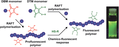
The development of a relatively simple system whose fluorescence can be reversible turned on and off is an exciting step forward in developing polymers, especially since the end groups of this functionalised polymer allows for its further incorporation into more complex polymeric systems. These polymers could have applications in not only the biomedical uses discussed, but also in a numerous other applications such as organic electronic devices, sensing materials and polymer materials like nanoparticles and hydrogels.

Read this HOT Chemical Science Edge article in full for free*!

Fluorescent and chemico-fluorescent responsive polymers from dithiomaleimide and dibromomaleimide functional monomers

Mathew P. Robin and Rachel K. O’Reilly
Chem. Sci.20145, 2717.
DOI: 10.1039/C4SC00753K, Edge Article

About the Writer

Anthea Blackburn is a guest web writer for Chemical Science. Anthea is a graduate student hailing from New Zealand, studying at Northwestern University in the US under the tutelage of Prof. Fraser Stoddart (a Scot), where she is exploiting supramolecular chemistry to develop multidimensional systems and study the emergent properties that arise in these superstructures. When time and money allow, she is ambitiously attempting to visit all 50 US states before graduation.

*Access is free untill 08.08.14 through a registered RSC account – click here to register

Digg This
Reddit This
Stumble Now!
Share on Facebook
Bookmark this on Delicious
Share on LinkedIn
Bookmark this on Technorati
Post on Twitter
Google Buzz (aka. Google Reader)

Small Molecule Mimics of Transport Proteins

Philip Gale, Chair of our sister Journal ChemSocRev, et al report the first use of thiosquaramides as anion receptors and pH-switchable anion transporters.

Many diseases are caused by faulty anion transport across cell membranes, such as faulty chloride transport leading to cystic fibrosis. Therefore, in recent years interest has grown in developing small molecule mimics of transport proteins that can be used to restore or disrupt the chemical processes within cells and thus cure or kill a range of diseases.

Researchers from the University of Southampton and the University of Sydney have produced a series of thiosquaramides which investigate pH dependent chloride transport properties. It was observed that the anion transport ability of the thiosquaramides was completely turned on below a pH of 7.2 but fully switched off at a pH value of 7.2 or higher. The developed thiosquaramides can promote chloride efflux mainly via a chloride/nitrate antiport process.

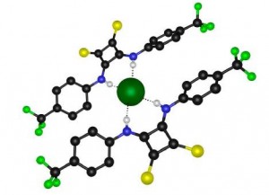
One of the thiosquaramide derivative with bound chloride anion

This paper provides one of the few examples of truly controllable and switchable anion transport by small synthetic molecules. Thiosquaramides form interesting targets for developing future biologically active anion transporters –  read the paper today to find out how to make them!

To read the details, check out the Chem Sci article in full for free:

Nathalie Busschaert, Robert B. P. Elmes, Dawid D. Czech, Xin Wu, Isabelle L. Kirby, Evan M. Peck, Kevin D. Hendzel, Scott K. Shaw, Bun Chan, Bradley D. Smith, Katrina A. Jolliffe and Philip A. Gale
DOI: 10.1039/C4SC01629G
About the Webwriter
Iain Larmour is a guest web writer for ChemSci. He has researched a wide variety of topics during his years in the lab including nanostructured surfaces for water repellency and developing nanoparticle systems for bioanalysis by surface enhanced optical spectroscopies. He currently works in science management. In his spare time he enjoys reading, photography, art and inventing.

Digg This
Reddit This
Stumble Now!
Share on Facebook
Bookmark this on Delicious
Share on LinkedIn
Bookmark this on Technorati
Post on Twitter
Google Buzz (aka. Google Reader)

Hot Chemical Science articles for June

All of the referee-recommended articles below are free to access until 21st July:

A full-adder based on reconfigurable DNA-hairpin inputs and DNAzyme computing modules
Ron Orbach, Fuan Wang, Oleg Lioubashevski, R. D. Levine, Francoise Remacle and Itamar Willner  
Chem. Sci., 2014, Advance Article
DOI: 10.1039/C4SC00914B, Edge Article


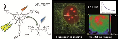
Millisecond lifetime imaging with europium complex using a commercial confocal microscope under one or two-photon excitations
Olivier Maury, Alexandre Haefele, Simon Pascal, Chantal Andraud, Alexei Grichine, Alain DUPERRAY and Richard MICHEL  
Chem. Sci., 2014, Accepted Manuscript
DOI: 10.1039/C4SC00473F, Edge Article


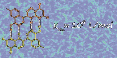
Supramolecular chemistry with ureido-benzoic acids
E W Meijer, Anja R. A. Palmans, Wilco Appel, Bas FM de Waal, Marko Nieuwenhuizen and Martin Lutz  
Chem. Sci., 2014, Accepted Manuscript
DOI: 10.1039/C4SC00871E, Edge Article


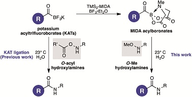
Synthesis and Chemoselective Ligations of MIDA Acylboronates with O-Me Hydroxylamines
Hidetoshi Noda and Jeffrey W Bode  
Chem. Sci., 2014, Accepted Manuscript
DOI: 10.1039/C4SC00971A, Edge Article


Digg This
Reddit This
Stumble Now!
Share on Facebook
Bookmark this on Delicious
Share on LinkedIn
Bookmark this on Technorati
Post on Twitter
Google Buzz (aka. Google Reader)

Artificial ion channels that work on a feedback loop

written by Cally Haynes, a Publishing Editor at the Royal Society of Chemistry

In nature, many cellular processes are controlled by structures that are only assembled when they are needed, in response to a chemical energy input. The structure (and hence the action of the structure) is maintained so long as the chemical energy input, or ‘fuel’ for the process is present. For example, microtubules can ‘grow’ or ‘shrink’ depending on the presence of guanosine triphosphate (GTP), and hence GTP serves as a ‘fuel’ for the function of the microtubules.

Inspired by this principle, Tom Fyles and his team from University of Victoria, Canada have used thiol–thioester exchange chemistry to assemble a metastable ion channel that disassembles over time in the absence of a chemical fuel.

In the scheme below, thiol 4 is not an active ion channel, but the addition of thioester 5 causes the formation of 3 by thiol-thioester exchange. 3 forms an active ion channel, but is metastable and therefore over time it disassembles into 4 + 6 which stops the ion transport. Adding more 5 (the fuel) will temporarily regenerate the ion transport as more C is formed.

While this system functions exactly as planned, its design was not without complications, says Fyles: “One setback was that these compounds do not work in vesicles so we had to develop reaction monitoring techniques using the bilayer clamp experiment.  That gives us exquisite sensitivity, but also allows us to detect very minor amounts of impurities.  So another setback was the observation that an oxidation impurity was vastly more active than the compounds we wanted to see.  This problem was easy enough to solve, but it took some time to recognize what was going on.”

This work also opens up exciting new avenues of investigation for the future for Fyles and his team. “The more general insight is that this system depends entirely on kinetic factors.  As chemists we are used to thinking about rates in terms of the reaction order and uniform reactant concentrations in the bulk.  Our system additionally shows the importance of local concentrations as opposed to the bulk concentration.  This introduces additional kinetic components related to diffusion.  The functioning of our system recorded using the bilayer clamp is largely controlled by diffusion processes, rather than the reaction kinetics we control in the design of the system.  This suggests that we might be able to make other systems that deliberately exploit non-uniform concentrations – yet another feature we can see in nature. Stay tuned.”

To download the full article for free*, click the link below:

Dissipative Assembly of a Membrane Transport System
A. K. Dambenieks, P. H. Q. Vu and T. M. Fyles
DOI: 10.1039/C4SC01258E

*Access is free untill the 17.07.14 through a registered RSC account – click here to register

Digg This
Reddit This
Stumble Now!
Share on Facebook
Bookmark this on Delicious
Share on LinkedIn
Bookmark this on Technorati
Post on Twitter
Google Buzz (aka. Google Reader)

Excellent visibility for your research

We want our authors’ research to get the visibility and recognition it deserves.

Chemical Science is dedicated to publishing findings of exceptional significance from across the chemical sciences.

The graph below shows the number of citations to articles published in 2013 in Chemical Science and other leading general chemistry journals. 

It’s clear to see from this data that articles in Chemical Science are less likely to receive no citations and after just a few months of publication articles will be highly cited – 45% of 2013 Chemical Science articles have already received 5 or more citations.

Publishing your research in Chemical Science means your article will have excellent visibility and will be read and cited quickly by your colleagues.

Chemical Science Citations

Citations to articles published in 2013 (Data downloaded from ISI Web of Science on 6 June 2014)

Browse the latest articles from Chemical Science here


Digg This
Reddit This
Stumble Now!
Share on Facebook
Bookmark this on Delicious
Share on LinkedIn
Bookmark this on Technorati
Post on Twitter
Google Buzz (aka. Google Reader)

Triptycene based hole transport materials for dye sensitized solar cells

Researchers from China have synthesized three novel hole-conducting molecules which may give rise to significant economic advantages for the production of dye sentisized solar cells. Web writer Kevin writes more…

Dye sensitized solar cells (DSSCs) are photovoltaic devices which use a sensitizer in combination with a photosensitive material and a wide band gap semiconductor to produce an electric current. Thin-film DSSCs can offer packaging and application opportunites, have potential as functional coatings, and exhibit high power conversion efficiencies (PCE) of up to 12-13%.

Current state-of-the-art dye sensitized solar cells are usually solid phase systems, removing any issues surrounding leakage of components or the corrosive nature of some electrolytes. In such configurations a constituent known as a hole transport material (HTM) is now commonly used instead of a liquid electrolyte.

In this Chemical Science Edge article, authors from the Energy Research Institute, Nanyang, and the Nanyang Technological University, Singapore, give details of a group of triptycene based molecules which, they claim, perform on a comparable level with the current best in class HTM, (spiro-OMeTAD). The materials described were prepared from a starting tri-iodotryptycene material which was derivatised using a variety of transformations including Stille and Suzuki coupling reactions. The materials contain diphenylamine moieties separated from the triptycene core by aromatic spacers, enabling charge transfer.

These materials were fabricated in a complex process into a typical DSSC, containing fluorine doped tin oxide, semiconducting titanium dioxide and lead iodide perovskite dye sensitizer material. Power conversion efficiencies are comparable to devices comprimising the best performing HTMs. For the T102 andT103 derivatives, PCEs were reported as 12.24 and 12.38%, comparable to devices fabricated with spiro-OMeTAD (12.78%). Without a HTM present the number falls to less than 5%.

This article presents details of promising HTM materials which may give rise to significant economic advantages for the production and commercialisation of solid state DSSCs, with current best in class performance levels.

Read this HOT Chemical Science Edge Article for free* today:

Novel hole transporting materials based on triptycene core for high efficiency mesoscopic perovskite solar cells

Anurag Krishna, Dharani Sabba, Hairong Li, Jun Yin, Pablo P. Boix, Cesare Soci, Subodh G. Mhaisalkar and  Andrew C. Grimsdale
DOI: 10.1039/C4SC00814F

About the webwriter

Kevin Murnaghan is a guest web-writer for Chemical Communications. He is currently a Research Chemist in the Adhesive Technologies Business Sector of Henkel AG & Co. KGaA, based in Düsseldorf, Germany. His research interests focus primarily on enabling chemistries and technologies for next generation adhesives and surface treatments. Any views expressed here are his personal ones and not those of Henkel AG & Co. KGaA.

*Access is free untill the 11.07.14 through a registered RSC account – click here to register

Digg This
Reddit This
Stumble Now!
Share on Facebook
Bookmark this on Delicious
Share on LinkedIn
Bookmark this on Technorati
Post on Twitter
Google Buzz (aka. Google Reader)

Fighting cancer with artificial antigen-presenting cells

Researchers from the Netherlands discuss the development of artificial antigen-presenting cells

Antigen-presenting cells (ACPs) are key players in the immune systems fight against cancer. On detection of tumor tissues, these ACPs pick up antigenic fragments of the tumor cell, migrate to the lymphoid organs and present the antigen to T-cells which play a central role in cell mediated immunity. Therefore, artificial ACPs (aAPCs) are often used in the development of immune-mediated anti-cancer therapies as they show great potential to mimic antigen-presentation, promoting T-cell activation.

Carl Figdor, a world-class immunologist, and colleagues from Radbound University discuss the current status of aAPC development in a Chemical Science Perspective. This review is partly focused on the developments in nanoscience which might improve future designs for immune-mediated anti-cancer therapies.

As a synthetic mimic, aAPCs must encompass the three signals that natural APCs use to encourage T-cell activation. This perspective discusses how these signals have been incorporated into aAPC designs, but also how physical properties such a size and shape are essential for targeting the aAPCs to T-cell rich areas in vivo.

Artificial antigen-presenting cells have the potential to develop into a widespread and powerful therapeutic tool. To download the full perspective, click on the link below – access is free for a limited time only!

Joep van der Weijden, Leonie Paulis, Martijn Verdoes, Jan C M van Hest and Carl Figdor
DOI: 10.1039/C4SC01112K
Looking for the best articles at the chemistry-biology interface. Check out our Chemical Biology Themed Collection showcasing some of the great research papers and reviews published in Chemical Science, in the areas of chemical biology and bioinorganic chemistry.
*Access is free through a registered RSC account – click here to register
Digg This
Reddit This
Stumble Now!
Share on Facebook
Bookmark this on Delicious
Share on LinkedIn
Bookmark this on Technorati
Post on Twitter
Google Buzz (aka. Google Reader)

A Faraday Discussion – Nanoparticle Synthesis and Assembly

Abstract submission is now open – submit your oral abstract by 21 July 2014

How does the nanoparticle-nanoparticle potential govern nanoparticle formation or assembly? Can one make more robust nanoparticle superlattice structures, which can be moulded, milled and machined into desired shapes? How does the assembly process vary with nanoparticle shape?

These questions cannot be answered in isolation – join the Nanoparticle Synthesis and Assembly, a Faraday Discussion which will be held on 20-22 April 2015 in Chicago, USA.

Abstract submission is now open, so take advantage of this excellent opportunity to showcase your latest research alongside leading scientists from across the globe. Don’t leave it too late – the deadline for oral abstracts is 21 July 2014.

Themes will include:

  • Nanoparticle synthesis – physical chemistry of nanoparticle shape and ligand control
  • Theoretical insights into nanoparticle synthesis and nanoparticle assembly
  • Nanoparticle self-assembly
  • Nanoparticle directed assembly

For more information and to see details of the speakers, visit the event website.

Digg This
Reddit This
Stumble Now!
Share on Facebook
Bookmark this on Delicious
Share on LinkedIn
Bookmark this on Technorati
Post on Twitter
Google Buzz (aka. Google Reader)

The Importance of Biometal Distribution in Neurodegenerative Disorders

Iain Larmour is a guest web writer for ChemSci. He has researched a wide variety of topics during his years in the lab including nanostructured surfaces for water repellency and developing nanoparticle systems for bioanalysis by surface enhanced optical spectroscopies. He currently works in science management. In his spare time he enjoys reading, photography, art and inventing.

Anthony White from the Department of Pathology, University of Melbourne and team have investigated the use of X-ray Fluorescence Microscopy to investigate subcellular biometal homeostasis in a mouse model of the childhood neurodegenerative disorder – Batten disease. This is a particularly nasty disease that causes progressive blindness and motor impairment leading to premature death. The team have previously highlighted the redistribution of brain zinc in diseased samples using subcellular fractionation techniques. However, the lack of a rapid, specific and sensitive technique to provide quantitative subcellular information in situ on biometal distributions is severely limiting the understanding of the effect that the changes in biometals distributions can have in neurodegenerative diseases.

Consider the next time you go food shopping. What if you arrived at the supermarket and all the shelves were empty. A shop assistant reassures you that there is plenty of stock, hundreds of thousands of items indeed, but it all just happens to be sitting in their central depot instead of the supermarket. This does not help you to make dinner that night. This scenario is an example of how important the distribution of items can be.

When it comes to neurodegenerative disorders the location of biometals, just like the location of the groceries in the above case, provides much more useful information than the total amount of biometal present. The amount of biometal may not change between normal and diseased cells, but the distribution does. Biometals are important cofactors to a large number of enzymes and are also recognised as second messengers in neuronal signalling

Intracellular zinc and calcium distributions mapped by x-ray fluorescence microscopy.

In their Chemical Science paper the team have utilised the advances in data collection speed of X-ray fluorescence detectors to map the zinc and calcium distributions in a statistically relevant number of cells. Despite a lack of global changes in biometal levels this approach revealed the perturbed trafficking of zinc and the significantly altered subcellular calcium distributions. The restorative properties of a therapeutic zinc-complex were also shown using this technique.

Techniques and methodology reported in the Chemical Science paper can be applied to other neurodegenerative diseases and importantly can provide insights to the mechanism of action of novel therapeutics at the single cell level.

Read the Chem. Sci. paper in full today for free* and see if this technique could be applied to a disease you are studying!

X-ray fluorescence imaging reveals subcellular biometal disturbances in a childhood neurodegenerative disorder
A. Grubman, S.A. James, J. James, C. Duncan, I. Volitakis, J.L. Hickey, P.J. Crouch, P.S. Donnelly, K.M. Kanninen, J.R. Liddell, S.L. Cotman, M.D. de Jonge and A.R. White*
DOI: 10.1039/C4SC00316K


*Access is free until 20.06.14 through a registered RSC account – click here to register

Digg This
Reddit This
Stumble Now!
Share on Facebook
Bookmark this on Delicious
Share on LinkedIn
Bookmark this on Technorati
Post on Twitter
Google Buzz (aka. Google Reader)

Photooxidation system that’s membrane-bound for success

Polly Wilson writes about a hot Chemical Science article for Chemistry World

Photosensitiser–water oxidation catalysts in heterogeneous (top), membrane-bound (middle) and homogeneous (bottom) systems

For the first time, a water oxidation catalyst and photosensitiser have been co-embedded into a membrane to make an artificial water photooxidation system. The arrangement can generate oxygen from water at far lower catalyst concentrations than ever before.

Chemical systems to turn solar energy into fuel are of paramount importance in the quest for sustainable green energy. Mimicking natural photosynthesis, the aim is to achieve sunlight-driven conversion of water and carbon dioxide into carbohydrates and oxygen. One branch of the process involves photocatalytic water splitting, comprising oxidative and reductive half reactions. The more complex photoxidation step involves a four-electron transfer reaction and very reactive oxygen intermediates. In nature, this extremely endothermic reaction is performed by Photosystem II (PSII), using membrane bound chromophores and catalytic units.


Read the full article in Chemistry World»

Read the original journal article in Chemical Science – it’s free to download until 30th June:
Photocatalytic water oxidation at soft interfaces
Malte Hansen, Fei Li, Licheng Sun and Burkhard König
Chem. Sci., 2014, Advance Article
DOI: 10.1039/C4SC01018C, Edge Article

Digg This
Reddit This
Stumble Now!
Share on Facebook
Bookmark this on Delicious
Share on LinkedIn
Bookmark this on Technorati
Post on Twitter
Google Buzz (aka. Google Reader)