Author Archive

Self-adjusting MOFs

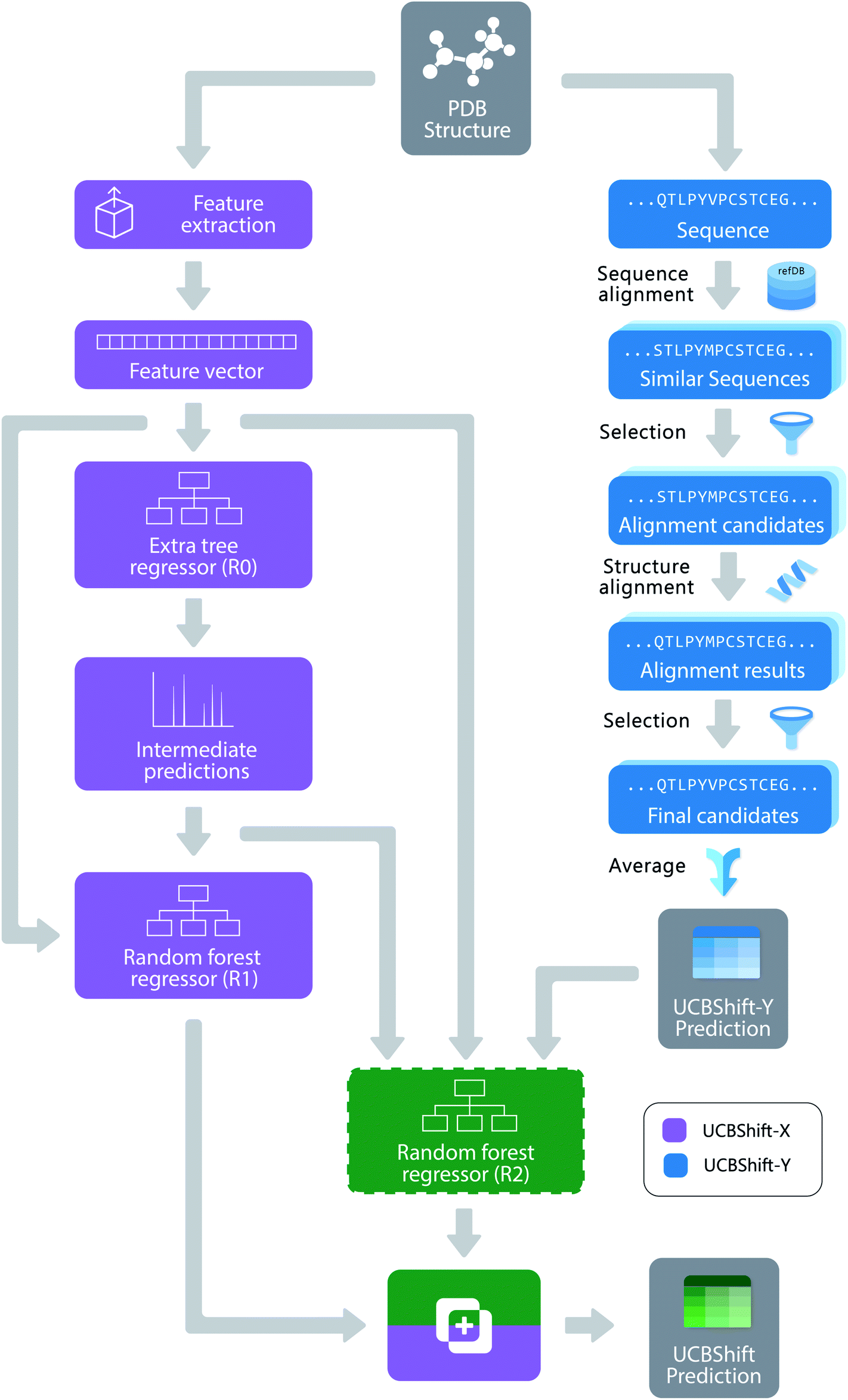
Recent decades have established that metal-organic frameworks (MOFs) are a pretty cool class of materials, with potential applications across a range of fields. In particular, their high porosities make them extremely attractive for storing a variety of gases, including possible fuels like methane and hydrogen. Two primary strategies have emerged to store H2 and CH4 in MOFs – synthesizing materials with unsaturated metals that can strongly bind to the target and synthesizing materials with small pores where multiple weak interactions combine to produce strong binding. Of course, these MOFs are designed with a single specific target in mind, making them synthetically complex and useful for storing only one type of molecule. Ideally, new MOFs with relatively straightforward syntheses that can bind multiple targets could be developed.

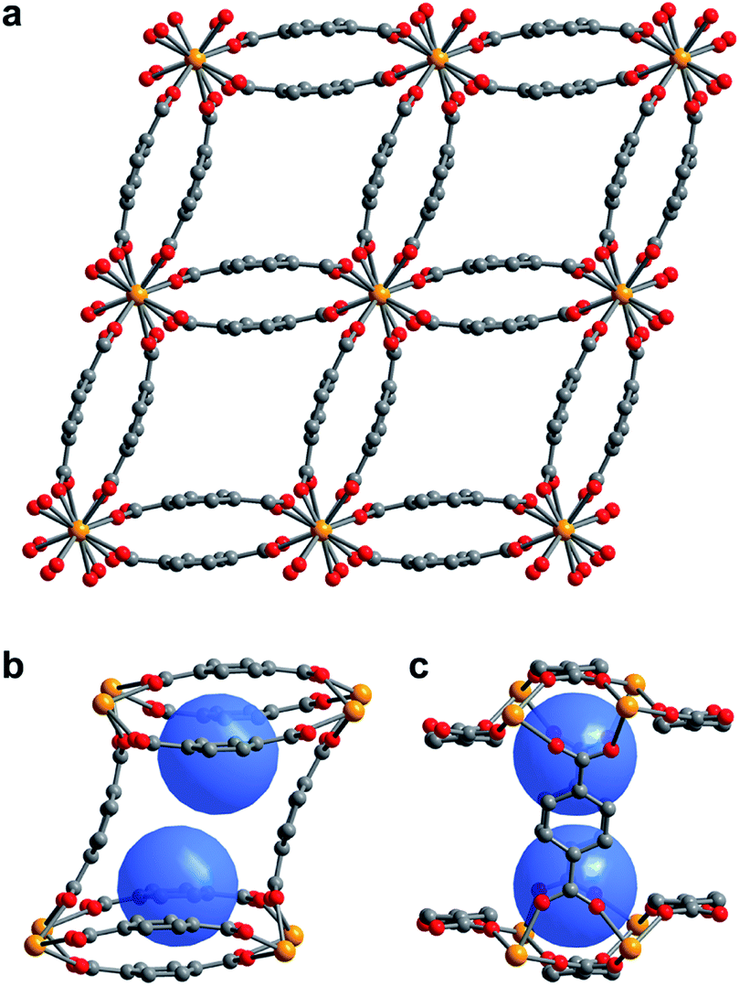
Figure 1. (a) Single crystal X-ray diffraction structure of 1–H2O. (b,c) View of the pores of 1–H2O showing binding pocket.

Scientists in the United States took a hybrid approach, creating MOFs with small, but flexible binding pockets. While the concept feels relatively straightforward and intuitive, it’s of course more complicated in practice. The MOF needs to hit a Goldilocks zone in terms of flexibility, where only a small number of select targets will bind rather than a wide range of gases. The researchers accomplished this by using an actinide (depleted uranium) as the metal nodes for their MOF as its tendency to adopt high coordination numbers should result in smaller pockets and limit possible rearrangements of the flexible linker. Also, the descriptor “as it is only mildly radioactive” is something I hadn’t read about a material before and rather caught my attention. The crystalline material, referred to as 1-H2O (Figure 1), was straightforwardly synthesized in an autoclave and isolated in relatively high yields. It features pores with two pockets that are capped by the bowl-shaped linkers. As synthesized, the pockets are occupied by water molecules that can be removed by heating the MOF under a dynamic vacuum.

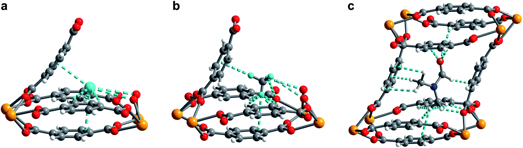
Figure 2. (a) Neutron powder diffraction structure of D2 adsorbed at site I in 1–D2. (b) Neutron powder diffraction structure of CD4 adsorbed at site I in 1–CD4. (c) Powder X-ray diffraction structure of DMF adsorbed inside the pore of U(bdc)2.

The MOF maintains its structural integrity after water removal, actually expanding slightly. This indicates that the MOF will contract upon binding, locking the target into place in the pocket. The researchers found that the MOF rapidly uptakes both H2 and CH4 at low temperatures, but the precise nature of the binding pocket adjustments can’t be determined by gas adsorption studies. To probe the structural details, the researchers turned to neutron powder diffraction to probe the binding of deuterated molecules to the MOF (Figure 2). The obtained structures show clear, cooperative effects that cause the adjustments to the binding pocket. The multiple different interactions allow the flexible structure to fit the two different adsorbates of interest, binding them both strongly. This work demonstrates the utility and versatility of flexible MOFs for adsorbing different gases with design principles that should be transferrable to non-radioactive materials.

To find out more, please read:

Self-adjusting binding pockets enhance H2 and CH4 adsorption in a uranium-based metal–organic framework

Dominik P. Halter, Ryan A. Klein, Michael A. Boreen, Benjamin A. Trump, Craig M. Brown and Jeffrey R. Long

Chem. Sci., 2020, Advance Article

About the blogger:

Dr. Beth Mundy is a recent PhD in chemistry from the Cossairt lab at the University of Washington in Seattle, Washington. Her research focused on developing new and better ways to synthesize nanomaterials for energy applications. She is often spotted knitting in seminars or with her nose in a good book. You can find her on Twitter at @BethMundySci.

Digg This
Reddit This
Stumble Now!
Share on Facebook
Bookmark this on Delicious
Share on LinkedIn
Bookmark this on Technorati
Post on Twitter
Google Buzz (aka. Google Reader)

Real World data for protein modeling

It should go without saying that NMR is an incredibly important characterization technique with profoundly broad applicability across the entirety of chemistry. Rarely do you find something that people who work on proteins and wacky main-group synthesis both consider crucial to their work. Given powerful enough magnets and high-quality samples, rich structural information can be obtained for all manner of molecules large and small. Large molecules do pose a problem with the sheer volume of information contained within a single spectrum. Because of this, there exists a need to develop computational programs that can translate spectra into detailed structural models. Currently, existing methods predict NMR spectra based on a combination of experimentally based databases with chemical shift heuristics. These simulations, while useful, lack high predictive rigor and often have difficulty simulating the messiness of real world data. This is particularly challenging because experimental spectra can often have significant chemical shift deviations from predicted values, with those peaks discarded as outliers.

Figure 1. The overall design of the novel UCBShift chemical shift prediction algorithm, combining both a transfer prediction module a machine learning module.

To face these challenges and generate more accurate results, researchers in the US developed a new algorithm that uses both machine learning and transfer prediction (Figure 1). Transfer prediction has been widely used and relies on the similarities of NRM peak sequences between known data, typically clean datasets, and the experimental sample in question. The advantage of the new approach is that it allows for data that would previously have been dismissed as anomalous to be utilized and to give more accurate predictions. The researchers used high-quality datasets that they modified for accuracy. In particular, they retained the water and ligand molecules that co-crystallized with the proteins that would likely be associated with the solvated forms of the proteins. As the interactions of these small molecules can alter the spectral shifts of NMR peaks, their inclusion increases the likelihood that peaks previously considered outliers will be incorporated and analyzed.

Figure 2. Difference between UCBShift-Y and SHIFTY+ (previous method) showing that overall the new algorithm is making better predictions.

Initial analysis with the new dataset produced some anomalous results, which were then mitigated by removing paramagnetic and other outlier proteins that would bias the results against the earlier algorithms. Once those were removed, the new algorithm still outperformed prior methods (Figure 2). While these advances are extremely useful for current researchers, they are approaching the limit of accuracy for systems that rely heavily on transfer predictions. In order to generate fully accurate models and structures intense work on combining deep learning with human expertise is necessary.

To find out more, please read:

Accurate prediction of chemical shifts for aqueous protein structure on “Real World” data

Jie Li, Kochise C. Bennett, Yuchen Liu, Michael V. Martin and Teresa Head-Gordon

Chem. Sci., 2020,11, 3180-3191

About the blogger:

Dr. Beth Mundy is a recent PhD in chemistry from the Cossairt lab at the University of Washington in Seattle, Washington. Her research focused on developing new and better ways to synthesize nanomaterials for energy applications. She is often spotted knitting in seminars or with her nose in a good book. You can find her on Twitter at @BethMundySci.

Digg This
Reddit This
Stumble Now!
Share on Facebook
Bookmark this on Delicious
Share on LinkedIn
Bookmark this on Technorati
Post on Twitter
Google Buzz (aka. Google Reader)

How to Identify Diverse Acids with High-Throughput Research

Have you ever heard of nonulosonic acids? As a materials chemist I hadn’t, as most of the acids I’ve dealt with are either acting as ligands or inorganic and I tend to forget that sugars are actually acids. These nine-carbon sugars are critical for a wide range of cellular functions throughout living organisms. Over 100 different members of this broad group of acids have been identified, all of which can undergo diversification at multiple positions, generating an even larger library of derivatives. What types of modifications can occur has yet to be fully realized, as novel compounds are continually discovered, particularly in prokaryotes. Even when examining a limited number of modifications, say 15, the number of possible derivatives is staggering, reaching several thousand without stereochemical considerations. Identifying these acids is key, as they have been linked to virulence in pathogenic bacteria and their previously mentioned diversity makes them challenging to genomically analyze.

Given these challenges, current detection practices rely on staining and fluorescent labeling which can be labor intensive and specific to a small number of acids. Early approaches based on mass spectroscopy were limited due to the complex backgrounds produced and the difficulties separating signal and noise. However, developments in high-resolution spectrometers and increased data processing abilities have led to successful analysis of increasing numbers of known metabolites. Researchers in the Netherlands and Russia extended this work to develop a screening approach to identify a diverse range of nonulosonic acids via small mass channel mass spectroscopy from previously unexplored environmental microbes (Figure 1).

Figure 1. Outline of the general procedure for the survey pipeline, beginning from the cells to the final structural analysis of the identified acids.

To successfully identify these unknown derivatives, the researchers identified specific ulosonic acid fragments invariant to functionalization. They used 1,2-diamino-4,5-methylene dioxybenzene, an alpha-keto-acid specific labeling reagent, to shift the double bond equivalents from the general cellular background and help conserve the core ulosonic acid structure. By screening a known range of acids with different functional group moieties, they were able to identify universal features for this category of molecules (Figure 2). From there, they created a full process for rapid-throughput studies that automated everything from initial identification of a potential acid molecule through filtering and structural analysis.

Figure 2. A) Universal fragmentation route used to identify nonulasonic acids in the screening, B) data output of various ulosonic acid derivatives identified in the study, and C) differences seen from the chemical labeling introduced in the procedure.

After the pipeline was established, the researchers used various standard cell lines, plant, animal, and algal, for a molecular level survey to confirm the validity of their approach for full-cell analysis. In general, the plants and yeast cells contained no nonulosonic acids, as expected, while the microalgae and animal cells had a range of different nonulosonic acids present. As these acids are thought to play a role in bacterial virulence, a bacteria known to display the acids was screened and several subtypes of animal and bacterial acids were identified as incorporated into the cells. With that validation, the researchers moved on to a wide range of non-pathogenic bacteria. They discovered that over half of these bacteria possess nonulosonic acids; in fact, some have only slightly lower acid abundance than mammalian cells. While much of this data was obtained from single cultures, it represents exciting validation of a broad new approach that could be used to identify potential targets for medical applications and continue to extend our understanding of the diversity of nonulosonic acids.

To find out more, please read:

Tackling the chemical diversity of microbial nonulosonic acids – a universal large-scale survey approach

Hugo B. C. Kleikamp, Yue Mei Lin, Duncan G. G. McMillan, Jeanine S. Geelhoed, Suzanne N. H. Naus-Wiezer, Peter van Baarlen, Chinmoy Saha, Rogier Louwen, Dimitry Y. Sorokin, Mark C. M. van Loosdrecht and Martin Pabst

Chem. Sci., 2020, 11, 3074-3080.

About the blogger:

Dr. Beth Mundy is a recent PhD in chemistry from the Cossairt lab at the University of Washington in Seattle, Washington. Her research focused on developing new and better ways to synthesize nanomaterials for energy applications. She is often spotted knitting in seminars or with her nose in a good book. You can find her on Twitter at @BethMundySci.

Digg This
Reddit This
Stumble Now!
Share on Facebook
Bookmark this on Delicious
Share on LinkedIn
Bookmark this on Technorati
Post on Twitter
Google Buzz (aka. Google Reader)

For catalysis, do you need one gold or two?

Doesn’t everyone love gold? Not only is it shiny and pretty in macroscopic form, but it’s one of the best-behaved nanoscale systems and the focus of extensive catalysis study. While much is known about the mechanisms of many gold-catalyzed reactions, a question of whether a number of organogold complexes are actual intermediates or off-cycle sinks remains. Catalysis of the nucleophilic addition of water to alkynes via gold complexes is a reaction with multiple hypothesized active intermediates and reaction pathways. Initially thought to occur via monoaurated species, subsequent work proposed activation by multiple gold catalysts. The problem, as seen in figure 1, is that the two pathways are connected and the presence of any specific intermediates can’t rule either pathway out.

Figure 1. Reaction scheme with potential intermediates for nucleophile addition to an alkyne via a gold-catalyzed pathway.

To solve this problem, researchers in the Czech Republic and the Netherlands developed a method to probe solution-phase intermediates with electrospray ionization mass spectrometry (ESI-MS) called Delayed Reactant Labeling. To do this, one of the reactants must be a mixture of isotopically labeled and unlabeled molecules, added separately with a time delay. This helps eliminate ionization artifacts and moves the reaction away from steady state conditions to allow for kinetic modeling. Using this technique, combined with other more standard characterization methods like NMR and infrared (IR) spectroscopy, the researchers studied gold-catalyzed water addition to alkynes. The catalyst is known to form digold hydroxides in the presence of water, which lends credence to the idea that a digold species is involved in the catalysis. Based on kinetic restrictions, they studied the addition of water to 1-phenylpropyne, which produces a mixture of regioisomers of intermediates that was a bit challenging to deconvolute. The initial ESI-MS spectra show the presence of both mono- and diaurated species and the different fragments were isolated, analyzed by IR photodissociation, and the spectra compared to theoretical models to corroborate their identity.

These results set the stage for the Delayed Reactant Labeling studies using deuterated 1-phenylpropyne. After the reaction reached equilibrium, in this case about 40 minutes, the labeled reactant was added to then allow for kinetic fitting of the intermediates. They determined that under standard conditions the monoaurated species has a half life of approximately 9 minutes and the diaurated species has a half life of 7 minutes. These decay constants could be altered by adding organic acids to degrade the complexes faster, while attempts to trap the species as salts were unsuccessful. Upon reaction with D2O a kinetic isotope effect doubling the lifetimes was observed and suggests that the mechanism is likely the same for all intermediates and that it involves a hydrogen/proton transfer. The two types of species also have slightly different rates of formation, with the diaurated species likely having a higher turn-over frequency. However, there isn’t a dramatic difference between the kinetics of these two types of intermediates.

Figure 2. Example of a) spectra obtained from the delayed reactant labeling method and b) fits of peak intensities over time used to extract kinetic information.

In order to determine which of the intermediates is catalytically relevant, the researchers changed the substrate to 3-hexyne. The symmetric alkyne has no regioisomeric intermediates to convolute the data, but the reaction kinetics are much faster and therefore not suited to the prior mechanistic studies. By adding an excess of acid, the rate determining step was moved from protodeauration.  Under these conditions, the rate has a linear dependence on the gold complex and likely proceeds primarily via monoaurated intermediates. This approach combining multiple analytical techniques elucidated the role of various gold-containing intermediates and demonstrated the utility of ESI-MS as a tool for determining reaction kinetics.

To find out more, please read:

Monoaurated vs. diaurated intermediates: causality or independence?

Mariarosa Anania, Lucie Jašková, Jan Zelenka, Elena Shcherbachenko, Juraj Jašk and Jana Roithová

Chem. Sci., 2020, Advance Article

About the blogger:

Beth Mundy is a PhD candidate in chemistry in the Cossairt lab at the University of Washington in Seattle, Washington. Her research focuses on developing new and better ways to synthesize nanomaterials for energy applications. She is often spotted knitting in seminars or with her nose in a good book. You can find her on Twitter at @BethMundySci.

Digg This
Reddit This
Stumble Now!
Share on Facebook
Bookmark this on Delicious
Share on LinkedIn
Bookmark this on Technorati
Post on Twitter
Google Buzz (aka. Google Reader)

Looking Inside Nanocomposites with Tomography

As a nanocrystal chemist, it pains me to say that sometimes nanoparticles aren’t enough. One strategy for engineering materials with complex functionalities is to embed nanoparticles into a larger host matrix structure. This has been widely studied for polymer-nanoparticle assemblies, but challenges abound for inorganic host matrices. Difficulties stem from problems controlling the microstructure of the host, as nanoparticles tend to accumulate at the borders of individual crystals in polycrystalline materials. Even when higher quality inorganic materials are made in the presence of nanocrystals loadings are below 1 wt%. A possible route to address these issues is by combining inorganic matrices with polymer-functionalized nanoparticles. The polymers can also form into vesicle structures that contain the nanoparticles which are larger and more compatible with characterization techniques.

One of those techniques is cryo-ptychographic X-ray computed tomography (cryo-PXCT), a fascinating and literally cool characterization method to image the internal structure of crystals. This is a variation on imaging techniques used heavily in medicine archeology to non-destructively visualize the interior of humans or artifacts. Cryo-PXCT cools the sample to -180 oC and has spatial resolution on the order of 50-70 nm. The researchers synthesized polymer vesicles and worms of approximately 232 nm in diameter and over 1 micron in length, respectively. The nanocomposites were made via an ammonia diffusion method with a solution of calcium chloride containing the polymer nano-structure exposed to gaseous ammonia and carbon dioxide to form CaCO3 crystals with nano-structure occlusions. The morphology of the nanocomposite crystal altered based on the type of occlusion – the vesicle/calcite combination retained a traditional calcite rhombohedral structure, while the worm/calcite composite crystals featured several rounded sides, an elongated shape, and only three flat faces. These composites possessed 15 – 25 wt% occlusions, significantly higher than prior work with pure nanoparticle incorporation.

Figure 1. SEM images of vessicle/calcite (left) and worm/calcite (right) nanocomposite single crystals.

Once prepared, the researchers examined the crystals by cryo-PXCT to determine the locations of the occlusions within the composites. In the vesicle/calcite composite the vesicles are non-uniformly distributed, with several layers of vesicle density, starting with a vesicle poor core, followed by a vesicle rich region, surrounded by another vesicle poor layer, with a slight vesicle enrichment near the surface. On average the vesicles are 300 nm apart and they maintain their shape, with the larger vesicles preferentially occluding in regions of higher occlusion densities.

Figure 2. Rendering of slice through the vesicle/calcite nanocomposite colored to show both components.

The worm/calcite composite crystals show a very different distribution of occlusions, with an hourglass of low density in the center of the crystal, surrounded by a worm rich zone, and an exterior worm poor layer. These zoning effects are likely determined by the interactions between the polymers and the growing crystal surfaces or the calcium cations in the solution. Cryo-PXCT offers a fascinating way to probe the internal structure of novel multicomponent crystals in three dimensions with nanoscale resolution, providing valuable information to eventually help determine structure-function relationships.

Figure 3. Tomographs of worm/calcite nanocomposites showing the localization of worms in an hourglass shape in the center of the crystal.

To find out more, please read:

Ptychographic X-ray tomography reveals additive zoning in nanocomposite single crystals

Johannes Ihli, Mark A. Levenstein, Yi-Yeoun Kim, Klaus Wakonig, Yin Ning, Aikaterini Tatani, Alexander N. Kulak, David C. Green, Mirko Holler, Steven P. Armes and Fiona C. Meldrum

Chem. Sci., 2020, 11, 355-363.

About the blogger:

Beth Mundy is a PhD candidate in chemistry in the Cossairt lab at the University of Washington in Seattle, Washington. Her research focuses on developing new and better ways to synthesize nanomaterials for energy applications. She is often spotted knitting in seminars or with her nose in a good book. You can find her on Twitter at @BethMundySci.

Digg This
Reddit This
Stumble Now!
Share on Facebook
Bookmark this on Delicious
Share on LinkedIn
Bookmark this on Technorati
Post on Twitter
Google Buzz (aka. Google Reader)

Molecular Switches from DNA

The idea of using DNA-based devices to create highly specific sensors, diagnostic tools, and therapeutics has inspired widespread research. DNA molecular switches, a basic class of DNA nanostructures, turn some process on and off depending on whether a substrate binds to the switch. They are typically coupled with some form of signal amplification process to increase sensitivity and should theoretically provide enhanced signal-to-noise. Unfortunately, current amplification procedures have a significant amount of background reactions, called leakage, that limit their current utility. One approach to design better switches is to find ways to observe single molecule dynamics. Combining transient binding of complimentary oligonucleotides with high resolution fluorescence microscopy allows for the development of highly sensitive molecular switches without leakage problems.

To do this, researchers in China developed a series of three-way junction DNA-based molecular switches. These TWJs possess a recognition domain, which interacts with the target of interest and induces a structural change in the TWJ, and a transduction domain, which then becomes accessible and binds to a fluorescent molecule (Figure 1).

Figure 1. General scheme of three-way junction molecular switch with two domains noted.

Experimentally, the TWJs were captured on the imaging surface and only fluorophores bound to a TWJ would remain in place long enough for signal to be acquired by the camera with a 500 ms integration time. The researchers found that shorter transduction domains with 5 or fewer base pairs were not stable enough to allow the fluorescent probes access in the absence of a bound target. The next generation of TWJs feature a hybridization probe to allow the switch to recognize specific DNA inputs. In the presence of inputs, the researchers observed transient binding behavior of the fluorophore, whereas they observed only nonspecific binding in the absence of inputs. The ability to differentiate between non-specific and transient binding in the single-molecule system gives a detection limit of 10 fM without concerns about leakage.

Building on this work, the researchers utilized the same general framework and substituted aptamer sequences for the hybridization probe in the recognition domain. They utilized split aptamer fragments that only draw together when bound to a target molecule. This motif was tested on ATP, a small molecule, and thrombin, a protein. These aptamer-coupled TWJs exhibited sensitivity to concentrations as low as 20 – 50 pm with high sensitivity (Figure 2). In the presence of molecular analogs to ATP or thrombin, the signal level showed no significant difference from that of a blank.

Figure 2. A) Split aptamer-based molecular switch schematic. B) Single-molecule fluorescence-time trajectory data in the presence (top) or absence (bottom) of targets. C) and D) Linear relationship between thrombin concentration and signal and specificity when compared to analogs. E) and F) Linear relationship between ATP concentration and signal and specificity when compared to analogs.

Another advantage of this system is its ability to provide information on the binding affinity of substrates, as it should impact the kinetics of the fluorescent probes. The dwell times of the fluorescence on and off states demonstrated exponential trends with changing input concentrations and could be fit to extract time constants. These time constants can then be used to derive the kinetics parameters and binding affinities of the target species. The general stability of the molecular switch framework allows for studying these types of interactions in a range of pH and salinity conditions, useful for mimicking different environments relevant to future applications. This provides a platform for studying the fundamental interactions that will allow DNA-based nanotechnology to move forward.

To find out more, please read:

Single-molecule dynamic DNA junctions for engineering robust molecular switches

Shuang Cai, Yingnan Deng, Shengnan Fu, Junjie Li, Changyuan Yu and Xin Su

Chem. Sci., 2019, 10, 9922-9927.

About the blogger:

Beth Mundy is a PhD candidate in chemistry in the Cossairt lab at the University of Washington in Seattle, Washington. Her research focuses on developing new and better ways to synthesize nanomaterials for energy applications. She is often spotted knitting in seminars or with her nose in a good book. You can find her on Twitter at @BethMundySci.

Digg This
Reddit This
Stumble Now!
Share on Facebook
Bookmark this on Delicious
Share on LinkedIn
Bookmark this on Technorati
Post on Twitter
Google Buzz (aka. Google Reader)

Metallohelices to kill microbes

I feel pretty safe saying that the development of effective antimicrobial drugs (looking at you penicillin!) was one of the most significant pharmacological events of recent history. Unfortunately, the widespread and often indiscriminate use of antibiotics has created an environment where bacteria with evolved drug resistances, colloquially known as “superbugs,” pose a serious threat to global health. While development of new small-molecule antimicrobial drugs is still ongoing, scientists are exploring alternative approaches as well. Of interest are antimicrobial peptides, found in plants and animals as a part of native immune systems. The most common are cationic antimicrobial peptides (CAMPs) comprised of 10-50 amino acids that possess both cationic and hydrophobic subunits and an overall positive charge. This allows them to strongly electrostatically interact with negatively charged bacterial cell membranes without disrupting neutral animal cells. Unfortunately, CAMPs have proved challenging to commercialize with generally unfavorable activities and issues with large-scale manufacture.

Figure 1. General synthetic scheme, structure, and key for components of metallohelices.

Researchers in the UK and Czech Republic recently developed a range of cationic metallohelices (Figure 1) that demonstrate structure-dependent activity against both Gram-positive and -negative bacteria. The diamine ligands form cleanly in the presence of [15]-crown-[5] and upon combination with 2-pyridinecarboxaldehyde and a metal salt self-assemble into the iron or zinc metallohelices. The metallohelices consist of a single enantiomer, as the ligands are optically pure, as determined by NMR and single-crystal X-ray diffraction when possible. Altering the aryl linker unit in the ligand the overall size and shape of the metallohelix in both zinc and iron derivatives. The iron metallohelices are water compatible, with lifetimes exceeding 10 days even in highly acidic conditions, and thus suitable for antimicrobial activity screening.

The researchers used ­in vitro studies to find minimum inhibitory concentrations (MICs), the lowest concentration necessary to see bacterial growth inhibition. While the metallohelices demonstrate the highest activity towards Gram-positive bacteria, some showed lethal effects on Gram-negative E. coli in 20-40 minutes. The 5b helices with a para-benzene bridging group acted most selectively on E. coli with the Λ-5b enantiomer acting as the champion compound and selected for further mechanistic study. The researchers exposed a notorious E. coli strain to inhibitory levels of Λ-5b in an attempt to isolate resistant mutants. The 17 isolates showed only slight tolerance increases rather than true resistance and could be classified into 4 genetic sub-types. Two of the sub-types developed mutations that altered the biophysical properties of their outer membrane, a third lost the ability to produce the vitamin B12 transporter protein, and the fourth lost the pO157 virulence plasmid (which makes this particularly E. coli strain particularly unpleasant). Taken in concert, these 4 sub-types show that tolerance can be derived from disrupting the ability of Λ-5b to interact with and cross the cellular membrane.

Figure 2. Fluorescence images of cells treated with Λ-5b coupled to a fluorescent probe, with arrows pointing to the localization of Λ-5b.

Given data suggesting the ability of Λ-5b to cross the cellular membrane, despite the relatively large of the metallohelices, the researchers used fluorescence microscopy to probe Λ-5b localization by coupling Λ-5b with a fluorescent label. The labeled metallohelix preferentially localized to regions in growing cells that contain anionic phospholipids (Figure 2). This indicates that Λ-5b can cross the cellular membrane and acts internally to the cell rather than simply acting via electrostatic interaction that disrupts the membrane. Overall, this work provides an exciting approach to developing novel anti-microbial drugs that mimic CAMPs with higher stability, activity, and easier synthesis.

To find out more please read:

Metallohelices that kill Gram-negative pathogens using intracellular antimicrobial peptide pathways

Daniel H. Simpson, Alexia Hapeshi, Nicola J. Rogers, Viktor Brabec, Guy J. Clarkson, David J. Fox, Ondrej Hrabina, Gemma L. Kay, Andrew K. King, Jaroslav Malina, Andrew D. Millard, John Moat, David I. Roper, Hualong Song, Nicholas R. Waterfield and Peter Scott

Chem. Sci., 2019,10, 8547-8557

About the blogger:

Beth Mundy is a PhD candidate in chemistry in the Cossairt lab at the University of Washington in Seattle, Washington. Her research focuses on developing new and better ways to synthesize nanomaterials for energy applications. She is often spotted knitting in seminars or with her nose in a good book. You can find her on Twitter at @BethMundySci.

Digg This
Reddit This
Stumble Now!
Share on Facebook
Bookmark this on Delicious
Share on LinkedIn
Bookmark this on Technorati
Post on Twitter
Google Buzz (aka. Google Reader)

Driving Molecular Motors with Visible Light

While nanobots are still a thing of science fiction, the development of the fundamental scientific concepts that could lead to highly complex molecular-scale machines has been an active area of research for over 2 decades. Molecular motors rely on isomerization through metastable intermediate states that leads to unidirectional rotation via a rotor portion of the molecule. Initial work utilized molecules that convert high-energy UV-light into this directional motion. However, these systems are limited in their future utility, particularly in biological applications, if they need UV-light for activation. Recent work focuses on driving molecular motors with visible light via a range of approaches, including altering the absorbance profile of the motor molecule itself. Unfortunately, changing the electronics of the molecule has previously substantially decreased the quantum yield while resulting in only slight red shifts.

Researchers in The Netherlands recently developed 2nd generation molecular motors featuring a mixture of electron-donating and electron-withdrawing groups that exhibit substantial red-shifts. Density functional theory (DFT) calculations predicted that adding cyano- and methoxy- groups to opposite halves of the motor would shift the absorbance past 410 nm. The new molecules were synthesized via a general procedure shown in Figure 1, with yields ranging from 3 – 10%.

Figure 1. General synthetic route to cyano- and methoxy- substituted molecular motors and photoisomerization reaction that results in unidirectional motion.

The addition of the cyano- and methoxy- groups shifted the absorbance maxima corresponding to the HOMO-LUMO transitions to from 422 – 453 nm, an increase of over 60 nm from the parent molecule. When irradiated, the molecules transitioned from their stable ground state to the metastable isomers. These were characterized by UV-Vis spectroscopy, with the emergence of further red-shifted features (Figure 2), and 1H NMR.  The 1H NMR spectra were obtained with in-situ irradiation (which sounds like a sweet experimental setup) at various wavelengths and the ratios of several specific protons on the rotor. The derivative 2 with a methoxy- group on the rotor and cyano- groups on the stator demonstrated activity with irradiation at wavelengths up to 530 nm.

Figure 2. UV-Vis absorption spectra of stable (solid) and metastable (dashed) isomers of the molecular motors.

The energy of activation of the rotation for all three derivatives was determined by Eyring analysis and corroborated by DFT calculations. All were around 90 kJ/mol, with 2 requiring the most energy to elongate the central alkene bond and isomerize. The quantum yields of the motors for the forward reaction range from 5.8 – 11.5%, comparable to state-of-the-art UV absorbing motors. The quantum yields for the back reactions were calculated to be significantly lower than those for the forward reaction, which corresponds to the excess of metastable isomers observed under active irradiation. These motors also exhibit high photostability, with no significant change in the ground state absorbance after irradiation and cycling. This is promising for smart materials applications where stability is crucial. This work pushes forward the design of molecular motor systems that utilize visible rather than UV light.

To find out more please read:

Photoefficient 2nd generation molecular motors responsive to visible light

Lukas Pfeifer, Maximilian Scherübl, Maximilian Fellert, Wojciech Danowski, Jinling Cheng, Jasper Pol and Ben L. Feringa

Chem. Sci., 2019, 10, 8768-8773.

About the blogger:

Beth Mundy is a PhD candidate in chemistry in the Cossairt lab at the University of Washington in Seattle, Washington. Her research focuses on developing new and better ways to synthesize nanomaterials for energy applications. She is often spotted knitting in seminars or with her nose in a good book. You can find her on Twitter at @BethMundySci.

Digg This
Reddit This
Stumble Now!
Share on Facebook
Bookmark this on Delicious
Share on LinkedIn
Bookmark this on Technorati
Post on Twitter
Google Buzz (aka. Google Reader)

Breaking C-H bonds with water, iron, and electricity

As we transition to an energy future composed primarily of intermittent, renewable technologies, finding ways to store the excess generated charges will be critical. While the general public typically thinks of batteries, another area of massive interest is storing the electrons in a chemical bond. This can be accomplished by creating electrocatalysts that can couple an electric current with chemicals like water and oxygen to generate stable new bonds in molecules for later use as fuels. These renewably generated fuels would then be available whenever needed, like at night. However, finding efficient electrocatalysts composed of earth-abundant materials has proven challenging. Many researchers have turned to nature for inspiration by designing molecules that mimic the active sites of enzymes.

Researchers in the United States used this approach, focusing on creating high-valent iron-oxo species, which others previously identified as the key catalytic intermediates in multiple enzymatic reactions. These types of species have traditionally been synthesized by reacting a reduced iron complex with an oxygen transfer reagent, but the researchers developed a system to generate highly reactive species using electricity as the reaction driving force and water as the oxygen source. The studied catalyst is a commercially available iron(III)-aquo complex with a tetraamido macrocyclic ligand (TAML) as the ancillary ligand (Figure 1B).

Figure 1. A. Cyclic voltammagram of (TAML)Fe in acetonitrile. B. Structure of (TAML)Fe and cyclic voltammagram showing increased current upon the addition of ethylbenzene.

When analyzed by cyclic voltammetry, an electrochemical technique where you cycle the voltage between set points and measure the current output, the (TAML)Fe shows two redox events at around 650 and 1250 mV that the researchers attributed to generating the FeIV-OH and FeV(O) species (Figure 1). Addition of ethylbenzene, which should react with the FeV species, increased the current at voltages of 1250 mV and higher, indicating (TAML)Fe turnover. However, isolating the Fev(O) species proved challenging as it reacts rapidly with the (TAML)Fe to form an FeIV dimer. This also limits the efficiency of the overall system by decreasing the amount of the most reactive species in solution.

Figure 2. A, B. Products generated by oxidation of various substrates screened with (TAML)Fe for electrocatalysis with isolated yield and calculated conversion in parenthesis. C. Substrates that did not react with the (TAML)Fe complex.

Both the FeIV dimer and FeV(O) species proved capable of oxidizing C-H bonds in ethylbenzene, but the FeV(O) is much more reactive and increases the oxidation rate at high electrochemical potentials. The researchers tested the scope of (TAML)Fe reactivity using a series of compounds with benzylic C-H bonds (Figure 2). They found that the (TAML)Fe performed well with electron-rich and electron-neutral derivatives, with an electron-deficient nitro-substituted derivative showing lower reactivity. Several substrates with non-benzylic C-H bonds showed high selectivity for oxidation at the benzylic C-H bond. (TAML)Fe also showed high electrocatalytic activity for oxidizing alcohols and converted substrates as simple as cyclohexanol and as complex as a steroid to ketones in high yields (up to 97%).

This study of an earth-abundant, stable, and commercially available electrocatalyst acts as a baseline for further studies with other similar metal complexes. Despite the efficiency limits attributed to dimerization, the high stability and selectivity of the (TAML)Fe could lead to its use with a broader range of substrates with varied functional groups.

To find out more please read:

Electrochemical C–H oxygenation and alcohol dehydrogenation involving Fe-oxo species using water as the oxygen source

Amit Das, Jordan E. Nutting and Shannon S. Stahl

Chem. Sci., 2019, 10, 7542-7548

About the blogger:

Beth Mundy is a PhD candidate in chemistry in the Cossairt lab at the University of Washington in Seattle, Washington. Her research focuses on developing new and better ways to synthesize nanomaterials for energy applications. She is often spotted knitting in seminars or with her nose in a good book. You can find her on Twitter at @BethMundySci.

Digg This
Reddit This
Stumble Now!
Share on Facebook
Bookmark this on Delicious
Share on LinkedIn
Bookmark this on Technorati
Post on Twitter
Google Buzz (aka. Google Reader)

Tryptophan, Featuring BN/CC Isosterism

Ever wanted to find a way to replace a carbon-carbon (CC) double bond with another bond that will change the physical and chemical properties of a molecule without significantly altering its sterics? Look no further than a boron-nitrogen bond! BN/CC isosterism involves substituting a CC double bond with a BN bond, which can substantially change the electronic properties of a molecule while keeping it the same size. This isosterism could be a powerful tool in biomedical studies of biologically relevant arene-containing organic molecules, which are plentiful. However, few studies report on the differences in functions cause by substituting a BN bond into an arene. Initial results suggest that the BN compounds can have similar or increased activity and availability when compared to the natural, all carbon molecules.

Figure 1. Image of naturally occuring tryptophan and the BN-tryptophan analogue.

Researchers in the United States synthesized a BN-analogue of tryptophan (Figure 1) for use as an unnatural amino acid (UAA) to study and intentionally alter the properties of proteins. Tryptophan, in addition to making Americans sleepy at Thanksgiving, is relatively rare, but participates in pi system interactions and is the primary source of native protein fluorescence. This makes it an important target for UAA research. The researchers synthesized the sodium salt of BN-tryptophan in a 6-step process, which can be modified to resolve the two enantiomers by chiral HPLC. The BN-tryptophan exhibits noticeably red-shifted absorbance and emission spectra, with the fluorescence maximum shifted by almost 40 nm in the BN compound.

In order to test whether the BN-tryptophan could be incorporated into proteins, researchers incorporated it into media without tryptophan and monitored whether E. coli cells that lacked the ability to produce tryptophan would grow. They found that the cells grew when in the presence of BN-tryptophan, but to a significantly lesser degree than with an equivalent quantity of natural tryptophan. However, cell growth increased when the media contained both BN-tryptophan and natural tryptophan. This suggests that cells will accept BN-tryptophan as a tryptophan analogue, but they don’t tolerate full replacement well.

Figure 2. Representation of the protein sequence, structures of other tryptophan analogues, and fluorescence plot for the studied substrates.

Further studies incorporated BN-tryptophan and three other previously utilized tryptophan analogues into a green fluorescent protein (GFP). For fluorescence to be detected, the analogue must be incorporated into the protein and then accurately read by cells. The BN-tryptophan performs as well or better than the established tryptophan analogues, proving its functionality (Figure 2). The proteins with BN-tryptophan also demonstrate several different properties than those containing natural tryptophan; their fluorescence is red shifted and they are more susceptible to oxidation by hydrogen peroxide. These alterations in activity could prove useful in future studies.

To find out more please read:

Synthesis and characterization of an unnatural boron and nitrogen-containing tryptophan analogue and its incorporation into proteins

Katherine Boknevitz, James S. Italia, Bo Li, Abhishek Chatterjee and Shih-Yuan Liu

Chem. Sci., 2019, 10, 4994–4998.

About the blogger:

Beth Mundy is a PhD candidate in chemistry in the Cossairt lab at the University of Washington in Seattle, Washington. Her research focuses on developing new and better ways to synthesize nanomaterials for energy applications. She is often spotted knitting in seminars or with her nose in a good book. You can find her on Twitter at @BethMundySci.

Digg This
Reddit This
Stumble Now!
Share on Facebook
Bookmark this on Delicious
Share on LinkedIn
Bookmark this on Technorati
Post on Twitter
Google Buzz (aka. Google Reader)