Researchers in Australia have devised a new way to test how well drugs penetrate the low-oxygen core of solid tumours. ‘Hypoxic’ regions of tumours are notoriously difficult to target with drugs and the new work could help in the development of new compounds that can effectively reach these areas and efficiently kill the cells.
Tumours often grow faster than the blood vessels that supply them, and parts of the tumour therefore become starved of oxygen and grow slowly. This makes it difficult for drugs carried in the blood to reach these areas; furthermore many drugs rely on the rapid proliferation of cancer cells, so slowly growing ones are less susceptible.
One approach has been to develop ‘prodrugs’, which become toxic to the cell only upon entering the low-oxygen environment. Some of these are based upon cobalt(III) attached to a toxic ligand. In a hypoxic environment the cobalt is reduced to cobalt(II) and the ligand is released. However, there are currently no reliable ways either to visualise the hypoxic region of a tumour or to measure penetration of the drugs.
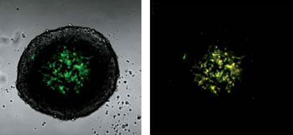
Now, Byung Kim, Trevor Hambley and Nicole Bryce at the University of Sydney have developed a three-dimensional model of a solid tumour with a hypoxic core that allows both the hypoxic region to be highlighted and the extent of penetration of prodrugs to be measured.
- Find out more in the Chemistry World news story and download the team’s Chemical Science Edge article for free.


")