Archive for March, 2017

Tomography keeps its cool to analyse ice cream

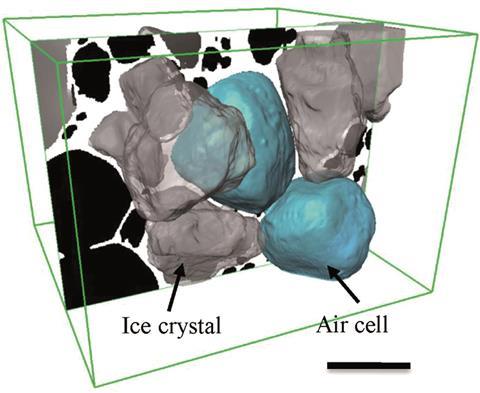
Source: © Royal Society of Chemistry This 3D rendered image shows a central air cell bounded by faceted ice crystals. Scale bar is 100mm

Researchers from the UK have developed a new 3D x-ray tomography (XRT) method to visualise the effects of changing temperature on the microstructure of ice cream.

Ice cream is a mixture of milk, fats, sugars, proteins, emulsifiers, stabilisers and flavours that are aerated and then frozen to form a soft solid comprising about 30% ice, 50% air, and 5–15% fat droplets suspended in a sugar solution. Its quality depends on the size of its ice crystals and air bubbles: smaller crystals and bubbles make it smoother and creamier. And since this complex colloid is unstable above –30˚C, its microstructure will change during shipping and storage (domestic freezers are usually at around –18˚C), which will affect its taste and texture.

Interested? The full article can be read in Chemistry World.

The original RSC Advances article can be read below and is open access:

Synchrotron X-ray tomographic quantification of microstructural evolution in ice cream – a multi-phase soft solid
Enyo Guo et al.,
RSC Adv.,2017, 7, 15561-15573
DOI: 10.1039/C7RA00642J

Digg This
Reddit This
Stumble Now!
Share on Facebook
Bookmark this on Delicious
Share on LinkedIn
Bookmark this on Technorati
Post on Twitter
Google Buzz (aka. Google Reader)

Can this quantum sized double-edged sword help diagnose and treat breast cancer?

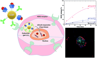
In a study led by Ko and colleagues at the Department of Dental Materials, School of Dentistry, Kyung Hee University, Korea, researchers armed graphene QDs with two therapeutic moieties: a HER-targeting antibody meant to help the therapeutic QDs find HER2-expressing breast cancer cells; and doxorubicin (DOX) – a chemotherapeutic drug used widely in treating breast cancer.

Consistent with previously established criteria (size, shape etc.) for drug carriers, the current study found that the estimated size of 222 nm makes the nanocarriers good candidates for further development toward diagnostic and therapeutic applications. Further, the nanocarriers had excitation and emission wavelengths of 370 nm and 450 nm respectively, making them glow in the ultraviolet range and as a result, optimal for medical imaging applications. The research team showed through chemical binding analysis that anti-HER antibodies were firmly bound to the QDs, and that the QDs were hydrophilic. The team conducted thermal stability studies and showed that the nanocarriers were stable at temperature ranges  much greater than the physiological body temperature range.


The study also analyzed whether the therapeutic nanocarriers were able to specifically target and enter breast cancer cells, release the DOX payload under specific pH and temperature conditions, and subsequently induce breast cancer cell death. Using a HER2-expressing breast cancer cell line, the team showed that the nanocarriers could kill cells in a dose dependent manner. A temperature of 37oC and pH of 5.5 were optimal for DOX release. Results in fluorescent microscopy studies suggested that DOX was released immediately after the nanocarriers entered HER2-expressing cells.


This study proposes that graphene-based QDs, when armed with anti-HER antibodies and DOX, have great potential for translation. In addition, with biomarker-based treatment decisions entering clinical practice in oncology settings, QD-based therapeutic nanocarriers are likely to have a notable impact on cancer therapy.
Read the full article here:

Graphene quantum dot-based theranostic agents for active targeting of breast cancer
N. R. Ko, M. Nafiujjaman, J. S. Lee, H.-N. Lim,a Y.-k. Lee and I. K. Kwon

Digg This
Reddit This
Stumble Now!
Share on Facebook
Bookmark this on Delicious
Share on LinkedIn
Bookmark this on Technorati
Post on Twitter
Google Buzz (aka. Google Reader)