Archive for the ‘Subject Areas’ Category

An Organometallic Toolbox for the Study and Synthesis of Unique N-Heterocyclic Carbenes

N-heterocyclic carbenes (NHCs) are an interesting example of chemical curiosity turned commonplace. NHCs are stable singlet carbenes located within an N-heterocycle, in which the carbon centre bears an sp2 hybridised pair of electrons. As early as 1835 chemists were thinking about carbenes, with Dumas’ optimistic (if unsuccessful) attempt to synthesise the methylene carbene by dehydrating methanol. For many years the intentional study of carbenes was considered too demanding because of their instability, and so they remained in relative obscurity. A number of seminal papers changed this preconception; in particular, a report by Wanzlick in 1968 reporting the synthesis of the first NHC-metal complex using mercury and the first synthesis of a stable and isolable NHC by Arduengo in 1991.

Intensification in research and interest in NHCs over the past thirty years may have originated with these seminal reports, but it continues because of the success of NHCs in catalysis: both as strongly σ-donating metal ligands and nucleophilic organocatalysts. One of the most valuable features of NHCs is the ability to tailor their steric and electronic properties by altering the heterocyclic ring and N-bound substituents. Accordingly, the study of NHC reactivity and the development of methods to functionalise NHCs are essential for continued innovation in this field.

Drs Marina Uzelac and Eva Hevia at the University of Strathclyde, Scotland, have written a review article summarising organometallic methods to metallate N-heterocyclic carbenes. The work summarises metallation of all three components of the NHC: the carbenic carbon, the heterocyclic backbone and the N-bound substituents.

The lithiated complex (1), synthesised by treatment of the N-heterocyclic carbene NHC with nBuLi, can be transmetallated at the C4 position by a number of main group elements to give a variety of bimetallic complexes (2). These complexes can be selectively quenched to generate NHC complexes with unconventional regiochemistry (3).

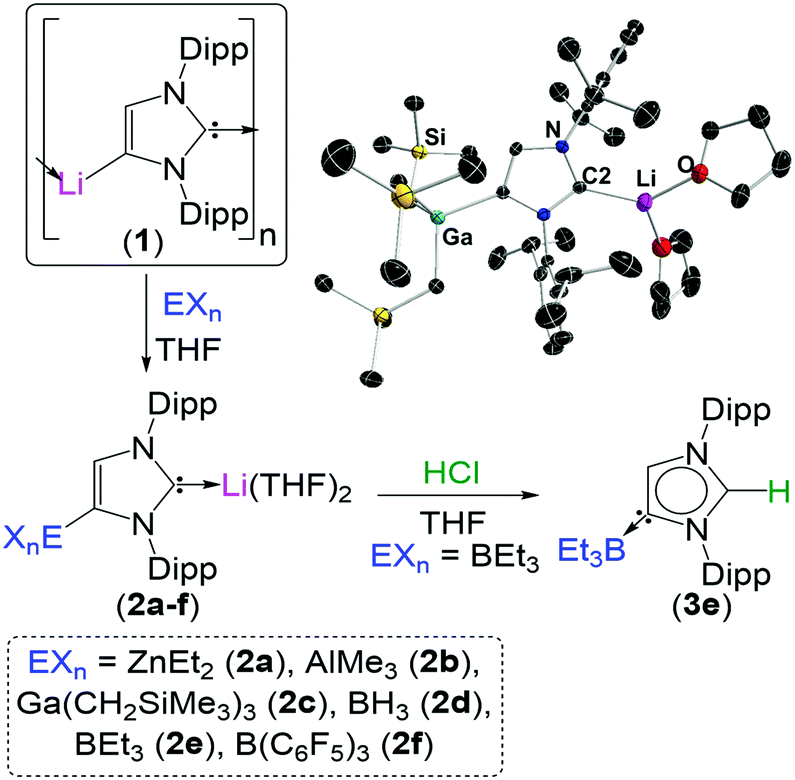
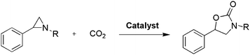
The lithiated complex (1) can be transmetallated at the C4 position by a number of main group elements to give a variety of bimetallic complexes (2). These complexes can be selectively quenched to generate NHC complexes with unconventional regiochemistry (3).

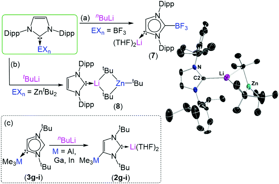
To exemplify the breadth of research discussed; beginning with 2,6-diisopropylphenyl (dipp) substituted imidazole-2-ylidenes, the reactivity of the NHC can be unlocked by initial addition of an alkali metal such as lithium, sodium or potassium (see figure). Metallation at the C4 position occurs by deprotonation of the vinyl protons in the NHC backbone, while a second metal coordinates to the carbene electron pair at the C2 position. From this species (1) it is possible to transmetallate the C4 position with a less-polar metal such as zinc, aluminium, gallium, boron or iron to furnish a bi-metallic NHC (2). Interestingly, addition of an electrophilic methyl or proton source to this species exclusively quenches the C2 position, generating a suite of unconventional complexes (3) with the carbene electron pair positioned on the C4 carbon.

Lithiation of NHC complexes: a) deprotonation of the backbone of NHC-borane complex; b) co-complexation of NHC-zinc complex with alkyllithium affording lithium zincate; c) deprotonation of the abnormal carbene complex.

Reactivity of main-group NHC complexes towards lithiation.

Further studies investigate how different reagents influence the regioselectively and extent of metallation, how metallated NHCs can activate small-molecules such as carbon dioxide, conditions which can lead to the metallation of N-dipp substitutents, as well as products and speciation following treatment of NHCs with a variety of bimetallic reagents.

In addition to expanding the knowledge of NHC reactivity, the work summarised in this review provides a reference and inspiration to researchers seeking to tailor NHCs for unique applications in synthesis and catalysis.

To find out more please read:

Polar organometallic strategies for regioselective C-H metallation of N-heterocyclic carbenes

Marina Uzelac and Eva Hevia.
Chem. Commun., 2018, Advance Article
DOI: 10.1039/c8cc00049b

About the author:

Zoë Hearne is a PhD candidate in chemistry at McGill University in Montréal, Canada, under the supervision of Professor Chao-Jun Li. She hails from Canberra, Australia, where she completed her undergraduate degree. Her current research focuses on transition metal catalysis to effect novel transformations, and out of the lab she is an enthusiastic chemistry tutor and science communicator.


Digg This
Reddit This
Stumble Now!
Share on Facebook
Bookmark this on Delicious
Share on LinkedIn
Bookmark this on Technorati
Post on Twitter
Google Buzz (aka. Google Reader)

Gold Rocks the Casbah

Researchers at the University of Texas have developed an inventive method to deliver molecules into the cell’s nucleus. Advances in gene therapy and the development of drugs that target DNA, the transcription machinery and other regulatory systems all rely on effective transport of molecules into the nucleus. Furthermore, achieving selective delivery of drugs reduces toxicity to non-target organs while maintaining the therapeutic effect.

Towards this aim, the authors delivered liposomes coated with clusters of gold nanoparticles into the cytoplasm. Laser irradiation of the cells heats the nanoparticles to high temperatures resulting in vapourisation of the water-based cytosol, and the transient formation of nanobubbles. The effect of this is an increase in the porosity of the nuclear envelope, enabling the transfer of various macromolecules from the cytoplasm into the nucleus. The authors describe this technique as ‘nanomechanical transduction’ because it is hypothesised that the mechanical effects brought on by the rapid growth and collapse (20 – 50 ns lifetimes) of the bubbles is responsible for the observed increase in porosity.

Local heating of gold nanoparticles and the subsequent formation of nanobubbles occurs due to ‘plasmon resonance’, whereby an electromagnetic field interacts with gold on the surface of the liposome and drives free-electron oscillation in resonance with the incident laser.

A diagram showing nanomechanical transduction. A gold-coated nanoparticle liposome enters the cell and, upon activation by a laser pulse, creates nanobubbles which causes mechanical disruptions in the cell and increased permeability of the nuclear membrane so molecules such as plasmids can enter.

A diagram showing nanomechanical transduction. A gold-coated liposome enters the cell and, upon activation by a laser pulse, creates nanobubbles and mechanical disruption within the cell, resulting in increased permeability of the nuclear membrane.

As a proof-of-concept the authors investigated whether nanomechanical transduction can improve the nuclear localisation of two different types of macromolecule: a dextran polymer labelled with a fluorescent dye, and a plasmid encoding the green fluorescent protein. In the first experiment, cells containing the dextran polymer were incubated with plasmonic liposomes and subjected to a near-infrared laser pulse. Up to 70% fluorescence intensity was observed in the nucleus compared to the cytoplasm, far exceeding the result from control experiments using electroporation to increase cell membrane permeability. In a similar way, nanomechanical transduction resulted in a 2.7 fold increase in the expression of the green-fluorescent protein compared to using electroporation, demonstrating efficient delivery of the plasmid into the nucleus.

The authors entitle their paper ‘rock the nucleus’ and, unintentional reference or not, I think a Casbah (one meaning is the central keep, or citadel, of a walled city) is a rather fitting analogy for the nucleus: the command post of the cell, and safeguard of genetic information. The authors of this work offer a sophisticated yet general method for molecules to breach the walls.

To find out more please read:

Rock the nucleus: significantly enhanced nuclear membrane permeability and gene transfection by plasmonic nanobubble induced nanomechanical transduction

Xiuying Li, Peiyuan Kang, Zhuo Chen, Sneha Lal, Li Zhang, Jeremiah J. Gassensmith and Zhenpeng Qin.
Chem. Commun., 2008, Advance Article
DOI: 10.1039/c7cc09613e

About the author:

Zoë Hearne is a PhD candidate in chemistry at McGill University in Montréal, Canada, under the supervision of Professor Chao-Jun Li. She hails from Canberra, Australia, where she completed her undergraduate degree. Her current research focuses on transition metal catalysis to effect novel transformations, and out of the lab she is an enthusiastic chemistry tutor and science communicator.

Digg This
Reddit This
Stumble Now!
Share on Facebook
Bookmark this on Delicious
Share on LinkedIn
Bookmark this on Technorati
Post on Twitter
Google Buzz (aka. Google Reader)

Anchoring Arynes on Graphene with Microwave but No Solvents

Recently in ChemComm, an international team from Italy and Spain reported a non-conventional way to anchor arynes onto graphene surface using microwave. Their developed method is fast, efficient, mild and solvent-free.

Attaching functional groups onto graphene surface, i.e. functionalization, allows the physical and chemical properties of graphene to be fine-tuned, such as electrical conductivity and solubility. Conventional solvent-based functionalization strategies usually involve time-consuming reactions and tedious purification steps. The poor suspension stability of graphene in solvents, particularly in polar organic solvents, greatly hinders the overall functionalization efficiency. Therefore, establishing easy and solvent-free functionalization protocols for graphene is highly needed.

M. Prato, A. Criado and coworkers made a breakthrough in addressing this challenge by developing a microwave-assisted functionalization method. Their method to functionalize graphene consists of cycloaddition reactions between few-layer graphene (FLG) and arynes (Figure 1). These reactions proceed by mixing the dry powder of FLG and arylene anhydrides, the precursors of arynes, followed by rapid heating under microwave irradiation. The whole process is solvent-free and occurs within half a minute. It is also applicable to a variety of arynes (Figure 2).

Figure 1. The schematic illustration of the microwave-assisted functionalization of graphene with arynes. This process can be carried out within half a minute and is solvent-free.

Figure 2. A variety of arynes capable of being anchored on graphene surface. 1~6 represent the arylene anhydrides and f-G(7)~f-G(12) are corresponding arynes attached onto graphene.

The most unique feature of the demonstrated method is the dual role of FLG. In addition to being one of the reactants, FLG is capable of absorbing microwave energy, and enables its surface to rapidly reach high temperatures that significantly accelerate the cycloaddition reactions.

This microwave-assisted functionalization method shows great promise as a stepping stone for the fast and efficient modulation of graphene surface and subsequently, the performance of graphene-based electronics.


To find out more please read:

Microwave-Induced Covalent Functionalization of Few-Layer Graphene with Arynes Under Solvent-Free Conditions

V. Sulleiro, S. Quiroga, D. Peña, D. Pérez, E. Guitián, A. Criado and M. Prato

Chem. Commun. 2018, DOI: 10.1039/C7CC08676H

About the blogger:

Tianyu Liu obtained his Ph.D. (2017) in Physical Chemistry from University of California, Santa Cruz in United States. He is passionate about scientific communication to introduce cutting-edge research to both the general public and scientists with diverse research expertise. He is an online blog writer for Chem. Commun. and Chem. Sci. More information about him can be found at http://liutianyuresearch.weebly.com/.

Digg This
Reddit This
Stumble Now!
Share on Facebook
Bookmark this on Delicious
Share on LinkedIn
Bookmark this on Technorati
Post on Twitter
Google Buzz (aka. Google Reader)

Hiding Carbon Dioxide in Oxazolidinones

Sometimes it feels as though the pinnacle of synthetic achievement is represented by 20 step total syntheses (with 10 contiguous stereocentres and 5 fused rings…). The level of chemical complexity that can be fashioned from simple building blocks is undoubtedly impressive, but amid such feats it is important not to lose sight of the elegance and worth of simple chemistry, especially when it aims to play a part in resolving profound challenges. One such challenge, which will increasingly confront future generations, is how to reduce the load of carbon dioxide in the atmosphere. One solution is to ‘fix’ carbon dioxide by integrating it into chemical building blocks of added complexity in a sustainable way.

The porosity and high surface area of metal organic frameworks (MOFs), a class of three-dimensional coordination networks, proffers them as ideal materials for capture and storage of carbon dioxide. A team of researchers have designed a MOF which consumes carbon dioxide in a different way: by transformation into value-added chemicals. The group have developed a catalytic MOF embedded with lewis-acidic copper centres capable of converting aziridines to oxazolidinones by the addition of carbon dioxide. Oxazolidinones are used as auxiliaries in chiral synthesis, and are structural components of some antibiotics.

The MOF, termed MMPF-10, is a metal-metalloporphyrin framework constructed from a copper-bound porphyrin ring chemically modified to incorporate 8 benzoic acid moieties, generating an octatopic ligand. These carboxylic acids groups form a second complex with copper in situ, termed a ‘paddlewheel’ for its appearance, with the formula [Cu2(CO2)4]. The resulting network contains hexagonal channels measuring 25.6 x 15.6 Å flanked by four of each of the two copper complexes. With 0.625 % of the catalyst at room temperature, 1 bar CO2 pressure, and in a solvent free environment, MMPF-10 catalyses the transformation of 1-methyl-2-phenylaziridine to yield 63% of the product.

metal-metalloporphyrin MOF catalyses catalyzes carbon dioxide fixation aziridines to oxazolidinones

Topology of MMPF-10 showing hexagonal channels in a) and c), and pentagonal cavities in b). Turquoise: copper, red: oxygen, grey: carbon, blue: nitrogen.

This work, a simple reaction to prepare oxazolidinones, shows that carbon dioxide can be fixed in specialised synthetic building blocks in a sustainable way. This is the way the first paragraph ended, ‘in a sustainable way’, because the challenge of developing such a reaction is two-fold: it must use carbon dioxide, and the reaction conditions must be sustainable. There will be no beneficial offset if the reaction uses a lot of energy, requires many resources, or generates larges quantities of waste. In this reaction the researchers have remained mindful of developing a mild, solvent-free reaction with low catalyst loading employing an earth abundant metal, reflecting an earnest aim to develop practical and sustainable chemistry.

To find out more please read:

A metal-metalloporphyrin framework based on an octatopic porphyrin ligand for chemical fixation of CO2 with aziridines

Xun Wang, Wen-Yang Gao, Zheng Niu, Lukasz Wojtas, Jason A. Perman, Yu-Sheng Chen, Zhong Li, Briana Aguila and Shengqian Ma
Chem. Commun., 2018, Advance Article
DOI: 10.1039/c7cc08844b

About the Author

Zoë Hearne is a PhD candidate in chemistry at McGill University in Montréal, Canada, under the supervision of Professor Chao-Jun Li. She hails from Canberra, Australia, where she completed her undergraduate degree. Her current research focuses on transition metal catalysis to effect novel transformations, and out of the lab she is an enthusiastic chemistry tutor and science communicator.

Digg This
Reddit This
Stumble Now!
Share on Facebook
Bookmark this on Delicious
Share on LinkedIn
Bookmark this on Technorati
Post on Twitter
Google Buzz (aka. Google Reader)

Single-Crystalline NiFe-Hydroxide Nanosheets for Catalyzing Oxygen Evolution

A group of scientists led by Prof. Shizhang Qiao has synthesized an oxygen evolution reaction (OER) catalyst combining the merit of low cost, excellent catalytic activity and long lifetime. This OER catalyst is composed of single-crystalline NiFe-hydroxide nanoflakes directly grown on nickel foams. The work has been published recently in ChemComm.

OER, the reaction of producing oxygen gas from water, is an indispensable component of electricity-generation devices using sustainable energy (e.g. fuel cells and photoelectrochemical water splitting cells). OER is usually the bottleneck limiting the overall energy conversion efficiency due to its sluggish kinetics and complex reaction pathways. As such, OER catalysts are needed to accelerate the OER reaction rate. Among the various OER catalysts, noble metal oxides stand out owing to their ultrahigh catalytic activity. However, the “shining” performance is dimmed by their high cost and short lifetime. Thus, obtaining alternatives with comparable OER catalytic activity as well as long-term stability is required to advance the utilisation of sustainable energy.

To address this challenge, the authors turned their attention to a low-cost transition metal, nickel. They developed a hydrothermal method using nickel foams to grow highly crystalline and near-vertically aligned NiFe-hydroxide nanosheets as OER catalysts (Figure 1a). The seamless integration between the hydroxide nanosheets and the nickel substrates reduces the contact resistance and facilitates interfacial electron transfer. The near-vertical orientation (Figure 1b) allows water molecules to fully contact the catalysts. Both of the characteristics render excellent OER catalytic activity. Additionally, the high crystallinity (Figure 1c) ensures the catalysts are robust enough to withstand extensive use without degradation in performance.

Figure 1. (a) The schematic illustration of the synthetic procedures of the NiFe-hydroxide [Fe-Ni(OH)2] nanosheets supported on nickel foams (NF). (b) The scanning electron microscopy image shows the near-vertically aligned nanosheets on a piece of nickel foam. (c) The transmission electron microscopy image reveals the crystallinity of the synthesized catalyst.

The NiFe-hydroxide nanosheets outperform most of the state-of-the-art OER catalysts, including those containing noble metal elements. Specifically, the nanosheets exhibit an onset potential of 1.497 V (Figure 2). The onset potential is a measure of the catalytic activity that equals the magnitude of potential required to yield a current density of 10 mA/cm2 (when appreciable amount of oxygen gas is evolved). Outstandingly, the onset potential of the NiFe-hydroxide is the smallest among the catalysts selected for comparison.

Figure 2. The polarisation curves of different OER catalysts. The onset potential is marked by the dotted line in the inset.

The catalytic activity is also highly stable, with no loss in performance after at least 100 h of measurement. Interestingly, the onset potential further shifts to a lower value of 1.465 V after 100 h. The authors attributed this observation to a “self-activation” process that involves the formation and accumulation of nickel oxyhydroxide (NiOOH) on the surface of the nanosheets.

The hydrothermal method demonstrated here could be used to synthesize other cost-effective crystalline catalysts to develop catalysts for reactions beyond OER, such as hydrogen evolution and carbon dioxide reduction.

To find out more please read:

Free-Standing Single-Crystalline NiFe-Hydroxide Nanoflake Arrays: A Self-Activated and Robust Electrocatalyst for Oxygen Evolution

Jinlong Liu, Yao Zheng, Zhenyu Wang, Zhouguang Lu, Anthony Vasileff and Shi-Zhang Qiao

Chem. Commun. 2017, DOI: 10.1039/c7cc08843d

About the blogger:

Tianyu Liu obtained his Ph.D. (2017) in Physical Chemistry from University of California, Santa Cruz in United States. He is passionate about scientific communication to introduce cutting-edge research to both the general public and scientists with diverse research expertise. He is a blog writer for Chem. Commun. and Chem. Sci. More information about him can be found at http://liutianyuresearch.weebly.com/.

Digg This
Reddit This
Stumble Now!
Share on Facebook
Bookmark this on Delicious
Share on LinkedIn
Bookmark this on Technorati
Post on Twitter
Google Buzz (aka. Google Reader)

Shoot the Messenger: Circular DNA-Graphene Oxide Material Targets mRNA in Living Cells

Schematic of the circular DNA cDNA/GO graphene oxide platform fabrication for intracellular mRNA messenger RNA imaging and gene therapy.

Scheme showing how cDNA/GO enters the cell and interacts with mRNA

Did you know that the combined length of DNA in your body’s cells is a number so large that the only references I could find use cosmic distances as a reference? Try twice the diameter of the solar system, or the distance to the moon and back 1500 times. Despite the complexity and infinite detail encountered when studying science, it is often something so simple as size that gives us pause. How can DNA be both uncomprehendingly huge and tiny at the same time?

The major function of DNA is to encode proteins, a process which begins with the transcription of genes into single-stranded messenger RNA (mRNA) molecules. It is mRNA that is directly translated into the strands of amino acids which fold to form proteins.

A team of researchers at Fuzhou University in China have developed a graphene oxide and circularised single-stranded DNA (cDNA/GO) hybrid material capable of penetrating living cells and binding mRNA. The material’s utility is shown in two practical applications: mRNA imaging and nucleotide therapeutics. The authors chose the mRNA of survivin and c-raf kinase as targets, because the enzymes are involved in carcinogenesis, and the mRNA are overexpressed in cancer cells and can be used as biomarkers.

cDNA was chosen for its increased stability over linear single-stranded DNA, which is rapidly degraded in vivo by exonucleases. For mRNA imaging the material is designed with a fluorescent dye coupled to the cDNA. GO was chosen as a hydrophilic delivery scaffold capable of adsorbing cDNA and quenching the dye. When cDNA/GO was incubated with HeLa cells (a cancer cell strain) a time-dependent increase in fluorescence was observed in the cytoplasm. Fluorescence is restored when cDNA encounters the target and desorbs from the GO to form a duplex with the mRNA.

CLSM images acquired for HeLa cells treated with both survivin and c-raf targeted cDNA/GO for duplexed intracellular mRNA imaging

The mRNA of both survivin and c-raf kinase can be imaged in living cells with cDNA/GO.

The researchers also probed whether the material might serve as a therapeutic agent: if formation of the cDNA-mRNA duplex blocks translation it may reduce the load of c-raf kinase and survivin in the cell and influence cancer cell growth. Accordingly, the researchers found that when the HeLa cells were incubated with cDNA/GO, cell proliferation was inhibited in a dose-dependent manner.

This research contributes a robust design which can be applied to diverse mRNA targets because optimisable properties such as stability, bioavailability and selectivity are largely independent of the sequence of nucleotides.

To find out more please read:

Circular DNA: a stable probe for highly efficient mRNA imaging and gene therapy in living cells

Jingying Li, Jie Zhou, Tong Liu, Shan Chen, Juan Li and Huanghao Yang
Chem. Commun., 2018, Advance Article
DOI: 10.1039/C7CC08906F

About the author

Zoë Hearne is a PhD candidate in chemistry at McGill University in Montréal, Canada, under the supervision of Professor Chao-Jun Li. She hails from Canberra, Australia, where she completed her undergraduate degree. Her current research focuses on transition metal catalysis to effect novel transformations, and out of the lab she is an enthusiastic chemistry tutor and science communicator.

Digg This
Reddit This
Stumble Now!
Share on Facebook
Bookmark this on Delicious
Share on LinkedIn
Bookmark this on Technorati
Post on Twitter
Google Buzz (aka. Google Reader)

In-Vivo Visualization of Glucose Metabolism with a Two-Color Imaging Technique

A group of scientists from Columbia University in United States have developed a state-of-the-art probing technique that can simultaneously map glucose uptake and incorporation activities in living cells.

Glucose is a ubiquitous “fuel” for most living organisms. Its metabolism, including uptake and incorporation, is vital to sustain the energy consumption of living organisms. Visualization of glucose metabolism is of critical importance for clinical diagnostics and fundamental biological researches. However, current imaging techniques are destructive to living cells, poorly resolved or incapable of probing uptake and incorporation at the same time.

Now in ChemComm, Prof. Min Wei’s research team demonstrates a breakthrough based on a vibrational imaging technique coupled with stimulated Raman scattering microscopy. This technique utilizes two glucose analogues to present the glucose metabolism, the 13C-labelled 3-O-propargyl-D-glucose (3-OPG-13C3) for glucose uptake and the D7-glucose for glucose incorporation. Conventional Raman spectroscopy is unable to distinguish the aforementioned two species due to their overlapping Raman peaks. The authors addressed this challenge by labelling 3-OPG with 13C that exhibits a blue shifted Raman peak, thus separating it from the peak of D7-glucose. Decoupling of the two peaks allows in-vivo imaging and simultaneous observation of glucose uptake and incorporation in cells with sub-cellular resolution.

Figure 1 shows the two-color mapping images collected for human cancer cells, PC-3. The blue (panel a) and red (panel b) areas display the regions where glucose incorporation and uptake are taking place, respectively. The two images can be easily obtained by tuning the wavenumber of the incident light to match with corresponding Raman peak positions. Use of light with other wavenumbers results in the black image (panel c) containing virtually no colored regions, showing the excellent selectivity of the technique. Additionally, this approach differentiates between cancer cells and healthy cells by comparing the blue to red color intensity ratio.

This novel and versatile imaging technique is expected to serve as a useful tool in advanced bio-imaging and future cancer diagnostics.

Figure 1. Two-color mapping images of PC-3 cells highlighting the (a) glucose-incorporation regions (Raman peak: 2133 cm-1) and (b) glucose-uptake regions (Raman peak: 2053 cm-1). (c) An image collected with a wavenumber (2000 cm-1) that does not match with either of the Raman peaks. Scale bar: 20 µm.


To find out more please read:

Two-color Vibrational Imaging of Glucose Metabolism Using Stimulated Raman Scattering

Rong Long, Luyuan Zhang, Lingyan Shi, Yihui Shen, Fanghao Hu, Chen Zeng and Wei Min

Chem. Commun. 2018, DOI: 10.1039/C7CC08217G

About the blogger:

Tianyu Liu obtained his Ph.D. in Physical Chemistry from University of California, Santa Cruz in United States. He is passionate about scientific communication to introduce cutting-edge research to both the general public and scientists with diverse research expertise. He is a web blog writer for Chem. Commun. and Chem. Sci. More information about him can be found at http://liutianyuresearch.weebly.com/.

Digg This
Reddit This
Stumble Now!
Share on Facebook
Bookmark this on Delicious
Share on LinkedIn
Bookmark this on Technorati
Post on Twitter
Google Buzz (aka. Google Reader)

Binder-free Integration of Bismuth Nanoflakes onto Nickel Foams for Sodium-ion Batteries

A new type of bismuth-based electrode material for sodium-ion batteries has been synthesized. This electrode consists of bismuth metal nanoflakes seamlessly integrated onto nickel foams. The electrode contains no polymer binders, a crucial component required to retain the structural integrity of most battery electrodes. This binder-free feature improves the amount of charge being stored (i.e. capacity) at fast charging rates.

Sodium-ion batteries are attracting worldwide research efforts as electric energy storage devices, in addition to the prevalent lithium-ion batteries, due to the abundance of sodium. Similar to the preparation of other battery electrodes, fabricating sodium-ion battery electrodes generally requires binders, e.g. polyvinylidene fluoride (PVDF), to hold powdered electrode materials together and glue them to metal supporting substrates. However, the electrically insulating nature of the binders impedes fast electron transport between electrode materials and supporting substrates, consequently degrading the capacity of the batteries at fast charging rates.

Now in ChemComm, researchers from Nankai University & the Collaborative Innovation Center of Chemical Science and Engineering in China demonstrate a bismuth-based electrode material that does not involve a binder. This characteristic is realized by the in-situ growth of bismuth nanoflakes onto nickel foams through a solution-based replacement reaction (Figure 1). Because the nanoflakes grow directly from the nickel foam surface and firmly anchor onto nickel (Figure 2a), the resultant Bi/Ni composite can be directly used as an electrode. Specifically, the bismuth nanoflakes and nickel foam serve as the active material and supporting substrate, respectively.

The Bi/Ni composite exhibited excellent electrochemical performance. It achieved a high capacity of 377.1 mAh/g at a current density of 20 mA/g. Significantly, when the current density increased 100-fold, its capacity could still retain 206.4 mAh/g, which is more than half of the capacity obtained at 20 mA/g (Figure 2b). This outstanding capacity retention is a benefit of the binder-free characteristic that reduces the resistance of electron transport.

The authors then elucidated the working mechanism of the bismuth nanoflakes by in-situ Raman spectroscopy. They concluded that a two-step alloying process was responsible for the charge storage activity.

Figure 1. A schematic illustration showing the synthetic process of the binder-free Bi/Ni electrode. By inserting a piece of nickel foam into an ethylene glycol (EG) solution containing bismuth(III) nitrate, Bi3+ can replace Ni metal, be reduced to Bi metal and deposit on the Ni metal surface.


Figure 2. (a) A scanning electron microscopy image of the bismuth nanoflakes. (b) A plot showing the capacity of the Bi/Ni electrode at different current densities.


The successful synthesis of the binder-free electrode is expected to encourage future works on the design and synthesis of integrated electrode materials to advance the performance of sodium-ion batteries.


To find out more please read:

In situ Synthesis of Bi Nanoflakes on Ni Foam for Sodium-ion Batteries

Liubin Wang, Chenchen Wang, Fujun Li, Fangyi Cheng and Jun Chen

Chem. Commun. 2017, DOI: 10.1039/c7cc08341f

About the blogger:

Tianyu Liu obtained his Ph.D. in Physical Chemistry from University of California, Santa Cruz in United States. He is passionate about scientific communication to introduce cutting-edge research to both the general public and scientists with diverse research expertise. He is a web blog writer for Chem. Commun. and Chem. Sci. More information about him can be found at http://liutianyuresearch.weebly.com/.

Digg This
Reddit This
Stumble Now!
Share on Facebook
Bookmark this on Delicious
Share on LinkedIn
Bookmark this on Technorati
Post on Twitter
Google Buzz (aka. Google Reader)

Creating Defects to Enhance Oxygen Evolution Activity: A Case Study using CoFe Layered Double Hydroxides

A group of scientists recently made a breakthrough in promoting the oxygen evolution activity of metal hydroxides. They developed a simple yet efficient strategy of immersing the metal hydroxides in diluted acid solutions.

The oxygen evolution reaction (OER) is a critical component for solar-driven water splitting that can sustainably acquire a clean fuel hydrogen gas by solar energy. Certain noble metal oxides, such as iridium dioxide (IrO2) and ruthenium dioxide (RuO2), work extremely well for catalyzing OERs. However, their scarcity restricts their potential for large-scale applications. To address the cost bottleneck, inexpensive alternatives such as metal hydroxides are being investigated worldwide. Unfortunately, their performance cannot compete with IrO2 or RuO2, partly due to their limited active sites for oxygen evolution. As such, there is a current need to develop strategies to promote the oxygen evolution activity of these metal hydroxides.

Figure 1. A schematic illustration showing the structural change of CoFe layered double hydroxide after being immersed in diluted nitric acid. Acid soaking creates Fe, Co and O defects (represented by VFe, VCo, and VO in the illustration, respectively) as well as separating the hydroxide layers.

Recently, Zhou et al. from Hunan University and Shenzhen University in China, demonstrated an easy acid-etching method that is capable of significantly improving the oxygen evolution activity of CoFe layered double hydroxide. When the hydroxide comes into contact with the nitric acid, protons remove some Co, Fe and O atoms and leave behind vacancies. These vacancies are named defects (Figure 2). Oxygen gas prefers to evolve at these defects and thus the defective hydroxide exhibits improved oxygen evolution activity. In addition, the nitrate anions can intercalate in between the metal hydroxide layers and break adjacent layers apart, exposing a large number of defect-containing surfaces and thus further boosting the oxygen evolution activity (Figure 1).

Figure 2. Transmission electron microscopy images of untreated CoFe layered double hydroxide (a, b) and acid-etched CoFe layered double hydroxide (c, d). After etching, the hydroxide nanoplates crack (due to layer separation) and surfaces become rough (due to creation of defects).

This method is expected to be applicable for a wide range of other metal hydroxides. The simplicity and efficiency of this method could make oxygen evolution catalysts cost-effective for commercialization.


To find out more please read:

Acid-etched Layered Double Hydroxides with Rich Defects for Enhancing the Oxygen Evolution Reaction

Peng Zhou, Yanyong Wang, Chao Xie, Chen Chen, Hanwen Liu, Ru Chen, Jia Huo and Shuangyin Wang

Chem. Commun. 2017, 53, 11778-11781

About the blogger:

Tianyu Liu obtained his Ph.D. in Physical Chemistry from University of California, Santa Cruz in United States. He is passionate about scientific communication to introduce cutting-edge research to both the general public and scientists with diverse research expertise. He is an online blog writer for Chem. Commun. and Chem. Sci. More information about him can be found at http://liutianyuresearch.weebly.com/.

Digg This
Reddit This
Stumble Now!
Share on Facebook
Bookmark this on Delicious
Share on LinkedIn
Bookmark this on Technorati
Post on Twitter
Google Buzz (aka. Google Reader)

Synthesis of Tin Dioxide Nanotubes for Lithium-ion Batteries with “A Grain of Oxalate Salt”

Preparation of tube-shaped electrode materials for lithium-ion batteries is a trending topic. Tubes with hollow cylindrical bodies allow exposure of the electrodes’ interior surface and can accommodate the large volumetric expansion commonly observed when lithium ions diffuse (either via intercalation or alloying) into the electrodes. The aforementioned two characteristics improve the specific capacity (a measure of how much electric energy one electrode can hold) and lifetime of electrodes.

Recently, the Mai research group from Wuhan University of Technology, China demonstrated a straightforward method for the synthesis of tin dioxide nanotubes as high-performance anodes for lithium-ion batteries. They adopted manganese(III) oxyhydroxide (MnOOH) nanowires as the sacrificial templates and immersed them in a batch of aqueous solutions containing tin(II) cations and oxalate anions (C2O42-). Afterwards, they warmed the mixture at 60 oC under constant magnetic stirring for 4 h and collected a white precipitate consisting of tin dioxide nanotubes. These nanotubes were then washed and coated with carbon thin films to improve their electrical conductivity and structural stability before being subjected to performance evaluations.

The presence of oxalate anions was crucial for producing the nanotubes with a well-defined shape. The function of these anions was revealed through a series of experiments. Oxalate anions first reduced MnOOH to manganese(II) cations and consumed protons in the vicinity of the MnOOH surface. The consumption of local protons increased the local pH and triggered precipitation and oxidation (by dissolved oxygen) of Sn2+ to tin dioxide. The two reactions proceeded, and eventually the MnOOH nanowires disappeared but tubes of tin dioxide formed around their surfaces (Figure 1). Samples obtained without oxalate salts were irregularly shaped.

Figure 1. (a) The schematic illustration of the synthesis steps of the tin dioxide nanotubes. (b) Scanning electron microscopy and (c) transmission electron microscopy images of the as-prepared tin dioxide nanotubes.

The carbon-coated tin dioxide nanotubes showed superior stability performance to bare tin dioxide nanotubes, as shown from the slower capacity-fading rate depicted in Figure 2a. In addition, carbon coating did not significantly sacrifice nanotubes’ charge-storage performance as both electrodes with and without a coating exhibited comparable capacity at all tested current densities (Figure 2b).

Figure 2. Performance comparison between carbon-coated tin dioxide nanotubes (SnO2@C NTs) and bare tin dioxide nanotubes (SnO2 NTs): (a) long-term stability and (b) capacity achieved at different current densities and charge-discharge cycle numbers.

To find out more please read:

Oxalate-assisted Formation of Uniform Carbon-confined SnO2 Nanotubes with Enhanced Lithium Storage

Chunhua Han, Baoxuan Zhang, Kangning Zhao, Jiashen Meng, Qiu He, Pan He, Wei Yang, Qi Li and Liqiang Mai

DOI: 10.1039/c7cc05406h

About the blogger:

Tianyu Liu is a Ph.D. in chemistry graduated from University of California, Santa Cruz in United States. He is passionate about scientific communication to introduce cutting-edge researches to both the general public and the scientists with diverse research expertise. He is a web blogger for the Chem. Commun. and Chem. Sci. blog websites. More information about him can be found at http://liutianyuresearch.weebly.com/.

Digg This
Reddit This
Stumble Now!
Share on Facebook
Bookmark this on Delicious
Share on LinkedIn
Bookmark this on Technorati
Post on Twitter
Google Buzz (aka. Google Reader)