Archive for the ‘Subject Areas’ Category

New template has potential for bone tissue engineering

British scientists at working at Imperial College, London have synthesised a macroporous hydroxyapatite bioceramic with potential uses in bone regeneration.

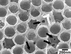
Porous hydroxyapatite (HA) bioceramics are widely studied for their potential use in bone regeneration. These materials are biocompatible and have a structure that contains many connected pores. This allows fluid to flow through the material and provides nucleation points for bone growth. The best results are obtained when the the pores are smaller than 1 μm and therefore synthetic methods that give good control over the pore size and material structure are highly desirable.

Prof. Molly Stevens and her team have now demonstrated a way of making an HA bioceramic with a 3D ordered (DOM) macroporous structure. They used an ordered macroporous carbon template (OMC)  in combination with a sol-gel method to form the desired bioceramic. They found that the DOM HA, when compared with HA material made without the OMC template, allowed the nucleation of calcium phosphate on the surface of the pores, thus demonstrating that the template method used affects the crystallisation of material.

hydroxyapatite bioceramics

The team hopes that these materials will find applications not only in the generation of bone but also in the generation of other materials. Want to find out more? Then download the ChemComm article for free today and leave a comment below to let us know what you think.

Digg This
Reddit This
Stumble Now!
Share on Facebook
Bookmark this on Delicious
Share on LinkedIn
Bookmark this on Technorati
Post on Twitter
Google Buzz (aka. Google Reader)

Probing cells’ power generators

UK scientists have developed a probe to monitor bicarbonate concentrations in mitochondria – components in living cells that generate chemical energy. Monitoring bicarbonate levels will improve researchers’ understanding of its role in cellular reaction mechanisms. 

A challenge when designing cellular probes is ensuring that the probe is not only selective for its target but can also be delivered to the site of interest within the cell. A team of scientists led by David Parker at the University of Durham has made a probe that can overcome this challenge. 

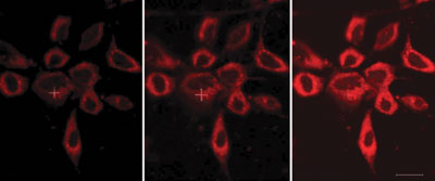
Stained HeLa cellsThe luminescent probe features an azaxanthone moiety, which is linked to a europium complex by an amide bond. The azaxanthone allows the probe’s uptake into cells and localisation within the mitochondria, and the europium complex has an affinity for bicarbonate ions. The ability to probe bicarbonate levels ‘can offer an unprecedented insight into signalling mechanisms’, says Parker.

Read the rest of this story in Chemistry World and download Professor Parker’s ChemComm communication, which is free to access for a limited period.

Also of interest:
Definition of the uptake mechanism and sub-cellular localisation profile of emissive lanthanide complexes as cellular optical probes
Elizabeth J. New, Aileen Congreve and David Parker, Chem. Sci., 2010, 1, 111-118

Digg This
Reddit This
Stumble Now!
Share on Facebook
Bookmark this on Delicious
Share on LinkedIn
Bookmark this on Technorati
Post on Twitter
Google Buzz (aka. Google Reader)

Across the barrier for tumour imaging

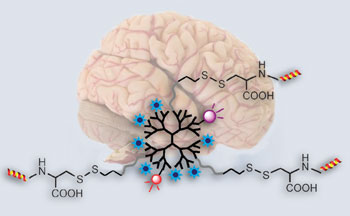
Brain and structure of nanoprobeA probe that can cross the blood-brain barrier to allow high sensitivity brain tumour imaging has been made by Chinese scientists. The probe could be used to pinpoint the location and extent of a tumour before an operation and be used for image-guided tumour removal. 

Establishing the position, extent and structure of brain tumours is crucial for their successful removal. But, current tumour imaging agents used in magnetic resonance imaging are limited by short circulation lifetimes, non-targeted specificity and poor blood-brain barrier permeability. The results of these limitations are that low grade tumours and 20-30 per cent of advanced brain tumours with an intact blood-brain barrier go unnoticed.

Cong Li from Fudan University, Shanghai, and his team made the probe starting with a dendrimer – a branched molecule with a long circulation lifetime – and attached functional groups with different tasks. One such group, a lipoprotein ligand angiopep-2, helps to deliver the probe across the blood-brain barrier and targets the lipoprotein’s receptors, which are present in increased amounts on tumour cells. High-resolution images can be generated thanks to imaging reporters, including fluorescence dyes, attached to the dendrimer. 

Read the rest of this story in Chemistry World and download Li’s ChemComm communication, which is free to access for the rest of the month.

Digg This
Reddit This
Stumble Now!
Share on Facebook
Bookmark this on Delicious
Share on LinkedIn
Bookmark this on Technorati
Post on Twitter
Google Buzz (aka. Google Reader)

New acetylene polymerisation catalyst is best yet

A team of chemists working in the UK and Australia have developed a new, highly active catalyst for acetylene polymerisation.

Polyacetylene is an important material as it has a degree of conjugation that leads to good conductive properties. It has found uses in areas such as synthetic metals and plastic electronics. New routes to make this polymer are constantly sought and, to date, the majority of catalysts for this task have low activities.

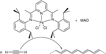
This problem has now been solved by two teams at the University of Tasmania and Imperial College London led by David McGuinness and George Britovsek, respectively. They took a bis(imino)pyridine iron catalyst, normally used for ethylene polymerisation, along with methylaluminoxane and successfully produced polyacetylene.

Graphical abstract: High activity acetylene polymerisation with a bis(imino)pyridine iron(ii) catalyst

An activity of 62 500 h-1 (turn over frequency; TOF) was obtained, which compares excellently with the 100  h-1 TOF obtained when using a standard existing acetylene polymerisation catalyst under the same conditions. This represents the most active acetylene polymerisation reported to date. Curently, the team are studying the effects of ligand modification and chain transfer reagents on the reaction.

Want to find out more? Then download  the full ChemComm article for free today.

Digg This
Reddit This
Stumble Now!
Share on Facebook
Bookmark this on Delicious
Share on LinkedIn
Bookmark this on Technorati
Post on Twitter
Google Buzz (aka. Google Reader)

New sensor for naked-eye fluoride detection

Fluoride, in its organic and inorganic forms, has been increasingly present in the food and beverage chain over the years. It has been added to toothpastes to prevent cavities; different forms of fluoride have been included in pesticides; several categories of food, including cereals, have been fortified with fluoride and even water for public consumption has been artificially fluorinated for decades.
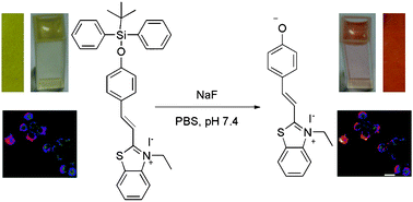
Graphical abstract: A highly selective colorimetric and ratiometric fluorescent chemodosimeter for imaging fluoride ions in living cellsDespite being a useful supplement to support the healthy growth of hair, nails and teeth and the strengthening of bones, an excessive intake of fluorine can lead to adverse effects on development. It can cause mottling of teeth and skeletal fluorosis (causing joint pain and abnormalities in the skeletal structure). Other effects have only been studied in animal models and at concentrations unlikely to be encountered by humans.

Regardless of the debate about its toxicity, it cannot be denied that as such a ubiquitous chemical in the human food chain, the development of simple methodologies and techniques to accurately detect the concentration of this anion in vivo plays an important role in biochemical research.

One of these techniques employs the use of fluorescent indicators that detect the presence of fluoride anions in solution.

Recent research by the Chinese group of Ma, Du and Zhang has been focused on the realisation of a novel “chemodosimeter” for fluoride that responds to the requisites of ease of synthesis, activity in highly aqueous solutions and buffers, cell permeability, quantitative response and high selectivity.

Their sensor, incorporating benzothiazolium hemicyanine as the fluorophore, was tested in fluorine detection in water:ethanol solutions (7:3) containing phosphate buffered saline at a pH of 7.4. In these conditions, the fluorescence of the sensor was not only quantitatively responsive to changes in fluoride concentration, but showed a change in the fluorescence spectrum, with emission at a different wavelength when in presence of the analyte and even noticeable to the naked eye. The versatility and selectivity of the system was also assessed by performing competition experiments in the presence of other anions, such as CO32-, SO42-, NO3, Cl, I and selected aminoacids and proteins like cysteine and human serum albumin, demonstrating a remarkable preferential response for fluoride.

Tests were also performed on living cells in order to determine the cytotoxicity of the chemodosimeter, using HeLa cells as the test substrate, showing low toxicity under the operational conditions.

The selectivity over different anions and analytes of biological relevance, the ability to operate in strongly aqueous solutions, the reliability and quantitative response and the applicability to living cells may make this new chemodosimeter a beneficial tool for biomedical researchers.

To find out more, read the full article:

A highly selective colorimetric and ratiometric fluorescent chemodosimeter for imaging fluoride ions in living cells
Baocun Zhu, Fang Yuan, Rongxia Li, Yamin Li, Qin Wei, Zhenmin Ma, Bin Du and Xiaoling Zhang
Chem. Commun., 2011, DOI: 10.1039/C1CC11308A

Posted on behalf of Dr. Giorgio De Faveri, Web Writer for Catalysis Science & Technology.
Digg This
Reddit This
Stumble Now!
Share on Facebook
Bookmark this on Delicious
Share on LinkedIn
Bookmark this on Technorati
Post on Twitter
Google Buzz (aka. Google Reader)

The importance of chemistry in sequencing nucleic acids


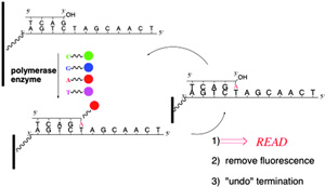
Solid phase DNA sequencing

Chemistry has played a vital role towards making routine, affordable sequencing of human genomes a reality. Leading UK scientist, Shankar Balasubramanian, provides a compelling review of the last 60 years, from the Sanger sequencing method through to the human genome project. In particular, Balasubramanian focuses on the achievements of Solexa (latterly, Illumina) on the modern developments of high throughput nucleic acid sequencing that originated in Cambridge, in the UK.
   

In association with the International Year of Chemistry (IYC), this Highlight in Chemistry emphasises the importance of chemistry and how it continues to contribute towards many other fields, most notably the biological and biomedical sciences. This sequencing approach is helping to transform science and offers intriguing prospects for the future of medicine.

Fancy reading more? Download the ChemComm Highlight, which will be free to access until the 1st July 2011.

 For your info, ChemComm is publishing Highlights in Chemistry articles throughout 2011, which has been recognised as an official activity for celebrating the IYC. Take a look at the IYC website to keep up-to-date on what else is happening throughout 2011 to celebrate the achievements of chemistry and its contributions to the well-being of humankind.

Digg This
Reddit This
Stumble Now!
Share on Facebook
Bookmark this on Delicious
Share on LinkedIn
Bookmark this on Technorati
Post on Twitter
Google Buzz (aka. Google Reader)

Totally synthetic over bryostatin

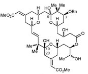
Bryostatins are macrocyclic natural products with various biological activities, including potent anti-cancer activity due to protein kinase C inhibition. Eric Thomas and his team from the University of Manchester have shown that it is possible to prepare 20-deoxybryostatin, using a modified Julia olefination to form the tricky 16,17-double-bond, followed by macrolactonisation, selective deprotection and oxidation.Fancy finding out more about the reaction conditions?

Download the ChemComm communication, which will be free to access until the 1st July 2011. 

20-deoxybryostatin

Digg This
Reddit This
Stumble Now!
Share on Facebook
Bookmark this on Delicious
Share on LinkedIn
Bookmark this on Technorati
Post on Twitter
Google Buzz (aka. Google Reader)

Cram Lehn Pedersen Prize – now open for nominations


Digg This
Reddit This
Stumble Now!
Share on Facebook
Bookmark this on Delicious
Share on LinkedIn
Bookmark this on Technorati
Post on Twitter
Google Buzz (aka. Google Reader)

Imaging brain tumours

A new probe for imaging brain tumours could offer increased hope for cancer patients, say Chinese chemists.

Den-Angio visualizes an orthotopic U87MG tumor non-invasively in vivo. (A) NIR fluorescence and X-ray/color coded NIR fluorescence images of a mouse head at 2 h PI of Den-Angio. Arrows point to the tumor. (B) T1-weighted MR images of a mouse brain (coronal plane) at 2 h PI of Den-Angio or Gd3+–DTPA with the same gadolinium dose (0.05 mmol kg−1). White arrows point to the tumor and red arrows point to the cerebral ventricle. (C) Histological H&E staining of identical brains in panel B. Scale bar, 2 mm.Cong Li, at Fudan University, Shanghai, and colleagues have made a dendrimer-based nanoprobe called Den-Angio that can cross the blood-brain barrier. It can be used in the magnetic resonance imaging of brain tumours and should make it easier for doctors to distinguish cancerous tissue from healthy cells when cutting out the tumour.

To find out more, read Li’s ChemComm communication.

Digg This
Reddit This
Stumble Now!
Share on Facebook
Bookmark this on Delicious
Share on LinkedIn
Bookmark this on Technorati
Post on Twitter
Google Buzz (aka. Google Reader)

Inducing protein heterodimerisation

Scientists in The Netherlands and Germany have shown that cucurbit[8]uril (CB[8]) induces selective heterodimerisation between two different proteins.
 

Supramolecular induced protein dimerisation

Luc Brunsveld and his team functionalised the two proteins with methylviologen (electron deficient) and napthalene (electron rich) guest molecules, which formed a charge transfer complex inside the CB[8] cavity.  Interestingly, the resulting dimerisation can be visually observed and has established that there is distinct interplay between the supramolecular components within the proteins.

Fancy reading more? Then download the ChemComm communication,which will be free to access until the 24th June 2011.

Digg This
Reddit This
Stumble Now!
Share on Facebook
Bookmark this on Delicious
Share on LinkedIn
Bookmark this on Technorati
Post on Twitter
Google Buzz (aka. Google Reader)