Archive for the ‘Subject Areas’ Category

Joint ChemComm–RSC Prizes and Awards Symposium in London

We are delighted to announce the forthcoming ChemComm–RSC Prizes & Awards Symposium in association with the RSC Analytical Division.

Date: Wednesday 22nd February 2012
Location: Imperial College London, UK
Time: 1300-1800

The purpose of this event is to bring together scientists in a stimulating and friendly environment to recognise the achievements of individuals in advancing the chemical sciences and also to foster collaborations. The symposium will appeal to academic and industrial scientists with an interest in analytical science, protein structure and interactions, and biosensors. Attendance at the symposium is FREE OF CHARGE and student participation is strongly encouraged.

The following distinguished scientists have agreed to speak:

For further details and to register your interest, please contact Anne Horan.

***
The closing date for RSC Prizes and Awards 2012 is 15th January 2012. Find out more >
***

Digg This
Reddit This
Stumble Now!
Share on Facebook
Bookmark this on Delicious
Share on LinkedIn
Bookmark this on Technorati
Post on Twitter
Google Buzz (aka. Google Reader)

Chemical Science and ChemComm poster prizes awarded at MASC

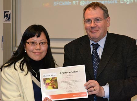
Congratulations to Hui-Chen Wang, from the University of Bath, who won the Chemical Science poster prize at the RSC Macrocyclic and Supramolecular Chemistry Meeting in Bath on 19th-20th December 2011. Her poster was entitled ‘New Boronic Acid Based Fluorescence Sensors’. 

Hui-Chen Wang receives the Chemical Science poster prize

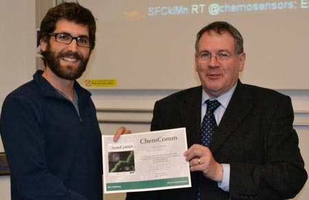
Well done also to Eric A. Appel (University of Cambridge) for his poster on ‘Aqueous Supramolecular Materials via Host-Guest Complexation with Cucurbit[8]uril’, for which he was awarded the ChemComm poster prize.

Eric Appel receiving the ChemComm poster prize

Hui-Chen and Eric receive a one-year personal subscription to Chemical Science and ChemComm respectively. Thank you to Paul Raithby, Professor of Inorganic Chemistry at the University of Bath, for awarding the prize certificates.

Did you know…? Paul Raithby has published more than 100 articles in ChemComm. He told me how his research has evolved into ‘making molecular movies’ in his ChemComm interview published in issue 1, 2012.

Digg This
Reddit This
Stumble Now!
Share on Facebook
Bookmark this on Delicious
Share on LinkedIn
Bookmark this on Technorati
Post on Twitter
Google Buzz (aka. Google Reader)

A radical approach towards the stephacidin alkaloids

Researchers from the University of Birmingham have developed a radical cascade process for rapid access to intermediates, which are important for the synthesis of alkaloids related to the stephacidin family.

Stephacidin A (1), along with other structurally related compounds, has been shown to possess activity against a number of tumour cell lines.

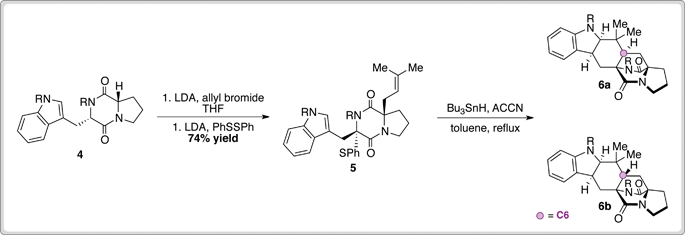
The group of Nigel Simpkins have demonstrated an elegant approach to the synthesis of the indoline core 3, present in a number of naturally occurring alkaloids.

Commencing from 4 ­­(available in five steps from tryptophan), a one-pot prenylation and sulfenylation sequence afforded cyclisation precursor 5 in good yield. Treatment of sulfenyl diketopiperazine 5 with Bu­3SnH and ACCN facilitated a double radical cyclisation to provide 6a and 6b. Pleasingly, isomers possessing the correct C6 stereochemistry represented the major products (4.6–3:1 depending on R group), and desired product 1 could be obtained following deprotection of 6a and 6b and subsequent purification.

This work represents an efficient entry into the stephacidin alkaloids and a potentially powerful method for the synthesis of other medicinally relevant analogues.

To read more on Simpkin’s indoline synthesis, download the ChemComm article.

Digg This
Reddit This
Stumble Now!
Share on Facebook
Bookmark this on Delicious
Share on LinkedIn
Bookmark this on Technorati
Post on Twitter
Google Buzz (aka. Google Reader)

Simple detection of RNA depurination by RIPs

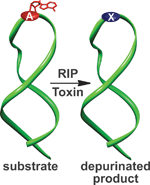
A class of protein toxins exists with an extremely apt acronym, RIP. This stands for Ribosome Inactivating Proteins but may as well stand for “Rest in Peace” when applied to cellular RNA. These proteins are known to attack the link between a purine base (adenine or guanosine) and its sugar causing depurination. This results in the formation of abasic sites (bases that are neither pyrimidines or purines) which in turn, causes the ribosome containing the sequence to have a lower affinity to elongation factors that are crucial for protein synthesis, ultimately leading to cell death.

Methods exist to detect RIPs such as ELISAs and antibody-based immunoassays or by monitoring the specific depurination activity through fluorescence, radiolabelling and immunoaffinity chromatography, amongst others. These techniques require sophisticated or elaborate set-ups, limiting the potential for high throughput (HTP) screening in a bid to discover potential inhibitors of these destructive toxins.

Seergazhi Srivatsan and co-workers have produced a label-free fluorescence hybridisation assay to detect the depurination activity of saporin, a RIP toxin, using a fluorescence ligand that specifically binds to the cytosine opposite an abasic site. If depurination takes place, then the ligand can bind and its fluorescence intensity will be quenched. This technique allows for a plethora of information to be obtained about the depurination activity of saporin as well as many other RIP toxins. This opens the door to using high throughput screening to find inhibitors of such toxins.

Download the article to read more…

Also of interest: Overcoming obstacles in labelling RNA

Posted on behalf of Sarah Brown, web science writer for ChemComm.

Digg This
Reddit This
Stumble Now!
Share on Facebook
Bookmark this on Delicious
Share on LinkedIn
Bookmark this on Technorati
Post on Twitter
Google Buzz (aka. Google Reader)

Scattering layer for dye-sensitised solar cells

We are all aware how important it is to find new improved ways to generate sustainable energy. One very promising candidate is the dye-sensitised solar cell which is particularly attractive due to its low production cost and mechanical robustness.

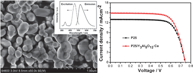
Scientists from East China Normal University have found a way to increase the efficiency of dye-sensitised solar cells by introducing an additional layer on top of the TiO2 photoanode. The extra layer, made up of Y3Al5O12:Ce down-converting microparticles, causes the conversion efficiency to increase from 6.97% to 7.91%.

SEM image of the Y3Al5O12:Ce layer and the J-V curves for the solar cell with and without the additional layer

Likun Pan and his team have attributed this improved performance to the increased light absorbing and scattering properties of the microparticle layer (meaning more suitable photons are available for absorption by the dye) and reduced electron transfer resistance.

Download the ChemComm article to read more about the fabrication of Pan’s solar cell device.

Digg This
Reddit This
Stumble Now!
Share on Facebook
Bookmark this on Delicious
Share on LinkedIn
Bookmark this on Technorati
Post on Twitter
Google Buzz (aka. Google Reader)

One nanoparticle, two nanoparticles, three nanoparticles, four!

Nanoparticles might be small but they frequent the pages of many a journal due to the ongoing boom in nanotechnology research. Whilst they are useful in a myriad of fields, it is still difficult to directly characterise these extraordinarily small entities. King among the visualisation techniques is electron microscopy but this often requires the isolation of the sample on a support – hardly sufficient for analysing a dynamic sample in solution! Dynamic light scattering is another potential technique but finds limitations when it comes to much smaller nanoparticle sizes.

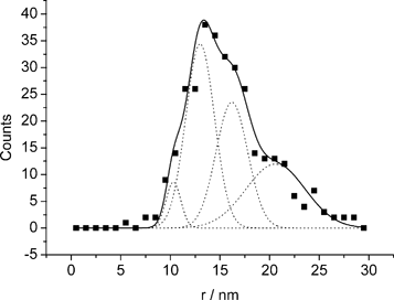
Ideally, you want to be able to count and size individual nanoparticles at a rate which produces reliable statistics. To address this challenge, Richard Compton and his team, including Neil Rees and Yi-ge Zhou who conducted the experiments alongside Jeseelan Pillay, Robert Tshikhudo and Sibulelo Vilakazi from Mintek, Randburg, have used anodic particle coulometry (APC) to measure gold nanoparticle collisions with a glassy carbon microelectrode and thus count and size individual nanoparticles.

With the electrode potential set above +1.0 V, they were able to record oxidative Faradaic transients from nanoparticle collisions and calculate an average nanoparticle radius which compared extremely well to the radius obtained from scanning electron microscopy measurements. They were also able to observe nanoparticle aggregation, which holds great promise for monitoring dynamic aggregation reactions.

It shouldn’t be long before this technique is routinely used to gain more information on all sizes of metallic nanoparticles which are currently being used in a variety of applications.

Read the ChemComm article by Compton and team for more.

Posted on behalf of Iain Larmour, web science writer for ChemComm.

Digg This
Reddit This
Stumble Now!
Share on Facebook
Bookmark this on Delicious
Share on LinkedIn
Bookmark this on Technorati
Post on Twitter
Google Buzz (aka. Google Reader)

Super-fast crystals

Identification of all solid forms of a pharmaceutical is important for drug delivery, due to the potential variability in the physical and chemical properties between amorphous and crystalline forms. The amorphous form of a compound is typically more soluble, but less stable than its crystalline counterpart. Not only that but different polymorphs of the crystalline compound can also have significantly different properties.  To accurately characterise drug action, these polymorphs need to be identified.

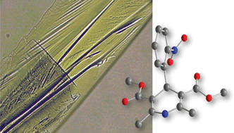
Using small-scale crystallisation and in-situ Raman spectroscopic analysis of the antihypertensive drug, nifedipine, Franziska Emmerling and colleagues discovered an extraordinarily fast transition from the glassy amorphous state to the metastable β polymorph in less than a minute. The β polymorph is stable for less than ten minutes before transforming again, to the thermodynamically stable α polymorph.

The speed at which the transformations take place implies that classical diffusion is not responsible for the different polymorphs but could instead be the result of small intramolecular changes arising from a pre-ordered physical arrangement of the molecules.

In an industrial world where screening for solid drug forms is always leaning towards scale-reduction and time-reduction, three physical forms on a glass slide in less than twenty minutes is pretty impressive!

To find out more, download the ChemComm article.

Also of interest…  Read Andrew Bond, U. Ramamurty, and Gautam Desiraju’s Chemical Science article on “Interaction anisotropy and shear instability of aspirin polymorphs established by  nanoidentation“.

Posted on behalf of Scott McKellar, web science writer for ChemComm.

Digg This
Reddit This
Stumble Now!
Share on Facebook
Bookmark this on Delicious
Share on LinkedIn
Bookmark this on Technorati
Post on Twitter
Google Buzz (aka. Google Reader)

Hybrid biofuel cell

Scientists from Israel have designed a biofuel cell that combines the advantages of both enzymatic and microbial fuel cells.

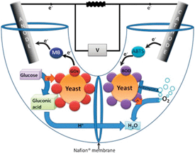
Biofuel cells use redox enzymes to convert chemical energy into electricity. These cells can be divided into two categories: enzymatic fuel cells which require the enzymes to be purified and microbial fuel cells which make use of an entire microorganism. There are pros and cons to both strategies – enzymatic fuel cells tend to have increased power output whilst microbial fuel cells enable full oxidation of a wider range of fuels.

Yet now, Lital Alfonta and co-workers demonstrate that by designing a hybrid cell, one can have the best of both worlds. The team have modified yeast to display redox enzymes on their surface and then introduced this into both the anode and cathode compartments. This approach removes the need to purify the enzymes and enables regeneration of both fuel compartments.

To find out more about Alfonta’s biofuel cell device, read the ChemComm article today.

Digg This
Reddit This
Stumble Now!
Share on Facebook
Bookmark this on Delicious
Share on LinkedIn
Bookmark this on Technorati
Post on Twitter
Google Buzz (aka. Google Reader)

Nitron, the new N-heterocyclic carbene

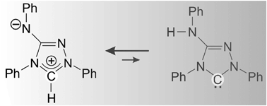
N-heterocyclic carbenes (NHCs) are extremely useful reagents in synthesis and catalysis, but unfortunately they are expensive with 1 gram costing several hundred US dollars.

However, as Ulrich Siemeling and his colleagues report in their latest ChemComm, it seems that a much cheaper alternative is on the horizon… 

The team of scientists based at the University of Kassel have discovered that Nitron, a low cost analytical reagent, exhibits surprising NHC reactivity, more akin to that of its tautomeric form than its conventional Lewis structure. By reacting Nitron with typical carbene trapping reagents such as elemental sulphur, CS2, and rhodium complexes, they have proven that it is indeed Nitron’s tautomer that it responsible for its NHC-like reactivity in solution, despite being present at concentrations undetectable by NMR spectroscopy.

At a fraction of the price and already commercially available, Nitron may soon become a very popular choice in NHC reactions.

To read more about how Siemeling and co-workers established the cause for Nitron’s unusual reactivity, download the ChemComm article.

Digg This
Reddit This
Stumble Now!
Share on Facebook
Bookmark this on Delicious
Share on LinkedIn
Bookmark this on Technorati
Post on Twitter
Google Buzz (aka. Google Reader)

Simple aptasensor for detecting protein

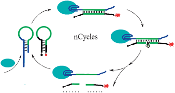
Graphical abstract: Nicking enzyme based homogeneous aptasensors for amplification detection of proteinChinese scientists have made a simple and sensitive sensor for detecting proteins, which could lead to improved disease detection.

Huang-Hao Yang and colleagues at Fuzhou University used single-stranded nucleic acids known as aptamers to detect thrombin, an important protein involved in blood clotting. 

Although other aptasensors are known, they are more complex than this new sensor, says Yang. And the sensitivity here is three orders of magnitude higher than traditional homogeneous aptasensors. 

The improvement is thanks to a nicking enzyme, which Yang used instead of the more usual polymerase. A nicking enzyme recognises a specific sequence in double-stranded DNA. It then cleaves only one strand, leaving a nick in the DNA.   

The aptasensor is capable of detecting thrombin in real samples and could be expanded to other proteins simply by changing the aptamer sequence. 

To find out more about how it works, download Yang’s ChemComm communication.

Digg This
Reddit This
Stumble Now!
Share on Facebook
Bookmark this on Delicious
Share on LinkedIn
Bookmark this on Technorati
Post on Twitter
Google Buzz (aka. Google Reader)