Archive for the ‘Analytical’ Category

Hybrid biofuel cell

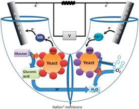
Scientists from Israel have designed a biofuel cell that combines the advantages of both enzymatic and microbial fuel cells.

Biofuel cells use redox enzymes to convert chemical energy into electricity. These cells can be divided into two categories: enzymatic fuel cells which require the enzymes to be purified and microbial fuel cells which make use of an entire microorganism. There are pros and cons to both strategies – enzymatic fuel cells tend to have increased power output whilst microbial fuel cells enable full oxidation of a wider range of fuels.

Yet now, Lital Alfonta and co-workers demonstrate that by designing a hybrid cell, one can have the best of both worlds. The team have modified yeast to display redox enzymes on their surface and then introduced this into both the anode and cathode compartments. This approach removes the need to purify the enzymes and enables regeneration of both fuel compartments.

To find out more about Alfonta’s biofuel cell device, read the ChemComm article today.

Digg This
Reddit This
Stumble Now!
Share on Facebook
Bookmark this on Delicious
Share on LinkedIn
Bookmark this on Technorati
Post on Twitter
Google Buzz (aka. Google Reader)

Simple aptasensor for detecting protein

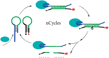
Graphical abstract: Nicking enzyme based homogeneous aptasensors for amplification detection of proteinChinese scientists have made a simple and sensitive sensor for detecting proteins, which could lead to improved disease detection.

Huang-Hao Yang and colleagues at Fuzhou University used single-stranded nucleic acids known as aptamers to detect thrombin, an important protein involved in blood clotting. 

Although other aptasensors are known, they are more complex than this new sensor, says Yang. And the sensitivity here is three orders of magnitude higher than traditional homogeneous aptasensors. 

The improvement is thanks to a nicking enzyme, which Yang used instead of the more usual polymerase. A nicking enzyme recognises a specific sequence in double-stranded DNA. It then cleaves only one strand, leaving a nick in the DNA.   

The aptasensor is capable of detecting thrombin in real samples and could be expanded to other proteins simply by changing the aptamer sequence. 

To find out more about how it works, download Yang’s ChemComm communication.

Digg This
Reddit This
Stumble Now!
Share on Facebook
Bookmark this on Delicious
Share on LinkedIn
Bookmark this on Technorati
Post on Twitter
Google Buzz (aka. Google Reader)

New electrochemiluminescence possibilities

Electrogenerated chemiluminiscence (ECL) is a promising detection technique but its application to certain targets, such as small ions, is compromised due to the necessity to use high concentrations of reagents which can contaminate the sample.

Eric Bakker and co-workers have devised a system which separates the sample compartment, where the analyte is introduced alongside the ruthenium-based ECL reagent, from the compartment which contains the co-reactant necessary for the chemiluminescence to be generated. The technique relies on a liquid membrane to selectively transport the ECL ruthenium compound from the sample towards the detector.

This electro-separation technique opens the door to even more targets capable of being detected using ECL.

To learn more about how Bakker and his team have implemented this strategy, download the ChemComm article.

Digg This
Reddit This
Stumble Now!
Share on Facebook
Bookmark this on Delicious
Share on LinkedIn
Bookmark this on Technorati
Post on Twitter
Google Buzz (aka. Google Reader)

ChemComm poster prize awarded at NMR-DG 2011

Congratulations to Rob Evans (University of Manchester, UK) who won the ChemComm poster prize at NMR-DG 2011 Postgraduate Meeting held earlier this summer at the University of Birmingham.

Rob presented his work entitled ‘Predicting Diffusion Coefficients for Small Molecules’. He receives a prize certificate and a one-year print subscription to ChemComm.

Rob Evans receiving his poster prize certificate from Iain Day
Rob Evans receiving his poster prize certificate from Iain Day, who organised the meeting
Digg This
Reddit This
Stumble Now!
Share on Facebook
Bookmark this on Delicious
Share on LinkedIn
Bookmark this on Technorati
Post on Twitter
Google Buzz (aka. Google Reader)

Chemosensor could lead to fewer deaths from bacterial infections

Millions of people die each year from bacterial infections. Scientists have been searching for a low-cost way to quickly identify bacteria so disease can be diagnosed and treated at an early stage. 

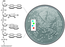
Graphical abstract: Fluorescent DNA chemosensors: identification of bacterial species by their volatile metabolitesEric Kool and colleagues at Stanford University, USA, have developed fluorescent DNA chemosensors which they claim can sense and distinguish bacteria by the volatile metabolites they release. They tested the sensor on bacteria responsible for tuberculosis, food poisoning, pneumonia and sepsis and showed that it could accurately differentiate the bacterial strains. 

The chemosensors could be developed into quick, cheap and reliable reporters for early identification of bacteria in both patient samples and contaminated food, say the authors.

Want to find out more? Download Kool’s ChemComm communication to read more about how the chemosensors work. You might also be interested in the group’s recent Chemical Science Edge article, where they use fluorescent DNA to sense toxic gases.

Digg This
Reddit This
Stumble Now!
Share on Facebook
Bookmark this on Delicious
Share on LinkedIn
Bookmark this on Technorati
Post on Twitter
Google Buzz (aka. Google Reader)

Yeast cell wall particles for multi-modal imaging

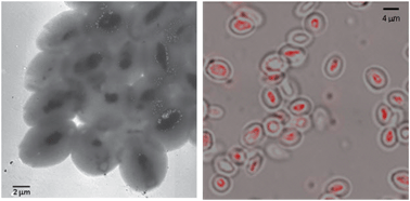
Scientists based in Italy and Portugal have developed a new carrier system for Magnetic Resonance Imaging (MRI) based on yeast cell wall particles (YCWPs).

YCWPs are well tolerated in vivo because they have a cell wall based on a glucan polymer. However, previous attempts at using it as a carrier of hydrophilic and amphiphilic chemicals have failed due to the porous and hydrophilic nature of the membrane.

In this work the team, led by Enzo Terreno at the University of Turin, realised that they could use the YCWPs as microreactors. Once loaded with an imaging agent the particles were exposed to a sudden change in solvent polarity therefore forming a micro-emulsion inside the particles. Importantly this traps the imaging agent in the particle core.

When loaded with gadolinium, the particles were found to have an increased paramagnetic density and also enhanced relaxivity per paramagnetic centre. In all, this should lead to better contrast when used for imaging. In the future Terreno envisages potential applications in cell tracking experiments and particularly for cells found in the immune system.

Want to find out more? Then download the full ChemComm article for free today.

Digg This
Reddit This
Stumble Now!
Share on Facebook
Bookmark this on Delicious
Share on LinkedIn
Bookmark this on Technorati
Post on Twitter
Google Buzz (aka. Google Reader)

Colourful toxin detection

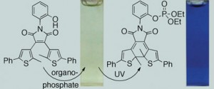
Scientists in Canada have developed a simple chemical detector that could be used to detect airborne neurotoxic organophosphorus chemical warfare agents.

Exposure to organophosphorus agents blocks the action of cholinesterase enzymes, which causes the neurotransmitter acetylcholine to accumulate in the brain. This rapid reaction causes bronchoconstriction (constriction in the airways in the lungs, owing to a tightening of surrounding smooth muscle), seizures, and finally death. Some agents, such as sarin and soman, are odourless and colourless, which makes them difficult to detect. Current detection methods require specialist equipment and trained personnel, so are of limited use in the field. 

The photoresponsive dithienylethene changes colour in the presence of an organophosphorus agent

Neil Branda at Simon Fraser University, Burnaby, and colleagues, have designed a chemical detector made of a dithienylethene compound that binds with organophosphorus agents in the same way that the agents bind to enzymes in the body. When the detector is bound to an agent, its structure alters, causing it to change from colourless to blue when exposed to UV light. This simple colour change provides a clear signal. Visible light resets the system by triggering the reverse reaction.

Read the full story in Chemistry World and download the ChemComm article to find out more about Branda’s research.

Digg This
Reddit This
Stumble Now!
Share on Facebook
Bookmark this on Delicious
Share on LinkedIn
Bookmark this on Technorati
Post on Twitter
Google Buzz (aka. Google Reader)

Detecting caffeine

A team of researchers from Switzerland have used a commercially available fluorophore for detecting caffeine in water.

Previous methods for caffeine detection have been based on hydrogen-bonding receptors but these suffer from the need to use organic solvents. Whilst water-based detection has been achieved, the sensitivity and selectivity for caffeine was low.

Inspired by the known affinity of caffeine for polyaromatic compounds, Kay Severin and colleagues discovered that HPTS, a polysulfonated pyrene dye, can be used to selectively probe caffeine in liquid and solid samples. The team used the probe to quantify caffeine levels in soft drinks, coffee and painkillers, proving it can be used as a simpler alternative to HPLC.

Download the ChemComm article to find out more.

Digg This
Reddit This
Stumble Now!
Share on Facebook
Bookmark this on Delicious
Share on LinkedIn
Bookmark this on Technorati
Post on Twitter
Google Buzz (aka. Google Reader)

Quantum dots selectively label endoplasmic reticulum

Graphical abstract: Selective labeling of the endoplasmic reticulum in live cells with silicon quantum dotsScientists are a step closer to understanding how an important cell organelle works, which could lead to new insight into disease such as diabetes and Alzheimer’s disease.

The endoplasmic reticulum (ER) plays a critical role in protein synthesis and transport. Its malfunction can lead to serious diseases so it is important to be able to observe how it works. 

Yukio Yamaguchi and colleagues at the University of Tokyo, Japan, have managed to selectively label the ER in live cells using quantum dots (QDs). Although organic dyes have previously been used for this purpose, Yamaguchi’s QDs are less toxic and more photostable. 

The QDs’ photoluminescence enabled the team to view the ER using a confocal microscope, making them a powerful tool for long-term real-time observation of the ER, Yamaguchi says.

Find out more by downloading the communication.

Digg This
Reddit This
Stumble Now!
Share on Facebook
Bookmark this on Delicious
Share on LinkedIn
Bookmark this on Technorati
Post on Twitter
Google Buzz (aka. Google Reader)

Magnetic nanoparticles detect low levels of cyanide

Chinese scientists have reported dumbbell-shaped nanoparticles capable of detecting cyanide concentrations as low as 4 x 10⁻⁷ M in drinking water. This low detection limit, which is below the acceptable limit advised by the World Health Organization, is attributed to a combination of fluorescence detection with a filtering process called ‘magnetic concentration-washing’.

The toxicity of cyanide and its potential prevalence in drinking water has made easy detection of cyanide an active area of research. Gold nanoparticles have attracted attention as cyanide sensors due to their selectivity for cyanide over other anions. Now, Shaojun Dong and colleagues at the Changchun Institute of Applied Chemistry, Chinese Academy of Sciences, have adapted a bifunctional nanoparticle made up of Au and magnetic Fe3O4 nanoparticles for sensitive detection of cyanide, even in the presence of interfering species. 

Schematic depiction of the turn-on fluorescent detection of cyanide based on the dual-functional Au–Fe3O4 dumbbell nanoparticles.

The group detected cyanide by relying on what is known as the ‘inner filter effect’, observed when two molecules present in a solution have overlapping absorption and emission wavelengths. The fluorescence emission from a fluorophore is blocked, or reabsorbed, by the second molecule, reducing or even quenching the overall fluorescence of the solution. In the present system, the excitation and emission energy for the fluorophore Rhodamine B is quenched by the Au-Fe3O4 nanoparticles. However, addition of cyanide to the sample reduces the gold component of the nanoparticles by forming an Au(CN)2 complex. This change in the structure alters the nanoparticles’ absorbance spectra and the Rhodamine B emissions are no longer fully absorbed. The team calculated the amount of cyanide present in a sample by monitoring the change in fluorescence.

To extend this detection method for use with environmental samples, which may contain dyes or other contaminants that absorb or emit at a similar wavelength to Rhodamine B, Dong and colleagues implemented a ‘magnetic concentration-washing process’. In it, the Au-Fe3O4 nanoparticles are magnetically separated from the sample solution after they have reacted with any cyanide present in the sample. This magnetic separation retains the bifunctional nanoparticles and leaves any interfering species behind. Pure buffered water containing additional Rhodamine B is added and the fluorescence is monitored. The authors showed that two cycles of this process were enough to remove any interfering species and accurately detect cyanide levels. 

To find out more, read the full article:

Dual-Functonal Au-Fe3O4 Dumbbell Nanoparticles for Sensitive and Selective Turn-on Fluorescent Detection of Cyanide Based on the Inner Filter Effect
Yueming Zhai, Lihua Jin, Ping Wang and Shaojun Dong, Chem. Commun., 2011, DOI: 10.1039/C1CC13149D 

Posted on behalf of Patricia Pantoș, web writer for ChemComm.

Digg This
Reddit This
Stumble Now!
Share on Facebook
Bookmark this on Delicious
Share on LinkedIn
Bookmark this on Technorati
Post on Twitter
Google Buzz (aka. Google Reader)