Archive for the ‘Hot Articles’ Category

Origami, without the papercut, visualised

Researchers in China have been able to visualize the intracellular location of DNA origami with a label-free fluorescent probe.

But let’s unfold a few things first and figure out what that means. DNA origami is the folding of a strand of DNA to make arbitrary 2 or 3 dimensional shapes; this serves as a ‘scaffold’ for shorter DNA strands that help hold the structure in its folded shape. These structures may be used for drug delivery, biosensors and more. I once made an origami frog; I wonder if there are any similarities…

Direct visualisation of the distribution and stability of DNA origami in live, cellular systems has not been achieved. Fluorescent labels can be attached to DNA strands but these have their drawbacks, such as weak emission intensity, photobleaching and expensive. Ding and co-workers looked at alternatives to visualize DNA origami in live cells.

The group were inspired by research on a series of carbazole-based cyanine fluorescence probes, which have a weak emission when they are monomolecularly dissolved but switch to a strong luminescent state upon binding to DNA or protein molecules.  The significant enhancement is attributed to restricted intramolecular rotational (RIR) motions by anchoring the DNA molecules, which causes the large reduction in the non-radiative decay of fluorescence molecules.

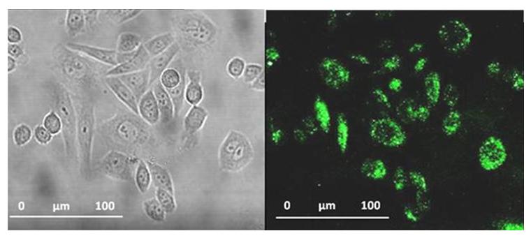
DNA-origami visualised in cells

Follow the instructions (a) and you too won’t make DNA-origami visualised in cells (b)

Ding and co-workers then took some tubular DNA origami, the cyanine fluorophore and found that the carbazole-based cyanine molecules could be used as a sensitive optical switch, turned on when DNA origami is detected and turned off when the nanostructure degrades. After incorporating the cyanine probe molecules, the DNA origami-probe complex was administered to live cells. Excitingly, the green-yellow frog… erm, I mean, fluorescence was visible inside the cells treated with the probe. The group went further to try and understand the internalization mechanism of the DNA origami and found the probe localized in lysosomes. Finally, degradation studies showed that most DNA origami were dissociated after 60 hours, also a bit like my origami frog.

Unlike my attempts at origami, Ding and co-workers have demonstrated an exciting step in scaffolded DNA origami and its future applications in nanomedicine.

Read this HOT Chem Comm article today (free to access until the  5th of December 2012):

Visualization of the intracellular location and stability of DNA origami with a label-free fluorescent probe
Xibo Shen, Qiao Jiang, Jinye Wang, Luru Dai, Guozhang Zou, Zhen-Gang Wang, Wei-Qiang Chen, Wei Jiang and Baoquan Ding
Chem. Commun., 2012, 48, 11301-11303

Published on behalf of Sarah Brown, Chemical Communications web science writer.

Digg This
Reddit This
Stumble Now!
Share on Facebook
Bookmark this on Delicious
Share on LinkedIn
Bookmark this on Technorati
Post on Twitter
Google Buzz (aka. Google Reader)

Flexible post-synthetic approach to functionalised MOFs

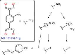
Metal-organic frameworks (MOFs), formed from the assembly of metal ions and organic bridging ligands, often have large pores and high surface areas.  Such properties are attractive for various potential applications from gas storage to catalysis.  The diverse structures and properties of MOFs can be extended by incorportaing functional groups onto the organic linker after MOF formation through post-synthetic modification.

MIL-101(Cr), a MOF first reported by Ferey et al. in 2005, exhibits mesoporous cages with accessible metal sites as well as high chemical and hydrothermal stability.  Amine functionalisation of the framework’s benzene dicarboxylate ligands has been reported for analagous iron and aluminium frameworks, but has so far proven elusive for MIL-101(Cr).

Burrows et al. from the University of Bath have synthesised MIL-101(Cr)-NH2 using a hydrothermal method, and found that the resultant framework is stable up to 250 °C and, most interestingly, is stable to acids.  This unusual stability has allowed them to post-synthetically transform the amine group into an arenediazonium salt which they used in situ to generate a variety of functional groups.  MIL-101(Cr)-azo, in particular, showed excellent CO2 selectivity at low pressure.

This new approach to post-synthetic modification has provided a flexible route to functionalised MIL-101 materials, with further studies concentrating on selected frameworks already underway.

Read this HOT Chem Comm article today (free to access until the  7th of December 2012):

Synthesis and post-synthetic modification of MIL-101(Cr-NH2) via a tandem diazotisation process

Dongmei Jiang, Luke L. Keenan, Andrew D. Burrows and Karen J. Edler
Chem. Commun., 2012, Advance Article
DOI: 10.1039/c2cc36344e

Digg This
Reddit This
Stumble Now!
Share on Facebook
Bookmark this on Delicious
Share on LinkedIn
Bookmark this on Technorati
Post on Twitter
Google Buzz (aka. Google Reader)

3D donor for organic photovoltaics with improved power efficiency

Scientists in China have developed a 3D donor for organic photovoltaics based on a spiro-fluorene molecule, which exhibited a power conversion efficiency of 4.82%.

Traditional 1D small molecule donors had only reached an efficiency of 1.69%. Small molecule donors have many advantages over conjugated polymer donors: high solubility allowing for high purification, good reproducibly and high charge-carrier mobility (because of its ordered, crystalline nature).

Donors with a 3D structure, including small molecular and dendritic macromolecular structures, have been expected to start a completely new self-assembly approach and more stable morphologies by increasing the dimension of the donor molecules.

Read the ChemComm ‘HOT’ article:

Spiro-fluorene based 3D donor towards efficient organic photovoltaics
Shuying Ma , Yingying Fu , Debin Ni , Jian Mao , Zhiyuan Xie and Guoli Tu
Chem. Commun., 2012, DOI: 10.1039/C2CC36301A

Digg This
Reddit This
Stumble Now!
Share on Facebook
Bookmark this on Delicious
Share on LinkedIn
Bookmark this on Technorati
Post on Twitter
Google Buzz (aka. Google Reader)

Site-Selective Protein Modification via Bioorthogonal Wittig Reaction

Synthetic organic chemists have long regarded the Wittig reaction as one of the most significant methods for the creation of new C–C bonds. Now, researchers at Peking University have reported that this time-honoured reaction can be used for the modification of proteins in a bioorthogonal process.

Bioorthogonal processes represent a growing area of interest, encompassing reactions which can take place under physiological conditions. That is, the reaction must proceed at neutral pH in an aqueous solvent at ambient temperature and low concentrations, with high selectivity. Given these challenging parameters, the range of bioorthogonal processes remains limited.

The Ye group, based at the Key State Laboratory of Natural and Biomimetic Drugs, have contributed to the expansion of this fascinating area of chemistry by successfully applying the Wittig reaction to their one-pot site-selective protein modification. The first step involves the incorporation of an aldehyde at the N-terminus of a peptide chain (1). This could be achieved by the periodate oxidation of N-terminal serine or threonine residues, or by PLP (pyridoxal-5-phosphate) oxidation of N-terminal glycines. The resulting aldehyde (2), without the need for isolation or purification, could be reacted with an ylide (3) to form a wide variety of functionalised peptide products (4). Di-, penta- and hexa-peptide substrates could be functionalised in this manner, using water and t-butanol as co-solvents at room temperature.

The strength of this site-selective reaction was further demonstrated by modifying myoglobin. Crucially, this was achieved with no damage to the protein’s secondary or tertiary structure and, furthermore, Prof. Ye’s group established that myoglobin’s oxygen storage and release function was unaffected.

The functionality introduced offers the potential for further structural modification, or for use in medical imaging. With protein-based pharmaceuticals becoming widely used, greater insight into protein function and behaviour is of paramount importance. This methodology has the potential to be a valuable tool in that understanding. 

Read the ‘HOT’ Chem Comm article today:

Enabling Wittig reaction on site-specific protein modification

Ming-Jie Han, De-Cai Xiong and Xin-Shan Ye
Chem. Commun., 2012, 48, 11079-11081
DOI: 10.1039/C2CC35738K

Published on behalf of Ruth Gilligan, Chemical Communications web science writer.

Digg This
Reddit This
Stumble Now!
Share on Facebook
Bookmark this on Delicious
Share on LinkedIn
Bookmark this on Technorati
Post on Twitter
Google Buzz (aka. Google Reader)

Ligand-enhanced lanthanide luminescence for anion sensing

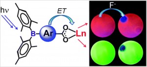
Lanthanide luminescenceLuminescence from lanthanide-based compounds is currently exploited in electroluminescent devices ranging from glucose monitoring sensors to thin-film displays. Triarylboron groups are known to greatly enhance the fluorescence or phosphorescence of transition metal complexes, but such compounds are very sensitive to oxygen quenching and are unsuitable for use as sensors under ambient conditions.  The combination of these groups with lanthanides may offer enhanced luminescence in more ambient-friendly lanthanide-based materials.

The Wang group from Queen’s University, Canada have synthesised the first examples of triarylboron functionalized Tb(III) and Eu(III) compounds, and have shown that the BMes2 group is extremely effective at activating lanthanide emissions.  The new compounds have been tested as sensors, showing that such triarylboron functionalized lanthanides may be promising new CN and F anion sensors.

To find out more, download this HOT article now (free to access until the 5th of December 2012).

Selective activation of lanthanide luminescence with triarylboron-functionalized ligands and visual fluoride indicators

Maria Varlan, Barry A. Blight and Suning Wang

Chem. Commun., 2012, Advance Article
DOI: 10.1039/C2CC36172H

Posted on behalf of Katie Renouf, Chemical Communications web science writer.

Digg This
Reddit This
Stumble Now!
Share on Facebook
Bookmark this on Delicious
Share on LinkedIn
Bookmark this on Technorati
Post on Twitter
Google Buzz (aka. Google Reader)

Prescription nanoreactors

Scientists in Switzerland have developed a nanoreactor that can synthesise and release the antibiotic cephalexin, which is used to treat bacterial infections.

Lowering the drug dose required to effectively treat a patient would save money and reduce side effects. To achieve this, one area of research is focusing on using a ‘prodrug’ approach in which less toxic substrates, or prodrugs, are given to a patient and are converted to the active drug form by an enzyme only at a specific site.

To maintain its catalytic activity, the enzyme needs to be protected from the surrounding environment, but also be accessible to the prodrug and able to release the final active drug.

Wolfgang Meier and colleagues at the University of Basel have designed a biocompatible nanoreactor that can effectively protect an enzyme. The nanoreactor comprises a copolymer, an outer membrane protein from bacteria cells (OmpF) and the catalyst penicillin acylase. When mixed together, the copolymer self assembles into vesicles forming, along with the membrane protein, a membrane around the catalyst that allows passage of molecules such as a drug and substrates.

Read the whole Chemistry World story or download the full ChemComm article:

Communication Polymer nanoreactors shown to produce and release antibiotics locally
Karolina Langowska , Cornelia G. Palivan and Wolfgang Meier
Chem. Commun., 2012, DOI: 10.1039/C2CC36345C

Digg This
Reddit This
Stumble Now!
Share on Facebook
Bookmark this on Delicious
Share on LinkedIn
Bookmark this on Technorati
Post on Twitter
Google Buzz (aka. Google Reader)

Selective formation of (Z)-enynes via Zirconium catalysed alkyne dimerisation

Researchers from the University of British Columbia have developed a ‘head to head’ dimerisation of alkynes to provide (Z)-enynes in high yields.

The conjugated enyne products are important, both as intermediates for organic synthesis and in optoelectronics – a type of electronics that detect, source and control light. Therefore, new methods for the selective synthesis of these compounds are in demand.

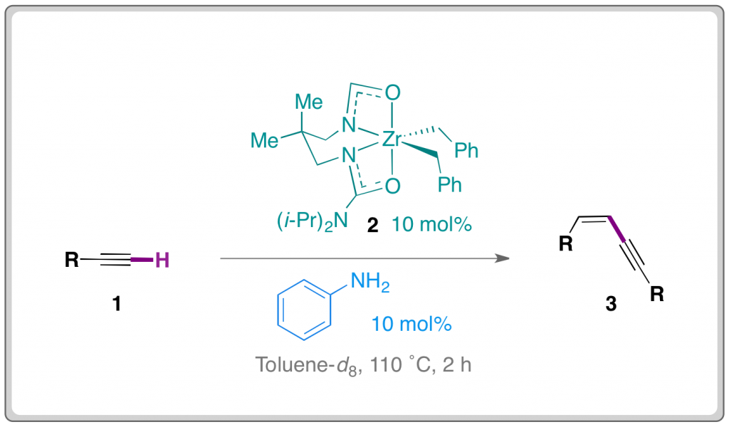
Platel and Schafer have overcome regioselectivity problems often associated with alkene dimerisation.  The researchers used a readily accessible Zirconium catalyst (2) in conjunction with aniline as co-catalytic proton source.

A number of alkynes (1) were successfully dimerised to provide (Z)-enynes (3) in mostly high yields. The reaction is tolerant of a number of differentially substituted aryl compounds and also some alkyl rings.

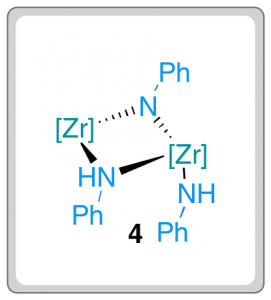
The exclusive formation of (Z)-enynes was at odds with previously established mechanistic pathways for metal-catalysed alkyne dimerisation and led the researchers to further probe the mechanism. Complex 4 was isolated when a stoichiometric amount of aniline and complex 2 were mixed, leading the researchers to consider the validity of 4 as a catalytic intermediate. When 4 was mixed with alkyne (1) under the reaction conditions, complete conversion to (Z)-enyne (3) was observed. This result suggests that the unusual dimeric imido-bridged species is a viable intermediate in the reaction pathway.

This method represents an interesting development in catalytic and regioselective C­–C bond formation.

Read the ‘HOT’ Chem Comm article today:

Zirconium catalyzed alkyne dimerization for selective Z-enyne synthesis
Rachel H. Platel and Laurel L. Schafer
Chem. Commun., 2012, 48, 10609-10611
DOI: 10.1039/C2CC35913H

Digg This
Reddit This
Stumble Now!
Share on Facebook
Bookmark this on Delicious
Share on LinkedIn
Bookmark this on Technorati
Post on Twitter
Google Buzz (aka. Google Reader)

Magnetic mixed-metal molecules

The first Re(IV) system incorporating a 4f ion has been reported by Martínez-Lillo and colleagues at the University of Edinburgh

At present, molecular magnetism in heavy 4d, 5d and 4f-type metal ions is heavily researched since most known systems only involve 3d ions.  Attempts to extend this research into the unexplored area of mixed 5d-4f systems has paid off with this discovery of a mixed Re(IV)-Gd(III) system. 

Magnetic mixed-metal molecules: Rhenium 4f ion mixed with Gadolinium

Single crystals of the new compound, (NBu4)5[Gd{ReBr4(μ-ox)}4(H2O)]·H2O, were analysed at the ESRF to obtain a crystal structure.  Magnetic studies were performed on the new compound, and significant intramolecular antiferromagnetic interactions were found to occur unambiguously between Re(IV) and Gd(III). 

Further work on the preparation of the parent Re(IV)-Ln(III) complex will be carried out in the near future.

To read more about this new compound, its crystal structure and its intramolecular antiferromagnetic coupling, check out this HOT article (free to access for a limited period).

A heteropentanuclear oxalate-bridged [ReIVGdIII] complex: synthesis, crystal structure and magnetic properties

José Martínez-Lillo, Laura Cañadillas-Delgado, Joan Cano,  Francesc Lloret, Miguel Julve and Juan Faus

Posted on behalf of Katie Renouf, Chemical Communications web science writer.

Digg This
Reddit This
Stumble Now!
Share on Facebook
Bookmark this on Delicious
Share on LinkedIn
Bookmark this on Technorati
Post on Twitter
Google Buzz (aka. Google Reader)

Supramolecular splicing: Catalytic fusion of supramolecules

Non-covalent interactions dictate the assembly of many of nature’s most elegant structures. Similarly, supramolecular chemists have long been intrigued by the challenge of designing functional structures that spontaneously self-assemble from simpler fragments which mutually recognise each other.

A popular self-assembly approach is to produce coordination compounds from transition metal salts with rigid organic ligands. Directional bonding around transition metal centres allows the production of predictable and controllable shapes. Michael Schmittel’s group at the University of Siegen have been exploring a newer approach. They prepared two assemblies, a 2-component triangle T1 and a 3-component rectangle R1. The transition metal “corner” arrangements in T1 and R1 are disfavoured, so if the two assemblies are combined the components re-shuffle to form a more favourable assembly- the 5-component triangle T2. The transformation occurs at room temperature, and can be completed in just 1 hour in the presence of a catalyst, which accelerates the re-shuffling by labilising the metal-ligand bonds.

Supramolecular splicing: Catalytic fusion of supramolecules: re-shuffling of a 2-component triangle and a 3-component rectangle to give a more favoured supramolecular 5-component triangle

Unlike previous examples, the conditions needed for the transformation are very mild. The authors compare the process to gene shuffling, the combination of dissimilar genes to form new genetic material. The strategy could be considered a first step towards the evolution of supramolecular architectures, and a great route to more complex supramolecular assemblies with higher information content.

The full communication can be downloaded here (free to access for a limited period).

Cally Haynes

Digg This
Reddit This
Stumble Now!
Share on Facebook
Bookmark this on Delicious
Share on LinkedIn
Bookmark this on Technorati
Post on Twitter
Google Buzz (aka. Google Reader)

Overcoat makes carbon quantum dots biocompatible

Quantum dots are currently being developed for a variety of applications, including as sensors and cellular tags. Semiconductor quantum dots are attractive for their high fluorescence quantum yields but the toxicity of some of the metals involved, such as cadmium, pose a problem for biological applications.

Carbon quantum dots (CQDs) offer an alternative however when transferred into aqueous solution they possess low quantum yields. The problem is how do you harness the higher fluorescence of CQDs prepared in an organic solvent for biological applications?

To answer this question John Callan and his team have employed an amphiphilic polymer to act as an overcoat and transfer agent for the CQDs. Surprisingly they found that the transfer actually improved the quantum yield rather than the normally expected repression when ligand exchange is used. These aqueous carbon quantum dots were taken up into cells and were found to be non-toxic.

Chinese Hamster Ovarian cells loaded with carbon quantum dots

The development of inexpensive and biocompatible quantum dots with an improved quantum yield holds great potential for a wide range of future biological applications.

To find out more, download the ChemComm article today (free to access for a limited period).

Digg This
Reddit This
Stumble Now!
Share on Facebook
Bookmark this on Delicious
Share on LinkedIn
Bookmark this on Technorati
Post on Twitter
Google Buzz (aka. Google Reader)