Author Archive

Elucidating the Molecular Behavior on the pH Responsiveness of Chitosan

Written by Tianyu Liu, University of California, Santa Cruz

Chitosan is a polysaccharide derived from chitin, a second most abundant bio-polymer on earth that consists of the shells of crustaceans (such as shrimps and crabs). It is a commonly used bio-compatible material for bio-medical applications such as drug delivery.

Previous studies have shown that chitosan is pH sensitive in water: it forms a viscous solution at low pH values and becomes insoluble at high pH values. Preliminary investigations suggest that such pH responsiveness is associated with the protonation and de-protonation processes of the amine groups on the chitosan polymer chain (the red box in Figure a). However, there still lacks fundamental understanding on how the pH affects the behavior of chitosan.

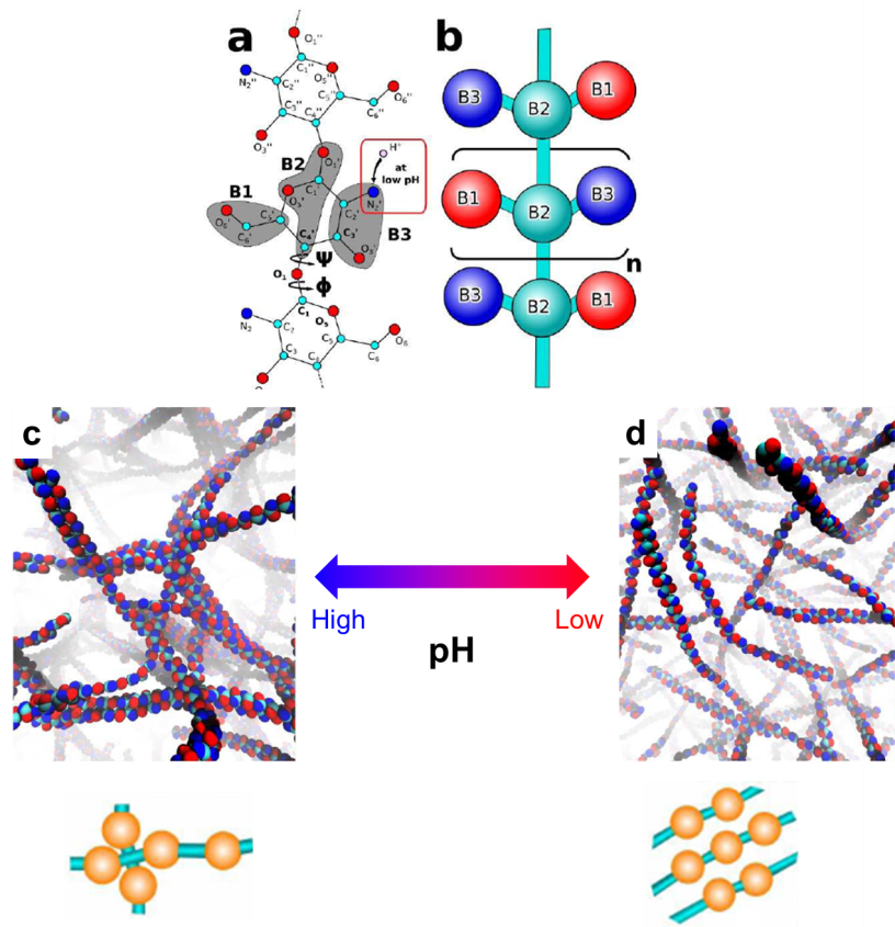
Now writing in Chem. Commun., Xu and Matysiak from University of Maryland, USA provided us new insights on the chitosan’s pH responsiveness at the molecular level. They adopted a method called “coarse-grained molecular simulation” to illustrate the self-assembly behaviors of chitosan polymer chains at different pH values. Unlike atomistic molecular simulations that focus on individual atoms of a molecule, the coarse-grained molecular simulation treats a group of atoms as one ensemble and probes the collective behavior of each ensemble (Figure a and b). This simulation technique demands less time than the atomistic counterparts without significantly reducing the simulation accuracy. It is suitable for characterizing polymers composed of thousands of atoms, such as chitosan.

The key discovery of this work is that the chitosan polymer chains can adopt different configurations at different pH values. At high pH values, each chain tends to crosslink perpendicularly with adjacent chains. The crosslinking reaction propagates and eventually builds up a three-dimensional dense chain network (Figure c). At low pH values, the protonated amine groups favor parallel crosslinking. Thus, each chain aligns in parallel with each other, which leads to a loosely-packed structure (Figure d). The perpendicularly cross-linked configuration reduces the solubility of chitosan in water but renders robustness and elasticity of the chitosan networks. The parallel cross-linked morphology increases water solubility but decreases the elasticity of the chitosan assembly. These conclusions obtained by the simulation are consistent with experimental results.

To find out more please read:
Effect of pH on Chitosan Hydrogel Polymer Network Structure
Hongcheng Xu and Silvina Matysiak
DOI: 10.1039/C7CC01826F

About the author:
Tianyu Liu is a Ph.D. in chemistry from University of California-Santa Cruz. He is passionate about scientific communication to introduce cutting-edge researches to both the general public and the scientists with diverse research expertise. He is a web writer for the Chem. Commun. and Chem. Sci. blog websites. More information about him can be found at http://liutianyuresearch.weebly.com/

Digg This
Reddit This
Stumble Now!
Share on Facebook
Bookmark this on Delicious
Share on LinkedIn
Bookmark this on Technorati
Post on Twitter
Google Buzz (aka. Google Reader)

Siliconrhodamine Probes Enable Bio-imaging with Super-resolution

Written by Tianyu Liu, University of California, Santa Cruz

Intracellular imaging is used to reveal fine details of live organisms. It is an indispensable component for the exploration of biomolecular processes in living cells. Super-resolution microscopy (SRM) is an emerging intracellular imaging technique which can acquire images of much higher resolution than those collected by conventional optical microscopy. Currently, the greatest challenge facing SRM is to develop imaging probes that are suitable for site-specific tagging of intracellular biomolecules. Such probes must be biocompatible, membrane-permeable, intensively fluorescent and photo-stable.

Writing in ChemComm., Dr. Peter Kele and coworkers at Research Center for Natural Sciences, Hungarian Academy of Sciences have developed a group of siliconrhodamine probes that permit the labelling of intracellular proteins with excellent selectivity as well as fast response time (within 10 min).

The synthesized siliconrhodamine probes consist of a siliconrhodamine backbone anchored with a carboxyl group. The carboxyl group is responsible for the polarity-responsive property of the probes. When bound to polar protein surfaces, the probes exist in a fluorescent form. While upon non-specific binding to hydrophobic surfaces, the probes change their configurations and consequently, the fluorescence is lost. This conversion process is based on an intra-molecular Diels-Alder reaction (Figure below) that can be readily initiated by a polarity change without interrupting native biochemical processes in cells. Such a mechanism provides the probe biocompatibility and fast response characteristics.

The probe has been demonstrated for site-specific super-resolution imaging for live cells. The figure below depicts the experimental results collected using a mammalian cell. The cyan colored image (left) presents the actual cell image (as the reference). The middle magenta colored image was obtained by using one of the synthesized imaging probes. The overlay image (right) exhibits near-perfect co-localization of the reference and labelling images, indicating the probe’s excellent selectivity. Moreover, the labelling process is efficient with the probe concentration as low as 1.5 μM, and the duration as short as 10 min.

These stable, efficient, and biocompatible probes could profoundly advance super-resolution imaging of various intracellular structures.

To find out more please read:

Bioorthogonal Double-Fluorogenic Siliconrhodamine Probes for Intracellular Super-resolution Microscopy
Eszter Kozma, Gemma Estrada Girona, Giulia Paci, Edward A Lemke and Peter Kele
DOI: 10.1039/C7CC02212C

Digg This
Reddit This
Stumble Now!
Share on Facebook
Bookmark this on Delicious
Share on LinkedIn
Bookmark this on Technorati
Post on Twitter
Google Buzz (aka. Google Reader)

Destruction and Reconstruction of Nanorods Controlled by Visible Light

Written by Tianyu Liu, University of California, Santa Cruz

Supramolecular smart materials are a family of materials composed of several molecules. They have the ability to change their configurations in response to external stimuli such as the presence of enzymes, light irradiation, and changes in pH. This property can be manipulated for a variety of applications including drug delivery and tissue engineering.

In recent years, pH-responsive supramolecular smart materials have been intensively investigated due to the simplicity of pH alteration. However, adjusting pH can have undesired consequences. First, chemical species other than the supramolecular materials (e.g., acid and base) are needed for tuning pH. The involvement of external reagents hinders the readiness of operation. Additionally, the use of acid and base inevitably introduces waste products, which could eventually suppress the stimulus-response activity of the smart materials. Therefore, developing alternative ways to initiate the configuration modification of the supramolecular smart materials is highly desirable.

In a recent ChemComm. publication, Professor Heng-Yi Zhang, Professor Yu Liu and coworkers from Nankai University, China have developed supramolecular smart nanorods consisting of β-cyclodextrin (β-CD) and 4,4’-bipyridine-coordinated zinc ions. In the presence of protonated merocyanine (MEH) in water, the nanorods are able to dissociate upon visible light illumination and reconstruct themselves when placed in the dark (Figure above).

The method by which these structures can reconfigure involves a light-driven proton transfer process (Figure below). MEH molecules absorb energy from visible light and subsequently release their protons to the surroundings. These free protons then combine with the 4,4’-bipyridine (DPD). The protonated DPD molecules lose their coordination ability and disassemble with zinc ions. As a result, the entire nanorod structure collapses. When no light is present, the aforementioned proton transfer process is reversed and the nanorods are reformed. Such a process is highly reversible with no observable light-responsive activity loss for at least five cycles.

The demonstrated light-responsive supramolecular nanorods enable facile operations with no additional chemicals. This technology opens up endless new opportunities in remote control of light-responsive processes.

To find out more please see:

Light-controlled reversible self-assembly of nanorod suprastructures

Jie Guo, Heng-Yi Zhang, Yan Zhou and Yu Liu

DOI:10.1039/C7CC03280C


Digg This
Reddit This
Stumble Now!
Share on Facebook
Bookmark this on Delicious
Share on LinkedIn
Bookmark this on Technorati
Post on Twitter
Google Buzz (aka. Google Reader)

High Content & Phenotypic Screening 2017

High Content and Phenotypic Screening 2017 will be held at Holiday Inn, Cambridge, UK from 25th – 26th April.

This conference brings together researchers from both academia and industry, and will discuss the development of techniques and tools implemented in High Content technologies and Phenotypic Screening applications. 

Hot topics to be covered include 3D cell based screening methods, high content screening and data management, the use of model organisms and novel approaches for phenotypic screening. 

Key date:

19 April – Poster Submission Deadline

Find out more here

Digg This
Reddit This
Stumble Now!
Share on Facebook
Bookmark this on Delicious
Share on LinkedIn
Bookmark this on Technorati
Post on Twitter
Google Buzz (aka. Google Reader)

Outstanding Reviewers for Chemical Communications in 2016

Following the success of Peer Review Week in September 2016 (dedicated to reviewer recognition) during which we published a list of our top reviewers, we are delighted to announce that we will continue to recognise the contribution that our reviewers make to the journal by announcing our Outstanding Reviewers each year.

We would like to highlight the Outstanding Reviewers for Chemical Communications in 2016, as selected by the editorial team, for their significant contribution to the journal. The reviewers have been chosen based on the number, timeliness and quality of the reports completed over the last 12 months.

We would like to say a big thank you to those individuals listed here as well as to all of the reviewers that have supported the journal. Each Outstanding Reviewer will receive a certificate to give recognition for their significant contribution.

Professor Martin Albrecht, Universität Bern

Dr Guanghui An, Heilongjiang University

Professor Rahul Banerjee, National Chemical Laboratory

Dr Justin Chalker, Flinders University

Dr Takashi Hirose, Kyoto University

Dr Astrid Müller, Caltech

Dr David Nelson, University of Strathclyde

Dr Kyungsoo Oh, Chung-Ang University

Dr Zhenlei Song, SiChuan University

Dr Xuehai Yan, Max Planck Institute of Colloids and Interfaces

We would also like to thank the Chemical Communications board and the General Chemistry community for their continued support of the journal, as authors, reviewers and readers.

If you would like to become a reviewer for our journal, just email us with details of your research interests and an up-to-date CV or résumé.  You can find more details in our author and reviewer resource centre

Digg This
Reddit This
Stumble Now!
Share on Facebook
Bookmark this on Delicious
Share on LinkedIn
Bookmark this on Technorati
Post on Twitter
Google Buzz (aka. Google Reader)

Molecular structure is teixobactin’s pièce de résistance

Study builds scientists’ arsenal against drug-resistant superbugs

Scientists in the UK, Belgium and the Netherlands have gained a crucial understanding of the structure–activity relationship of new antibiotic, teixobactin. Since reports of its discovery in early 2015, researchers have shown it can kill a number of pathogens without them developing resistance to it.

The University of Lincoln’s Ishwar Singh explains that there are several reasons for teixobactin’s potency: ‘It uses multiple modes of action to kill resistant bacteria, this makes it very attractive since, if it worked by only one mode, bacteria could modify more easily. It is much more challenging for bacteria to mutate on multiple levels.’ Teixobactin also targets lipids in the bacteria’s cell walls, which are considered to be less able to mutate and develop resistance.

Read the full story by Hannah Dunckley on Chemistry World.

Source: © Royal Society of Chemistry
Structure of teixobactin and with the D-amino acids highlighted in red

Digg This
Reddit This
Stumble Now!
Share on Facebook
Bookmark this on Delicious
Share on LinkedIn
Bookmark this on Technorati
Post on Twitter
Google Buzz (aka. Google Reader)

Congratulations to the Poster Prize winners at The 20th Symposium of the Society of Silicon Chemistry

The Royal Society of Chemistry were delighted to sponsor Chemical Communications Poster Prizes at The 20th Symposium of the Society of Silicon Chemistry. This event was held from 7th – 8th October 2016 in Hiroshima, Japan and hosted by The Society of Silicon Chemistry Japan.

We would like to take the time to congratulate our prize winners!

Ms Mirei Motomatsu from Gakushuin University‘s poster was titled “Reaction of a cationic metallogermylene with nitrogen containing compounds“.

Mr Tomohiro Sugahara from Kyoto University‘s poster was titled “Synthesis and structure of cyclic compounds containing germanium atoms utilizing a digermacyclobutene derivative“.

Dr Hiromitsu Urakami, RSC Manager for Japan, awarded the winners with their certificates.

Ms Mirei Motomatsu with Dr Hiromitsu Urakami

Mr Tomohiro Sugahara with Dr Hiromitsu Urakami

Digg This
Reddit This
Stumble Now!
Share on Facebook
Bookmark this on Delicious
Share on LinkedIn
Bookmark this on Technorati
Post on Twitter
Google Buzz (aka. Google Reader)