Written by Tianyu Liu, University of California, Santa Cruz
Intracellular imaging is used to reveal fine details of live organisms. It is an indispensable component for the exploration of biomolecular processes in living cells. Super-resolution microscopy (SRM) is an emerging intracellular imaging technique which can acquire images of much higher resolution than those collected by conventional optical microscopy. Currently, the greatest challenge facing SRM is to develop imaging probes that are suitable for site-specific tagging of intracellular biomolecules. Such probes must be biocompatible, membrane-permeable, intensively fluorescent and photo-stable.
Writing in ChemComm., Dr. Peter Kele and coworkers at Research Center for Natural Sciences, Hungarian Academy of Sciences have developed a group of siliconrhodamine probes that permit the labelling of intracellular proteins with excellent selectivity as well as fast response time (within 10 min).
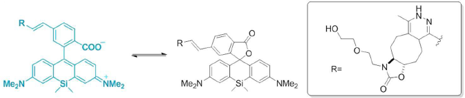
The synthesized siliconrhodamine probes consist of a siliconrhodamine backbone anchored with a carboxyl group. The carboxyl group is responsible for the polarity-responsive property of the probes. When bound to polar protein surfaces, the probes exist in a fluorescent form. While upon non-specific binding to hydrophobic surfaces, the probes change their configurations and consequently, the fluorescence is lost. This conversion process is based on an intra-molecular Diels-Alder reaction (Figure below) that can be readily initiated by a polarity change without interrupting native biochemical processes in cells. Such a mechanism provides the probe biocompatibility and fast response characteristics.
The probe has been demonstrated for site-specific super-resolution imaging for live cells. The figure below depicts the experimental results collected using a mammalian cell. The cyan colored image (left) presents the actual cell image (as the reference). The middle magenta colored image was obtained by using one of the synthesized imaging probes. The overlay image (right) exhibits near-perfect co-localization of the reference and labelling images, indicating the probe’s excellent selectivity. Moreover, the labelling process is efficient with the probe concentration as low as 1.5 μM, and the duration as short as 10 min.
These stable, efficient, and biocompatible probes could profoundly advance super-resolution imaging of various intracellular structures.

To find out more please read:
Bioorthogonal Double-Fluorogenic Siliconrhodamine Probes for Intracellular Super-resolution Microscopy
Eszter Kozma, Gemma Estrada Girona, Giulia Paci, Edward A Lemke and Peter Kele
DOI: 10.1039/C7CC02212C


")