Author Archive

Grinding gathers momentum for coordination polymers

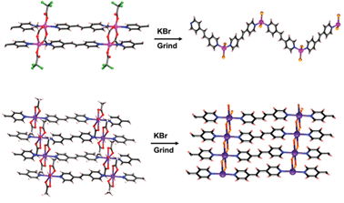
It is a fairly common assumption that the sample you are characterising is the same sample that you made at the lab bench. While it may indeed be the same sample, it may not be the same structure as Peter Stephens and Jagadese Vittal discovered.

By grinding coordination polymers with KBr (as is standard practise in solid state sample preparation for infrared characterisation), they generated coordination polymers with completely new structures – and as a result, completely different optical properties as well.

The team have attributed this to an exchange between the bridging ligands and the bromide ions from KBr. While this is an interesting avenue to explore for the preparation of new coordination polymers, Stephens and Vittal warn researchers working with such materials that observed changes in sample colour or texture during pre-characterisation preparation may not always be a physical phenomenon and to tread with caution…

Read the ChemComm article today.

Digg This
Reddit This
Stumble Now!
Share on Facebook
Bookmark this on Delicious
Share on LinkedIn
Bookmark this on Technorati
Post on Twitter
Google Buzz (aka. Google Reader)

Solvent effects in supramolecules

We know the importance of water in protein folding but what about organic solvent effects in self-assembling structures?

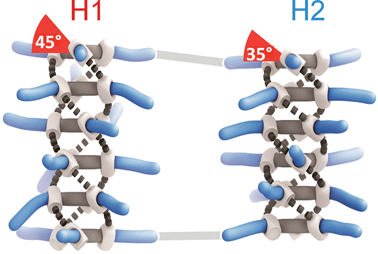
A team of scientists from the Netherlands decided to find out what happens to the supramolecular structure of deuterated benzene tricarboxamide (D-BTA) when the molecular structure of the organic solvent is changed. You may not think that swapping methylcyclohexane for heptane may make much difference – both are non-polar with similar properties. Think again.

D-BTA conformers exhibiting M helicity

Paul van der Schoot, Bert Meijer, Anja Palmans and their team discovered that substitution of one solvent for another was enough to influence the helical sense preference and conformation of D-BTA supramolecular polymers. It seems that linear solvents, such as heptane used here, actively participate in the self-assembly of the D-BTA units, causing the supramolecular aggregates to favour one helicity over the other, whereas solvents with branched or cyclic molecular structures do not permit such solvent–molecule interactions.

Once again, chemistry shows us that the smallest of changes on the molecular scale can influence more than first thought.

Keep an eye out for many more chirality-related articles to come as part of our forthcoming Chirality web theme issue. To read more about Palmans and colleagues’ findings, download the ChemComm article.

Digg This
Reddit This
Stumble Now!
Share on Facebook
Bookmark this on Delicious
Share on LinkedIn
Bookmark this on Technorati
Post on Twitter
Google Buzz (aka. Google Reader)

Monooxygenase blends

When deciding which material to use for a particular application, it’s often necessary to weigh up the pros and cons of each candidate. Wouldn’t it be great if you could combine the best bits from each one to produce the ideal material?

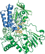
This is exactly what Marco Fraaije and his team from the University of Groningen did to create a new monooxygenase enzyme capable of performing Baeyer–Villiger oxidations with ultimate catalytic properties. For biocatalytic applications, enzymes need to be robust and should ideally be able to catalyse a broad range of substrates. Unfortunately, the only monooxygenase shown to be thermally stable (phenylacetone monooxygenase, PAMO) has narrow substrate specificity. On the other hand, there is cyclohexanone monooxygenase, CHMO, which can oxidise hundreds of substrates yet cannot be used at elevated temperatures.

The monooxygenase. Original PAMO structure is shown in green; the replaced sub-domain is shown in blue.

By replacing the substrate-binding domain of PAMO with that of CHMO or steroid monooxygenase (STMO), Fraaije was able to engineer an enzyme that was thermally robust and able to accept a wide range of substrates. Not only were the team able to combine the best of both worlds but in some cases, supersede them as they found when evaluating the conversions and enantiomeric excesses. It seems that the enzyme blend is not necessarily an average of the parent enzymes but can exhibit new properties.

Read the ChemComm article to find out more on how the team were able to improve the properties of Baeyer–Villiger monooxygenases.

Also of interest… ChemComm‘s Enzymes and Proteins web theme issue guest edited by Professors Nicholas Turner, Wilfred van der Donk and Herbert Waldmann.

Digg This
Reddit This
Stumble Now!
Share on Facebook
Bookmark this on Delicious
Share on LinkedIn
Bookmark this on Technorati
Post on Twitter
Google Buzz (aka. Google Reader)

Polydiacetylenes: Functional supramolecular smart materials

Polydiacetylene polymers (PDAs) are a popular research topic for polymer and materials chemists due to their interesting optical, spectral, electronic and structural properties. Jong-Man Kim and colleagues’ recent Feature Article gives a detailed overview of the diverse range of structural morphologies and the related functional properties featured by PDAs in recent years and highlight their importance in sensor and display technologies.

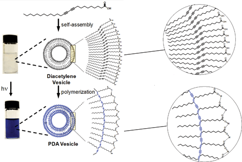
Interactions between the diacetylene (DA) monomers before polymerisation can directly influence the final polymerised structure. The monomers can be functionalised to contain motifs that encourage non-covalent interactions such as hydrogen bonding, π-stacking, electrostatics and hydrophobic interactions, allowing the DAs to self-assemble into nanostructures. Subsequent polymerisation of the acetylene groups results in cross-linking within the nanostructure, forming new materials with striking properties. One particularly interesting example of this is shown below – the DA monomers are substituted with long hydrophobic chains and polar head groups which assemble in water to form vesicles. Shining UV light on the vesicles causes the diacetylene groups to polymerise, generating PDA vesicles which are blue in colour.

For an in-depth and fascinating overview of the recent conceptual and technological advances in the chemistry of PDAs, download the full Feature Article here.

Posted on behalf of Cally Haynes, web science writer for ChemComm.

Digg This
Reddit This
Stumble Now!
Share on Facebook
Bookmark this on Delicious
Share on LinkedIn
Bookmark this on Technorati
Post on Twitter
Google Buzz (aka. Google Reader)

Look into my MRI: Non-invasive detection of melanin formation

Magnetic Resonance Imaging (MRI) is used as a medical tool to image soft tissue. It can give insights into the physical state of brain, identify sports injuries and diagnose cancer without ionising radiation. Its main drawback is the low sensitivity of the contrast enhancing probes it uses. These are the paramagnetic metal complexes, often containing gadolinium or manganese, that distinguish between healthy and damaged tissue by altering the relaxation times of the water protons in the body. The challenge is to increase the concentration of the contrast reagents in the tissues.

One solution is to use nanocarriers to deliver a high concentration of the contrast agent to the specific site of interest. Silvio Aime and his team recently used the nanocarrier, apoferritin, to transport solid MnOOH, which was subsequently reduced to paramagnetic Mn2+. Their recent ChemComm details how they have now taken this one step further to find a way of generating the Mn2+ contrast agent using a naturally occurring reductant. The result: a probe for melanin.

Melanin is produced from the successive oxidation of tyrosine – a process that is up-regulated in malignant melanoma. Aime exploited this oxidation process by introducing MnOOH-loaded apoferritin into melanoma cells – the MnOOH was reduced, generating the contrast agent and enabling the cells to be successfully imaged by MRI.

in vivo MRI images of tumour-bearing mice before and after administration of the contrast agent loaded nanocarrier

The team tested the sensor on melanogenic cells (melanin-forming cells) against non-melanogenic cells. They demonstrated that the signal generated belonged only to the melanin-producing cells which had internalised the Mn(III)OOH–apoferriton payloads. Animal studies revealed enhanced signal intensity in melanogenic tumours.

This interesting research has the potential to provide non-invasive, early diagnoses of skin cancers and evaluate the development of tumours, critical for saving people’s lives. The in vivo sensor may also be used to monitor other processes involving massive oxidative processes. The work of Silvio Aime is one to watch.

To find out more, download the ChemComm article today…

Posted on behalf of Sarah Brown, web science writer for ChemComm.

Digg This
Reddit This
Stumble Now!
Share on Facebook
Bookmark this on Delicious
Share on LinkedIn
Bookmark this on Technorati
Post on Twitter
Google Buzz (aka. Google Reader)

24 days until 100 issues…

…and did you know..?

24% of the articles published in ChemComm this year were from China.

This is almost three times as much as the proportion 5 years ago – in 2007, Chinese articles only made up 9% of publications in the journal. This represents the explosion of chemistry research in China over the last decade.

Digg This
Reddit This
Stumble Now!
Share on Facebook
Bookmark this on Delicious
Share on LinkedIn
Bookmark this on Technorati
Post on Twitter
Google Buzz (aka. Google Reader)

Simple detection of RNA depurination by RIPs

A class of protein toxins exists with an extremely apt acronym, RIP. This stands for Ribosome Inactivating Proteins but may as well stand for “Rest in Peace” when applied to cellular RNA. These proteins are known to attack the link between a purine base (adenine or guanosine) and its sugar causing depurination. This results in the formation of abasic sites (bases that are neither pyrimidines or purines) which in turn, causes the ribosome containing the sequence to have a lower affinity to elongation factors that are crucial for protein synthesis, ultimately leading to cell death.

Methods exist to detect RIPs such as ELISAs and antibody-based immunoassays or by monitoring the specific depurination activity through fluorescence, radiolabelling and immunoaffinity chromatography, amongst others. These techniques require sophisticated or elaborate set-ups, limiting the potential for high throughput (HTP) screening in a bid to discover potential inhibitors of these destructive toxins.

Seergazhi Srivatsan and co-workers have produced a label-free fluorescence hybridisation assay to detect the depurination activity of saporin, a RIP toxin, using a fluorescence ligand that specifically binds to the cytosine opposite an abasic site. If depurination takes place, then the ligand can bind and its fluorescence intensity will be quenched. This technique allows for a plethora of information to be obtained about the depurination activity of saporin as well as many other RIP toxins. This opens the door to using high throughput screening to find inhibitors of such toxins.

Download the article to read more…

Also of interest: Overcoming obstacles in labelling RNA

Posted on behalf of Sarah Brown, web science writer for ChemComm.

Digg This
Reddit This
Stumble Now!
Share on Facebook
Bookmark this on Delicious
Share on LinkedIn
Bookmark this on Technorati
Post on Twitter
Google Buzz (aka. Google Reader)

28 days until 100 issues…

… and did you know..?

ChemComm‘s impact factor (Journal Citation Reports®) has increased by 28% over the past 5 years. Our latest 2010 impact factor is 5.787 – a clear indication of the high quality research that ChemComm publishes.

Digg This
Reddit This
Stumble Now!
Share on Facebook
Bookmark this on Delicious
Share on LinkedIn
Bookmark this on Technorati
Post on Twitter
Google Buzz (aka. Google Reader)

Scattering layer for dye-sensitised solar cells

We are all aware how important it is to find new improved ways to generate sustainable energy. One very promising candidate is the dye-sensitised solar cell which is particularly attractive due to its low production cost and mechanical robustness.

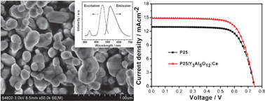
Scientists from East China Normal University have found a way to increase the efficiency of dye-sensitised solar cells by introducing an additional layer on top of the TiO2 photoanode. The extra layer, made up of Y3Al5O12:Ce down-converting microparticles, causes the conversion efficiency to increase from 6.97% to 7.91%.

SEM image of the Y3Al5O12:Ce layer and the J-V curves for the solar cell with and without the additional layer

Likun Pan and his team have attributed this improved performance to the increased light absorbing and scattering properties of the microparticle layer (meaning more suitable photons are available for absorption by the dye) and reduced electron transfer resistance.

Download the ChemComm article to read more about the fabrication of Pan’s solar cell device.

Digg This
Reddit This
Stumble Now!
Share on Facebook
Bookmark this on Delicious
Share on LinkedIn
Bookmark this on Technorati
Post on Twitter
Google Buzz (aka. Google Reader)

One nanoparticle, two nanoparticles, three nanoparticles, four!

Nanoparticles might be small but they frequent the pages of many a journal due to the ongoing boom in nanotechnology research. Whilst they are useful in a myriad of fields, it is still difficult to directly characterise these extraordinarily small entities. King among the visualisation techniques is electron microscopy but this often requires the isolation of the sample on a support – hardly sufficient for analysing a dynamic sample in solution! Dynamic light scattering is another potential technique but finds limitations when it comes to much smaller nanoparticle sizes.

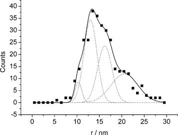
Ideally, you want to be able to count and size individual nanoparticles at a rate which produces reliable statistics. To address this challenge, Richard Compton and his team, including Neil Rees and Yi-ge Zhou who conducted the experiments alongside Jeseelan Pillay, Robert Tshikhudo and Sibulelo Vilakazi from Mintek, Randburg, have used anodic particle coulometry (APC) to measure gold nanoparticle collisions with a glassy carbon microelectrode and thus count and size individual nanoparticles.

With the electrode potential set above +1.0 V, they were able to record oxidative Faradaic transients from nanoparticle collisions and calculate an average nanoparticle radius which compared extremely well to the radius obtained from scanning electron microscopy measurements. They were also able to observe nanoparticle aggregation, which holds great promise for monitoring dynamic aggregation reactions.

It shouldn’t be long before this technique is routinely used to gain more information on all sizes of metallic nanoparticles which are currently being used in a variety of applications.

Read the ChemComm article by Compton and team for more.

Posted on behalf of Iain Larmour, web science writer for ChemComm.

Digg This
Reddit This
Stumble Now!
Share on Facebook
Bookmark this on Delicious
Share on LinkedIn
Bookmark this on Technorati
Post on Twitter
Google Buzz (aka. Google Reader)