Author Archive

Superpowers ahoy! Electric field causes DNA mutations

What can cause a mutation in DNA?  Well, if you were to ask the Incredible Hulk (nicely), he would probably say– well not a lot, he’s more of a doer, but Bruce Banner might tell you gamma rays.  But that is so 20th century.

In a Communication recently published in ChemComm, José Pedro Pedro Cerón-Carrasco (Université de Nantes) and Denis Jacquemin (Institut Universitaire de France) have shown that DNA can mutate permanently if an appropriate external electric field is applied.

Application of the right level of electric field can lead to proton transfer, which can cause the formation of tautomers, i.e. isomers of the DNA bases.  By interfering with the bases and their interaction, a mismatch or mutation can be induced.

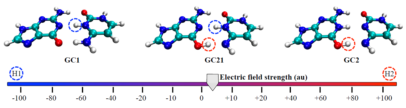
Turn the power up a little more and soon I will become Science Girl!: The DNA tautomers form under the influence of an external electric field. Circles indicate the protons that have been shifted compared to the canonical structure: H1 in blue and H2 in red.

Cerón-Carrasco and Jacquemin used a computational model to assess the effects of both positive and negative external electric fields on a DNA model to achieve an in vivo-like outcome.  When applying an increasing strength of negative electric fields, they saw the more acidic H1 proton shift to the other base; intense positive fields activated the H2 proton.

The authors conclude that intense electric fields might damage DNA in a partially controlled way.  This could have exciting applications for biochemistry or medicine– for example, selectively mutating a disease-causing cell.  Or maybe, bestowing me with superpowers…

Interested in more?  Read this HOT ChemComm article in full!

Electric field induced DNA damage: an open door for selective mutations
José Pedro Pedro Cerón-Carrasco and Denis Jacquemin
Chem. Commun., 2013, Accepted Manuscript
DOI: 10.1039/C3CC42593B

Sarah Brown is a guest web-writer for Chemical Communications.  Sarah hung up her lab coat after finishing her PhD and post-doctorate in nanotechnology for diagnostics and therapeutics to become an assistant editor at the BMJ Publishing Group.  When not trying to explain science through ridiculous analogies, you can often find her crocheting, baking and climbing, but not all at once.


Digg This
Reddit This
Stumble Now!
Share on Facebook
Bookmark this on Delicious
Share on LinkedIn
Bookmark this on Technorati
Post on Twitter
Google Buzz (aka. Google Reader)

If you like it, then you should put two rings on it

Microring resonators are pretty amazing things, offering label-free biosensing by coupling with light and then circulating the photons inside the cavity of the ring, enhancing the interaction between the light and the analytes.

However, I like to think of traditional microring resonators as tents: difficult to construct. They require a submicrometer gap between the input waveguide and the resonator ring structure to allow the coupling of light and before you can even get to that stage, you need to make the microring resonator, which requires a series of complex fabrication steps (FIG 1a).  In this Communication, which is part of ChemComm‘s Microfluidics themed web collection, Professor Jonathan Cooper and his colleagues at Glasgow University and at the International Islamic University Malaysia’s CTS Department have made (what I think of as) the double pop-up tent equivalent– or as they call it, the dual disk resonator (DDR). Made from SU8, an epoxy-based polymer used in microfluidics chips, it can be patterned in a single lithographic step. Not only that, but the DDR uses a gapless design and two rings, increasing the sensitivity of the device (FIG 1b).

FIG 1: The hard way or the easy way (a) the traditional microring resonator with submicrometer gap (b) gapless dual disk resonator

Once they had constructed the DDR, the team characterised the optical sensitivity of the device using sucrose solutions to vary the refractive index of the sample above the waveguide. They then went on to show that the sensor could be used to evaluate the dynamics of antibody interactions on surfaces, exploring avidin-biotin-based immobilisations; sharp resonance shifts confirmed the assembly and disassembly of constructs.

The simpler fabrication shows great promise, as the authors suggest that the sensitivity of the device could be greatly improved by coupling more disks to it– in which case Beyoncé might soon be singing ‘if you like it, then you should put a chain on it.’

Read this ‘HOT’ ChemComm article today:

Polymer dual ring resonators for label-free optical biosensing using microfluidics

Muhammad H. M. Salleh, Andrew Glidle, Marc Sorel, Julien Reboud and Jonathan M. Cooper

Chem. Commun., 2013, Advance Article

DOI: 10.1039/C3CC38228A

Digg This
Reddit This
Stumble Now!
Share on Facebook
Bookmark this on Delicious
Share on LinkedIn
Bookmark this on Technorati
Post on Twitter
Google Buzz (aka. Google Reader)

pHantastic developments in lysosomal storage disease research

Lysosomes are cellular organelles that contain enzymes which break down cellular waste, a bit like stomachs. They have an internal pH of 4.8 and maintain this acidic pH (compared to the cytosol, pH 7.2) by pumping protons across the membrane. Changes in lysosomal pH can indicate the onset of disease.  In fact, elevated lysosomal pH has been noted in several lysosomal storage diseases. This group of around 50 rare, inherited, metabolic disorders result in symptoms such as seizures, deafness and/or blindness.

Now David Parker and colleagues at the University of Durham have designed responsive, low molecular weight probes which can permeate the target organelle and report the pH using a ratiometric signal. The probes could help evaluate the impact of drugs created to treat lysosomal storage diseases.

The team made europium and terbium complexes of two structurally related ligands that contain a sulfonamide moiety which acts like a switch, reversibly binding to the lanthanide and changing the metal coordination environment. The change is signalled by variation of emission spectral form and relative intensity and also modulates the circular polarisation of luminescence as the local helicity at the metal centre switches.  

Testing in a range of cell lines and altering the pH of the cellular and intracellular environment, the researchers developed an emission intensity ratio method using lanthanide luminescence which can be used to assess lysosomal pH variation for the first time.

Find out more by reading their ChemComm communication, free to download for a limited period.

Also of interest:
Times have changed since David Parker wrote his first ChemComm on a typewriter. He discusses his research path, chemical prostitution and targeted devastation in his ChemComm interview.

Digg This
Reddit This
Stumble Now!
Share on Facebook
Bookmark this on Delicious
Share on LinkedIn
Bookmark this on Technorati
Post on Twitter
Google Buzz (aka. Google Reader)

Tomayto, tomato? Enantiospecific kinking of DNA

A few years ago, when I discovered what an intercalator was, I thought it would be a great name for a burger bar (probably best situated near a chemistry department). In scientific terms (and not catering as sadly the idea didn’t take off), intercalators have attracted a great deal of attention and are best known for their use in anticancer treatments.

Chelate compounds of polycyclic heteroaromatics with transition metals can bind to DNA. The polycyclic moeities intercalate between the base pairs of the DNA, a little like the burger in a bun.  This can have a profound effect on the DNA’s structure, separating the base pairs and causing the helix to kink. However, the extent of this effect is dependent on parameters such as the ligand and which enantiomer of the instrinsically chiral compounds is involved.

A study by Anna Reymer and Bengt Nordén into the ruthenium compound, [Ru(phenanthroline)3]2+,  investigates its two enantiomers (Δ and Λ) and the effect each one has on binding specificity with DNA. Using molecular dynamics simulations, they demonstrated that the Δ-form induced a kink of 53° whilst the Λ-form produced a more typical bend of only 16°. They also reveal information about binding affinities of the compounds and how ‘deeply’ they can insert themselves into the base stack.

This interesting simulation is significant in the context of chiral recognition and evolutionary selection. It also gives further insight into the behaviour of DNA-protein interactions; an analogous kink as produced by Δ- [Ru(phenanthroline)3]2+ have been observed for several classes of operatory proteins.

To find out more download Reymer and Nordén’s communication.

Digg This
Reddit This
Stumble Now!
Share on Facebook
Bookmark this on Delicious
Share on LinkedIn
Bookmark this on Technorati
Post on Twitter
Google Buzz (aka. Google Reader)

Proteins perform (useful) tricks via DNA-based self assembly

Proteins are very useful molecules and when they work together, or assemble, they can display biocatalytic cascades, performing sequential multistep transformations of substrates. Scientists have tried to mimic nature for years, by creating artificial multi-enzyme complexes to replicate these biomolecules’ ability to catalyse reactions for use in biofuels, bioelectronics, bioproduction etc.

The arrangement of the proteins’ active sites relative to one another is intrinsic to the success of these reactions. One method of synthetically engineering these arrangements is through the use of DNA nanostructures.  DNA aptamers can be used as scaffolds to encourage the proteins’ assembly and even introduce other functional properties – imagine this as the bottom layer of a human pyramid in Cirque du Soleil.

However, the DNA scaffolds are reported to degrade and the protein assemblies decompose. (Now, imagine someone telling a really good joke to the bottom layer of the human pyramid and it all falling apart.)  The scaffolds and proteins are difficult to separate and this has limited the application of this strategy. Until now….

Masahiro Goto and co-workers have managed to arrange protein molecules (in this case, thrombin) on a DNA scaffold with the use of a DNA aptamers. With the addition of a chemical cross-linker, the neighbouring protein molecules were covalently cross-linked and retained their activity.

Programmable protein-protein conjugation via DNA-based self-assembly

Using a DNA template for thrombin binding aptamers, and hybridising that with three thrombin binding aptamers with sticky ends, they formed a comb-like structure with branched arms. The thrombin molecules bind with these arms and a chemical cross-linker encourages the neighbouring thrombins to cross-link. This has been intonated on the diagram with ‘holding hands’. (Told you they were inspired by Cirque du Soleil).

Using polyacrylamide gel electrophoresis (PAGE), the group elegantly illustrated their results, successfully demonstrating that DNA scaffolds can produce successful protein-protein conjugation. The group continue to develop and improve their work to overcome limitations in the size of conjugate proteins, efficiency and applications.

Find out more – download the ChemComm communication, free for 4 weeks.

Digg This
Reddit This
Stumble Now!
Share on Facebook
Bookmark this on Delicious
Share on LinkedIn
Bookmark this on Technorati
Post on Twitter
Google Buzz (aka. Google Reader)