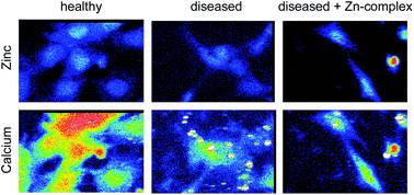
Anthony White from the Department of Pathology, University of Melbourne and team have investigated the use of X-ray Fluorescence Microscopy to investigate subcellular biometal homeostasis in a mouse model of the childhood neurodegenerative disorder – Batten disease. This is a particularly nasty disease that causes progressive blindness and motor impairment leading to premature death. The team have previously highlighted the redistribution of brain zinc in diseased samples using subcellular fractionation techniques. However, the lack of a rapid, specific and sensitive technique to provide quantitative subcellular information in situ on biometal distributions is severely limiting the understanding of the effect that the changes in biometals distributions can have in neurodegenerative diseases.
Consider the next time you go food shopping. What if you arrived at the supermarket and all the shelves were empty. A shop assistant reassures you that there is plenty of stock, hundreds of thousands of items indeed, but it all just happens to be sitting in their central depot instead of the supermarket. This does not help you to make dinner that night. This scenario is an example of how important the distribution of items can be.
When it comes to neurodegenerative disorders the location of biometals, just like the location of the groceries in the above case, provides much more useful information than the total amount of biometal present. The amount of biometal may not change between normal and diseased cells, but the distribution does. Biometals are important cofactors to a large number of enzymes and are also recognised as second messengers in neuronal signalling
In their Chemical Science paper the team have utilised the advances in data collection speed of X-ray fluorescence detectors to map the zinc and calcium distributions in a statistically relevant number of cells. Despite a lack of global changes in biometal levels this approach revealed the perturbed trafficking of zinc and the significantly altered subcellular calcium distributions. The restorative properties of a therapeutic zinc-complex were also shown using this technique.
Techniques and methodology reported in the Chemical Science paper can be applied to other neurodegenerative diseases and importantly can provide insights to the mechanism of action of novel therapeutics at the single cell level.
Read the Chem. Sci. paper in full today for free* and see if this technique could be applied to a disease you are studying!
X-ray fluorescence imaging reveals subcellular biometal disturbances in a childhood neurodegenerative disorder
A. Grubman, S.A. James, J. James, C. Duncan, I. Volitakis, J.L. Hickey, P.J. Crouch, P.S. Donnelly, K.M. Kanninen, J.R. Liddell, S.L. Cotman, M.D. de Jonge and A.R. White*
DOI: 10.1039/C4SC00316K
*Access is free until 20.06.14 through a registered RSC account – click here to register


