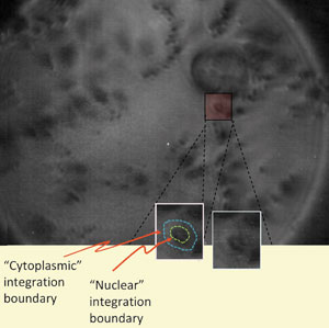
A fast, high-resolution infrared imaging technique that can ‘freeze’ living specimens has been designed by UK scientists and tested on human ovarian cancer cells. The technique could lead to a better understanding of how cancer drugs work.
Infrared spectroscopy of cell images can be used in a number of fields including forensic science and cancer research. However, taking pictures of samples can take up to 12 hours. Chris Phillips and his team at Imperial College London have developed a technique to produce 2D images that takes a fraction of a second. By combining a purpose-built pulsed IR laser source with a charge-coupled device camera, rather like a digital camera, they were able to generate pictures 1011 times faster than current IR spectroscopic imaging methods. The IR source generates very short pulses (~100 psec) that keep the illumination levels below cell phototoxicity limits and allow moving specimens to be frozen in a way that mimics conventional flash photography.
Previous attempts to image cells in this way have required long illumination times, which causes the cells to move away from the light source or can kill them. ‘Because you can do it so quickly, you can freeze the action in living things, and because you have so much more light signal, you can get right inside the cells to take chemical maps,’ says Phillips.
Link to journal article
Ultrafast infrared chemical imaging of live cells
Hemmel Amrania, Andrew P. McCrow, Mary R. Matthews, Sergei G. Kazarian, Marina K. Kuimova and Chris C. Phillips
Chem. Sci., 2011, DOI: 10.1039/c0sc00409j


