Archive for the ‘Chemical Biology’ Category

Sugar injection to beat hospital infection

BacteriaA carbohydrate from the surface of the most virulent strain of the bacterium Clostridium difficile has been synthesised by chemists in Germany. The molecule could be used to develop a vaccine against the infection.

C. difficile infections are the most common cause of hospital acquired diarrhoea and can lead to the death of elderly patients and those with weakened immune systems.’C. difficile is on the rise in industrialised countries,’ says Peter Seeberger, who led the team that carried out the research at the Max Planck Institute of Colloids and Interfaces, Potsdam. ‘There is a need for a vaccine but it’s a big challenge.’

Find out more about Seeberger’s progress towards developing a vaccine in the full Chemistry World news story and download his ChemComm communication, free for a limited period.

Digg This
Reddit This
Stumble Now!
Share on Facebook
Bookmark this on Delicious
Share on LinkedIn
Bookmark this on Technorati
Post on Twitter
Google Buzz (aka. Google Reader)

Quantum dots selectively label endoplasmic reticulum

Graphical abstract: Selective labeling of the endoplasmic reticulum in live cells with silicon quantum dotsScientists are a step closer to understanding how an important cell organelle works, which could lead to new insight into disease such as diabetes and Alzheimer’s disease.
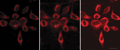
The endoplasmic reticulum (ER) plays a critical role in protein synthesis and transport. Its malfunction can lead to serious diseases so it is important to be able to observe how it works. 

Yukio Yamaguchi and colleagues at the University of Tokyo, Japan, have managed to selectively label the ER in live cells using quantum dots (QDs). Although organic dyes have previously been used for this purpose, Yamaguchi’s QDs are less toxic and more photostable. 

The QDs’ photoluminescence enabled the team to view the ER using a confocal microscope, making them a powerful tool for long-term real-time observation of the ER, Yamaguchi says.

Find out more by downloading the communication.

Digg This
Reddit This
Stumble Now!
Share on Facebook
Bookmark this on Delicious
Share on LinkedIn
Bookmark this on Technorati
Post on Twitter
Google Buzz (aka. Google Reader)

Probing cells’ power generators

UK scientists have developed a probe to monitor bicarbonate concentrations in mitochondria – components in living cells that generate chemical energy. Monitoring bicarbonate levels will improve researchers’ understanding of its role in cellular reaction mechanisms. 

A challenge when designing cellular probes is ensuring that the probe is not only selective for its target but can also be delivered to the site of interest within the cell. A team of scientists led by David Parker at the University of Durham has made a probe that can overcome this challenge. 

Stained HeLa cellsThe luminescent probe features an azaxanthone moiety, which is linked to a europium complex by an amide bond. The azaxanthone allows the probe’s uptake into cells and localisation within the mitochondria, and the europium complex has an affinity for bicarbonate ions. The ability to probe bicarbonate levels ‘can offer an unprecedented insight into signalling mechanisms’, says Parker.
Read the rest of this story in Chemistry World and download Professor Parker’s ChemComm communication, which is free to access for a limited period.

Also of interest:
Definition of the uptake mechanism and sub-cellular localisation profile of emissive lanthanide complexes as cellular optical probes
Elizabeth J. New, Aileen Congreve and David Parker, Chem. Sci., 2010, 1, 111-118

Digg This
Reddit This
Stumble Now!
Share on Facebook
Bookmark this on Delicious
Share on LinkedIn
Bookmark this on Technorati
Post on Twitter
Google Buzz (aka. Google Reader)

Across the barrier for tumour imaging

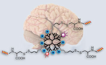
Brain and structure of nanoprobeA probe that can cross the blood-brain barrier to allow high sensitivity brain tumour imaging has been made by Chinese scientists. The probe could be used to pinpoint the location and extent of a tumour before an operation and be used for image-guided tumour removal. 
Establishing the position, extent and structure of brain tumours is crucial for their successful removal. But, current tumour imaging agents used in magnetic resonance imaging are limited by short circulation lifetimes, non-targeted specificity and poor blood-brain barrier permeability. The results of these limitations are that low grade tumours and 20-30 per cent of advanced brain tumours with an intact blood-brain barrier go unnoticed.

Cong Li from Fudan University, Shanghai, and his team made the probe starting with a dendrimer – a branched molecule with a long circulation lifetime – and attached functional groups with different tasks. One such group, a lipoprotein ligand angiopep-2, helps to deliver the probe across the blood-brain barrier and targets the lipoprotein’s receptors, which are present in increased amounts on tumour cells. High-resolution images can be generated thanks to imaging reporters, including fluorescence dyes, attached to the dendrimer. 

Read the rest of this story in Chemistry World and download Li’s ChemComm communication, which is free to access for the rest of the month.

Digg This
Reddit This
Stumble Now!
Share on Facebook
Bookmark this on Delicious
Share on LinkedIn
Bookmark this on Technorati
Post on Twitter
Google Buzz (aka. Google Reader)

The importance of chemistry in sequencing nucleic acids


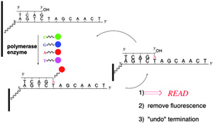
Solid phase DNA sequencing

Chemistry has played a vital role towards making routine, affordable sequencing of human genomes a reality. Leading UK scientist, Shankar Balasubramanian, provides a compelling review of the last 60 years, from the Sanger sequencing method through to the human genome project. In particular, Balasubramanian focuses on the achievements of Solexa (latterly, Illumina) on the modern developments of high throughput nucleic acid sequencing that originated in Cambridge, in the UK.
   

In association with the International Year of Chemistry (IYC), this Highlight in Chemistry emphasises the importance of chemistry and how it continues to contribute towards many other fields, most notably the biological and biomedical sciences. This sequencing approach is helping to transform science and offers intriguing prospects for the future of medicine.

Fancy reading more? Download the ChemComm Highlight, which will be free to access until the 1st July 2011.

 For your info, ChemComm is publishing Highlights in Chemistry articles throughout 2011, which has been recognised as an official activity for celebrating the IYC. Take a look at the IYC website to keep up-to-date on what else is happening throughout 2011 to celebrate the achievements of chemistry and its contributions to the well-being of humankind.

Digg This
Reddit This
Stumble Now!
Share on Facebook
Bookmark this on Delicious
Share on LinkedIn
Bookmark this on Technorati
Post on Twitter
Google Buzz (aka. Google Reader)

Imaging brain tumours

A new probe for imaging brain tumours could offer increased hope for cancer patients, say Chinese chemists.

Den-Angio visualizes an orthotopic U87MG tumor non-invasively in vivo. (A) NIR fluorescence and X-ray/color coded NIR fluorescence images of a mouse head at 2 h PI of Den-Angio. Arrows point to the tumor. (B) T1-weighted MR images of a mouse brain (coronal plane) at 2 h PI of Den-Angio or Gd3+–DTPA with the same gadolinium dose (0.05 mmol kg−1). White arrows point to the tumor and red arrows point to the cerebral ventricle. (C) Histological H&E staining of identical brains in panel B. Scale bar, 2 mm.Cong Li, at Fudan University, Shanghai, and colleagues have made a dendrimer-based nanoprobe called Den-Angio that can cross the blood-brain barrier. It can be used in the magnetic resonance imaging of brain tumours and should make it easier for doctors to distinguish cancerous tissue from healthy cells when cutting out the tumour.

To find out more, read Li’s ChemComm communication.

Digg This
Reddit This
Stumble Now!
Share on Facebook
Bookmark this on Delicious
Share on LinkedIn
Bookmark this on Technorati
Post on Twitter
Google Buzz (aka. Google Reader)

Inducing protein heterodimerisation

Scientists in The Netherlands and Germany have shown that cucurbit[8]uril (CB[8]) induces selective heterodimerisation between two different proteins.
 

Supramolecular induced protein dimerisation

Luc Brunsveld and his team functionalised the two proteins with methylviologen (electron deficient) and napthalene (electron rich) guest molecules, which formed a charge transfer complex inside the CB[8] cavity.  Interestingly, the resulting dimerisation can be visually observed and has established that there is distinct interplay between the supramolecular components within the proteins.

Fancy reading more? Then download the ChemComm communication,which will be free to access until the 24th June 2011.

Digg This
Reddit This
Stumble Now!
Share on Facebook
Bookmark this on Delicious
Share on LinkedIn
Bookmark this on Technorati
Post on Twitter
Google Buzz (aka. Google Reader)

Ibuprofen: anticancer drug

Scientists in the UK have moved a step closer to understanding how ibuprofen could help treat cancer. The findings could lead to the drug being used as a preventative treatment for prostate cancer, in the future.

Ibuprofen – a common painkiller – can help reduce the risk of prostate cancer, but the mechanism by which it inhibits tumour cells is still not fully understood. Now, Matthew Lloyd and his team from the University of Bath in the UK, in collaboration with Cancer Research UK, have uncovered a mechanism suggesting that the chiral inversion of ibuprofen inhibits the activity of the protein alpha-methylacyl-CoA racemase (AMACR), levels of which are increased in the presence of prostate, some colon and other cancers.

To find out more, read the full news story in Chemistry World and download Lloyd’s ChemComm communication.

Digg This
Reddit This
Stumble Now!
Share on Facebook
Bookmark this on Delicious
Share on LinkedIn
Bookmark this on Technorati
Post on Twitter
Google Buzz (aka. Google Reader)

Antibody acts as rudder to steer DNA into a pore

Scientists from China and Canada have studied the effect of a DNA-binding antigen-binding fragment (Fab) of an antibody on the translocation of a DNA polymer. Both poly(dT)45:Fab HED10 and poly(dT)45 produce unique double step current traces, which were analysed in detail to get information about the translocation events.

Representation of an α-HL pore and the mechanism for the poly(dT)45 specific binding with Fab HED10. A biological α-HL nanopore is embedded in a lipid bilayer. The narrowest section of α-HL is 1.4 nm. The potential across the bilayer membrane is applied through Ag/AgCl electrodes. In 10 mM Tris-HCl (pH = 7.8) buffer, the antibody recognizes four consecutive thymine residues of poly(dT)45.

The results have important implications for understanding the translocation behaviour of polymers, which could help to develop nanopore biosensors with high sensitivity and specificity, says Yitao Long, from East China University of Science and Technology. In particular, the addition of polymer-binding antibodies may facilitate the use of nanopores in sequencing technologies.

The presence of the FAB HED10 decreases the time of the first level of the step but not the second. The Fab appears to behave as a rudder, which significantly decreases the energy barrier for poly(dT)45 translocation. A more rigid or extended conformation of poly(dT)45 would decrease the time required to find the entrance to the narrow constriction in the pore. The entropic barrier required to linearise the DNA strand may be the dominant contribution to the entire energy barrier.

Find out more by downloading the communication, recently published in ChemComm.

Digg This
Reddit This
Stumble Now!
Share on Facebook
Bookmark this on Delicious
Share on LinkedIn
Bookmark this on Technorati
Post on Twitter
Google Buzz (aka. Google Reader)

Novel chemical tag illuminates protein cholesterylation in cells

UK chemists have for the first time used a chemical probe to study the post- translational cholesterylation of proteins in living cells.

Post-translational modification (PTM) in cells plays an important role in the function of proteins in vivo. One example is the mammalian Hedgehog (Hh) protein family: the post-translational cholesterylation of Sonic hedgehog (shh) protein regulates its secretion. However, mis-regulation of this protein can promote different types of cancers. Therefore a simple way of studying this type of modification is important.

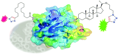
Graphical abstract: Bioorthogonal chemical tagging of protein cholesterylation in living cells

Edward Tate and colleagues at Imperial College, London have done just this. They first modified cholesterol molecules to bear an azide group and then gave this to their target cells, where it was used in PTM. They next managed to attach, via ‘click’ chemistry, a dye molecule called TAMRA to the modified proteins that carried the synthetic cholesterol. The team used this dye for ‘fluorescence visualisation’ of the target protein.

When compared to traditional techniques for studying cholesterylated proteins, this new method stacks up well. It makes significant savings in both time and expense, as well as avoiding the use of potentially harmful radiation. Furthermore, Tate suspects that in the future an optimised version of this process might be used to search for new cholesterylated proteins.

Want to find out more? Then download the ChemComm article for free today. You can also check out coverage of this article in C&EN.

Digg This
Reddit This
Stumble Now!
Share on Facebook
Bookmark this on Delicious
Share on LinkedIn
Bookmark this on Technorati
Post on Twitter
Google Buzz (aka. Google Reader)