Archive for the ‘Chemical Biology’ Category

Drug delivery: implications of gold-protein interactions

Researchers in Italy have shown that medicinal gold compounds interact strongly with the proteins of the copper trafficking system, which could have implications for drug delivery.

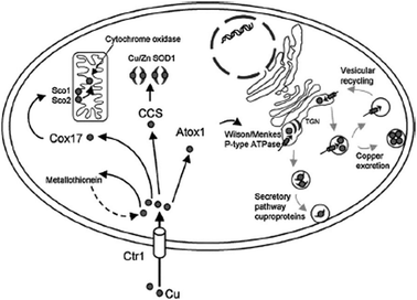
The copper trafficking system consists of proteins that help the uptake of copper into cells and then promote its transfer and delivery to copper-dependent cellular proteins.  One of these ‘chaperones’ is known as Atox-1.

Copper trafficking within a mammalian cell

Previous work has shown that platinum-based anticancer drugs strongly interact with copper trafficking system proteins and Messori and co-workers hypothesised that medicinal gold compounds might also do the same, especially in the +1 oxidation state; soft lewis acids, such as gold (I) ions react eagerly with Atox-1.

The interactions of three gold (III) compounds with Atox-1 were analysed through ESI-MS and revealed the formation of metal-protein adducts. The same major adduct was invariantly formed, matching the protein binding of a single gold (I) ion. Formation of this adduct implied that the gold (III) complex had broken down, a loss of ligands and reduction to a gold (I) species. ESI-MS also displayed peaks that corresponded to protein binding with two gold (I) ions. A stability study showed that one of the three gold-protein adducts was stable over 72 hours.

From their findings, the authors conclude that the cytotoxic gold compounds investigated form stable adducts with copper chaperone, Atox-1. These results have implications for medicinal drug design and our little friend, Atox-1 stays in a job.

Read this HOT Chem Comm article today (free to access until the 14th of December 2012):

Medicinal gold compounds form tight adducts with the copper chaperone Atox-1: biological and pharmacological implications
Chiara Gabbiani, Federica Scaletti, Lara Massai, Elena Michelucci, Maria A. Cinellu and Luigi Messori
Chem. Commun., 2012, 48, 11623-11625

Published on behalf of Sarah Brown, Chemical Communications web science writer.

Digg This
Reddit This
Stumble Now!
Share on Facebook
Bookmark this on Delicious
Share on LinkedIn
Bookmark this on Technorati
Post on Twitter
Google Buzz (aka. Google Reader)

Origami, without the papercut, visualised

Researchers in China have been able to visualize the intracellular location of DNA origami with a label-free fluorescent probe.

But let’s unfold a few things first and figure out what that means. DNA origami is the folding of a strand of DNA to make arbitrary 2 or 3 dimensional shapes; this serves as a ‘scaffold’ for shorter DNA strands that help hold the structure in its folded shape. These structures may be used for drug delivery, biosensors and more. I once made an origami frog; I wonder if there are any similarities…

Direct visualisation of the distribution and stability of DNA origami in live, cellular systems has not been achieved. Fluorescent labels can be attached to DNA strands but these have their drawbacks, such as weak emission intensity, photobleaching and expensive. Ding and co-workers looked at alternatives to visualize DNA origami in live cells.

The group were inspired by research on a series of carbazole-based cyanine fluorescence probes, which have a weak emission when they are monomolecularly dissolved but switch to a strong luminescent state upon binding to DNA or protein molecules.  The significant enhancement is attributed to restricted intramolecular rotational (RIR) motions by anchoring the DNA molecules, which causes the large reduction in the non-radiative decay of fluorescence molecules.

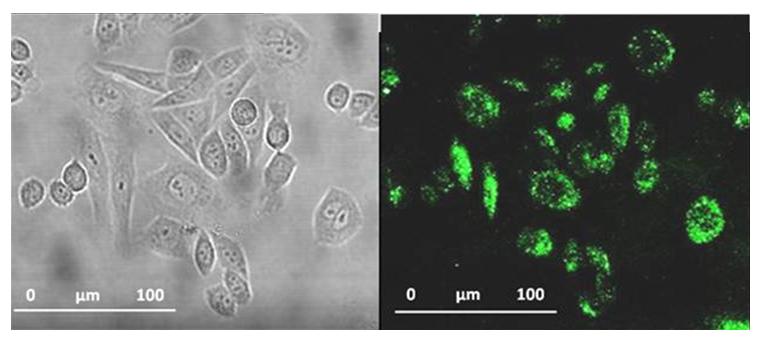
DNA-origami visualised in cells

Follow the instructions (a) and you too won’t make DNA-origami visualised in cells (b)

Ding and co-workers then took some tubular DNA origami, the cyanine fluorophore and found that the carbazole-based cyanine molecules could be used as a sensitive optical switch, turned on when DNA origami is detected and turned off when the nanostructure degrades. After incorporating the cyanine probe molecules, the DNA origami-probe complex was administered to live cells. Excitingly, the green-yellow frog… erm, I mean, fluorescence was visible inside the cells treated with the probe. The group went further to try and understand the internalization mechanism of the DNA origami and found the probe localized in lysosomes. Finally, degradation studies showed that most DNA origami were dissociated after 60 hours, also a bit like my origami frog.

Unlike my attempts at origami, Ding and co-workers have demonstrated an exciting step in scaffolded DNA origami and its future applications in nanomedicine.

Read this HOT Chem Comm article today (free to access until the  5th of December 2012):

Visualization of the intracellular location and stability of DNA origami with a label-free fluorescent probe
Xibo Shen, Qiao Jiang, Jinye Wang, Luru Dai, Guozhang Zou, Zhen-Gang Wang, Wei-Qiang Chen, Wei Jiang and Baoquan Ding
Chem. Commun., 2012, 48, 11301-11303

Published on behalf of Sarah Brown, Chemical Communications web science writer.

Digg This
Reddit This
Stumble Now!
Share on Facebook
Bookmark this on Delicious
Share on LinkedIn
Bookmark this on Technorati
Post on Twitter
Google Buzz (aka. Google Reader)

ChemComm celebrates its first Gold for Gold communication

Eugen Stulz (University of Southampton) and colleagues are the first ChemComm authors to publish a communication as part of our Gold for Gold initiative.Gold Image

Their communication, entitled ‘A DNA based five-state switch with programmed reversibility’ is now free to access for all.

‘I’m delighted that Eugen’s communication is the first open access communication to be published in ChemComm using the RSC’s Gold for Gold programme,’  says Phil Gale, Head of Chemistry at the University of Southampton. ‘This open access programme will allow us to showcase our research to a much wider audience.’

Gold for Gold is an innovative initiative rewarding UK RSC Gold customers with credits to publish a select number of papers in RSC journals via Open Science, the RSC’s Gold Open Access option.

More information on Gold for Gold is available on our website. If you have any questions on the procedure, or are an interested customer from outside the UK, please contact goldforgold@rsc.org.

Also of interest:
Gold for Gold – First Open Access credit used by University of Hull

Digg This
Reddit This
Stumble Now!
Share on Facebook
Bookmark this on Delicious
Share on LinkedIn
Bookmark this on Technorati
Post on Twitter
Google Buzz (aka. Google Reader)

Biocatalysis: an article collection

Beers, wines and cheeses are enjoyed around the world today and have been for millennia. In fact the practices of brewing and cheese-making pre-date recorded history so it is difficult to accurately determine when we first started using naturally occurring enzymes and microorganisms to create valuable (and in this case, tastier!) products.

Biocatalysts are of course used in far more diverse applications than the creation of food-stuffs, including in many organic syntheses and in the generation of fine chemicals. Due to their natural design, they can offer superior selectivity for particular products and have a far lower environmental impact than many traditional catalysts. Our knowledge and understanding of biocatalysts has increased dramatically in the last few decades, which has allowed us to develop biologically modified and biomimetic catalysts for a range of applications. 

To keep you up to date with the latest advances in this rapidly expanding field we have collected together these high impact articles and made them free to access until the 31st October!

Click here for the full list of free articles

Digg This
Reddit This
Stumble Now!
Share on Facebook
Bookmark this on Delicious
Share on LinkedIn
Bookmark this on Technorati
Post on Twitter
Google Buzz (aka. Google Reader)

ChemComm and the chemistry-biology interface

The chemical sciences make a huge contribution to solving challenges in the biological sciences. 

So quite rightly, articles at the chemistry–biology interface make up an important part of ChemComm.   

Here’s a selection of some recent articles, all free to access until 19th October

Nucleic acid aptamers: an emerging frontier in cancer therapy
Guizhi Zhu, Mao Ye, Michael J. Donovan, Erqun Song, Zilong Zhao and Weihong Tan
Chem. Commun., 2012, DOI: 10.1039/C2CC35042D 

Picomolar level profiling of the methylation status of p53 tumor suppressor gene by label-free electrochemical biosensor
Po Wang, Hai Wu, Zong Dai and Xiaoyong Zou
Chem. Commun., 2012, DOI: 10.1039/C2CC35615E 

Oriented Immobilization of Oxyamine-Modified Proteins
Long Yi, Yong-Xiang Chen, Po-Chiao Lin, Hendrik Schroeder, Christof M. Niemeyer, Yaowen Wu, Roger S. Goody, Gemma Triola and Herbert Waldmann
Chem. Commun., 2012, DOI: 10.1039/C2CC35237K 

Colorimetric detection of single-nucleotide polymorphisms with a real-time PCR-like sensitivity
Wei Shen, Huimin Deng, Alan Kay Liang Teo and Zhiqiang Gao
Chem. Commun., 2012, DOI: 10.1039/C2CC35070J

A bioresponsive controlled-release biosensor using Au nanocages capped with an aptamer-based molecular gate and its application in living cells
Wei Wang, Tao Yan, Shibin Cui and Jun Wan
Chem. Commun., 2012, DOI: 10.1039/C2CC33165A

Cascade imaging of proteolytic pathway in cancer cell using fluorescent protein-conjugated gold nanoquenchers
Kyoungsook Park, Jinyoung Jeong and Bong Hyun Chung
Chem. Commun., 2012, DOI: 10.1039/C2CC35687B

Eager for more? 

Check out the Nucleic acids: new life, new materials web theme, jointly organised with OBC and RSC Advances.

Digg This
Reddit This
Stumble Now!
Share on Facebook
Bookmark this on Delicious
Share on LinkedIn
Bookmark this on Technorati
Post on Twitter
Google Buzz (aka. Google Reader)

Glucometers altered to detect HIV

Glucometers used by diabetic patients can be altered to detect HIV-related DNA sequences, say scientists in China.

The commercially available personal glucometer has been the most successful point-of-care (POC) device up to date. But the glucometer only responds to glucose. Extending its use to monitoring different types of targets would potentially revolutionise POC technology.

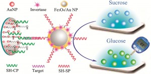
The team used invertase, an enzyme that catalyses the hydrolysis of sucrose into glucose, to interpret DNA recognition events into readouts measurable by the glucometer.

They loaded nanoparticle amplification labels with invertase, which, through target/probe DNA hybridisations, catalysed the conversion of sucrose on the sensing surface to glucose. They could detect as low as 0.5pM of target DNA. While they demonstrate the method with HIV DNA, it could potentially used to detect different DNAs.

Graphical Abstract


Link to journal article
Sensitive point-of-care monitoring of HIV related DNA sequences with a personal glucometer
J Xu et al
Chem. Commun., 2012, DOI: 10.1039/c2cc35941c

Digg This
Reddit This
Stumble Now!
Share on Facebook
Bookmark this on Delicious
Share on LinkedIn
Bookmark this on Technorati
Post on Twitter
Google Buzz (aka. Google Reader)

Overcoat makes carbon quantum dots biocompatible

Quantum dots are currently being developed for a variety of applications, including as sensors and cellular tags. Semiconductor quantum dots are attractive for their high fluorescence quantum yields but the toxicity of some of the metals involved, such as cadmium, pose a problem for biological applications.

Carbon quantum dots (CQDs) offer an alternative however when transferred into aqueous solution they possess low quantum yields. The problem is how do you harness the higher fluorescence of CQDs prepared in an organic solvent for biological applications?

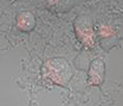
To answer this question John Callan and his team have employed an amphiphilic polymer to act as an overcoat and transfer agent for the CQDs. Surprisingly they found that the transfer actually improved the quantum yield rather than the normally expected repression when ligand exchange is used. These aqueous carbon quantum dots were taken up into cells and were found to be non-toxic.

Chinese Hamster Ovarian cells loaded with carbon quantum dots

The development of inexpensive and biocompatible quantum dots with an improved quantum yield holds great potential for a wide range of future biological applications.

To find out more, download the ChemComm article today (free to access for a limited period).

Digg This
Reddit This
Stumble Now!
Share on Facebook
Bookmark this on Delicious
Share on LinkedIn
Bookmark this on Technorati
Post on Twitter
Google Buzz (aka. Google Reader)

pHantastic developments in lysosomal storage disease research

Lysosomes are cellular organelles that contain enzymes which break down cellular waste, a bit like stomachs. They have an internal pH of 4.8 and maintain this acidic pH (compared to the cytosol, pH 7.2) by pumping protons across the membrane. Changes in lysosomal pH can indicate the onset of disease.  In fact, elevated lysosomal pH has been noted in several lysosomal storage diseases. This group of around 50 rare, inherited, metabolic disorders result in symptoms such as seizures, deafness and/or blindness.

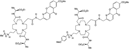
Now David Parker and colleagues at the University of Durham have designed responsive, low molecular weight probes which can permeate the target organelle and report the pH using a ratiometric signal. The probes could help evaluate the impact of drugs created to treat lysosomal storage diseases.

The team made europium and terbium complexes of two structurally related ligands that contain a sulfonamide moiety which acts like a switch, reversibly binding to the lanthanide and changing the metal coordination environment. The change is signalled by variation of emission spectral form and relative intensity and also modulates the circular polarisation of luminescence as the local helicity at the metal centre switches.  

Testing in a range of cell lines and altering the pH of the cellular and intracellular environment, the researchers developed an emission intensity ratio method using lanthanide luminescence which can be used to assess lysosomal pH variation for the first time.

Find out more by reading their ChemComm communication, free to download for a limited period.

Also of interest:
Times have changed since David Parker wrote his first ChemComm on a typewriter. He discusses his research path, chemical prostitution and targeted devastation in his ChemComm interview.

Digg This
Reddit This
Stumble Now!
Share on Facebook
Bookmark this on Delicious
Share on LinkedIn
Bookmark this on Technorati
Post on Twitter
Google Buzz (aka. Google Reader)

Silica sheets to deliver DNA into cells for disease prevention and treatment

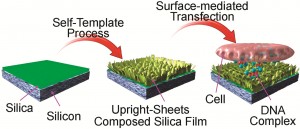
Scientists in Japan have made a new DNA delivery substrate based on networks of bio-friendly upright silica sheets. The network of sheets forms a porous film on to which they can immobilise DNA, ready for delivery (transfection) into cells. The transfection efficiency of the silica film is approximately double that of solution-based transfection. Gene transfection is a potential method for preventing and treating diseases and analysing cell functions.


Silica sheets to deliver DNA into cells for disease prevention and treatment

Link to journal article
Silica-based Gene Reverse Transfection: Upright Nanosheet Network for Promoted DNA Delivery to Cell

Q Ji et al

Chem. Commun., 2012, DOI: 10.1039/c2cc34289h


Digg This
Reddit This
Stumble Now!
Share on Facebook
Bookmark this on Delicious
Share on LinkedIn
Bookmark this on Technorati
Post on Twitter
Google Buzz (aka. Google Reader)

Film to aid drug metabolism study

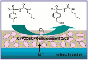
A way to study drug metabolism using cytochrome P450 enzymes (which are involved in the metabolism of over 60% of clinically used drugs) has been developed by scientists in China.

The team made a film of indium tin oxide nanoparticles (they have good conductivity) and cytochrome P450s encapsulated by chitosan (which are biocompatible) on a carbon electrode. They were able to bioelectronically initiate cytochrome P450 catalysis by replacing electron donation from expensive nicotinamide adenine dinucleotide phosphate with electrodes.

The system has potential for applications in drug discovery and development by monitoring substrate metabolism and enzyme inhibition. Other applications include biosensors for toxicity analysis and bioreactors for chemical synthesis.

Film to aid drug metabolism study


Link to journal article
Electrochemically Driven Drug Metabolism via Cytochrome P450 2C9 Isozyme Microsomes with Cytochrome P450 Reductase and Indium Tin Oxide Nanoparticle Composites

X Xu et al
Chem. Commun.,
2012, DOI: 10.1039/c2cc33575a

Digg This
Reddit This
Stumble Now!
Share on Facebook
Bookmark this on Delicious
Share on LinkedIn
Bookmark this on Technorati
Post on Twitter
Google Buzz (aka. Google Reader)