Archive for the ‘Materials’ Category

Synthesis of Tin Dioxide Nanotubes for Lithium-ion Batteries with “A Grain of Oxalate Salt”

Preparation of tube-shaped electrode materials for lithium-ion batteries is a trending topic. Tubes with hollow cylindrical bodies allow exposure of the electrodes’ interior surface and can accommodate the large volumetric expansion commonly observed when lithium ions diffuse (either via intercalation or alloying) into the electrodes. The aforementioned two characteristics improve the specific capacity (a measure of how much electric energy one electrode can hold) and lifetime of electrodes.

Recently, the Mai research group from Wuhan University of Technology, China demonstrated a straightforward method for the synthesis of tin dioxide nanotubes as high-performance anodes for lithium-ion batteries. They adopted manganese(III) oxyhydroxide (MnOOH) nanowires as the sacrificial templates and immersed them in a batch of aqueous solutions containing tin(II) cations and oxalate anions (C2O42-). Afterwards, they warmed the mixture at 60 oC under constant magnetic stirring for 4 h and collected a white precipitate consisting of tin dioxide nanotubes. These nanotubes were then washed and coated with carbon thin films to improve their electrical conductivity and structural stability before being subjected to performance evaluations.

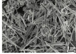
The presence of oxalate anions was crucial for producing the nanotubes with a well-defined shape. The function of these anions was revealed through a series of experiments. Oxalate anions first reduced MnOOH to manganese(II) cations and consumed protons in the vicinity of the MnOOH surface. The consumption of local protons increased the local pH and triggered precipitation and oxidation (by dissolved oxygen) of Sn2+ to tin dioxide. The two reactions proceeded, and eventually the MnOOH nanowires disappeared but tubes of tin dioxide formed around their surfaces (Figure 1). Samples obtained without oxalate salts were irregularly shaped.

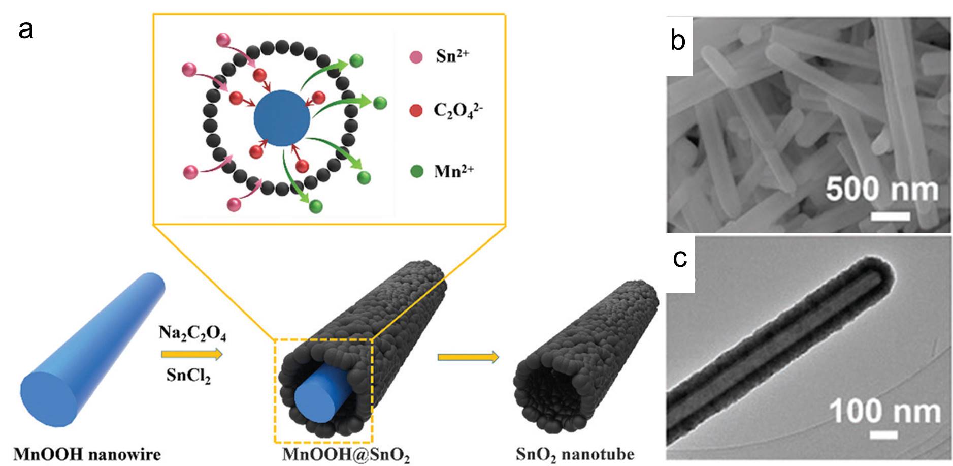
Figure 1. (a) The schematic illustration of the synthesis steps of the tin dioxide nanotubes. (b) Scanning electron microscopy and (c) transmission electron microscopy images of the as-prepared tin dioxide nanotubes.

The carbon-coated tin dioxide nanotubes showed superior stability performance to bare tin dioxide nanotubes, as shown from the slower capacity-fading rate depicted in Figure 2a. In addition, carbon coating did not significantly sacrifice nanotubes’ charge-storage performance as both electrodes with and without a coating exhibited comparable capacity at all tested current densities (Figure 2b).

Figure 2. Performance comparison between carbon-coated tin dioxide nanotubes (SnO2@C NTs) and bare tin dioxide nanotubes (SnO2 NTs): (a) long-term stability and (b) capacity achieved at different current densities and charge-discharge cycle numbers.

To find out more please read:

Oxalate-assisted Formation of Uniform Carbon-confined SnO2 Nanotubes with Enhanced Lithium Storage

Chunhua Han, Baoxuan Zhang, Kangning Zhao, Jiashen Meng, Qiu He, Pan He, Wei Yang, Qi Li and Liqiang Mai

DOI: 10.1039/c7cc05406h

About the blogger:

Tianyu Liu is a Ph.D. in chemistry graduated from University of California, Santa Cruz in United States. He is passionate about scientific communication to introduce cutting-edge researches to both the general public and the scientists with diverse research expertise. He is a web blogger for the Chem. Commun. and Chem. Sci. blog websites. More information about him can be found at http://liutianyuresearch.weebly.com/.

Digg This
Reddit This
Stumble Now!
Share on Facebook
Bookmark this on Delicious
Share on LinkedIn
Bookmark this on Technorati
Post on Twitter
Google Buzz (aka. Google Reader)

Janus Particle Chains that Can Rotate, Dissipate and Recombine

Janus is a god in ancient Roman mythology with two opposing faces. Its name has been brought to materials science to label particles with two or more distinct faces as “Janus particles”. Integrating multiple functions into one physical entity, Janus particles with various properties are extensively adopted as catalysts, electronic components and other applications.

Reporting in Chemical Communications, Bart Jan Ravoo and co-workers from Westfälische Wilhelms-Universität Münster in Germany developed a Janus particle colloidal assembly using a sandwich micro-contact printing method, a strategy reported previously by the same group. The Janus particle assembly consists of Janus particle chains, with the structure of one chain illustrated in Figure 1b. The authors first capped a batch of silica micro-beads with tri-block co-polymers on opposing ends (green parts shown in Figure 1). These copolymers serve as arms that extend and attach to functionalized magnetite (Fe3O4) nanoparticles. Two Janus particles will become magnetically glued together if they connect to the same nanoparticle at the two caps. This connection propagates and eventually forms Janus particle chains mainly consisting of two to four particles.

Figure 1. The schematic illustration depicting the structure of a Janus particle chain.

The artificial chains are responsive to an external magnetic field and photons with different wavelengths. Owing to the magnetic nanoparticles, the chains tend to arrange themselves according to the direction of the applied magnetic field. As shown in Figure 2a, the authors successfully rotated a chain by moving around a magnet.

Moreover, radiating the chains using UV light and green visible light will alter the chain configuration. The light sensitivity is rooted in a light-induced isomerization reaction of the co-polymer linkers: green light yields adhesive trans-isomers, whereas UV light produces cis-isomers that detach from magnetite. Hence, dissipation of the chains into individual Janus particles and then rejoining the particles together can be readily accomplished (Figure 2b).

Figure 2. Optical microscopy images showing (a) the magnetic and (b) the photo-switching properties of one Janus particle chain. All scale bars are 10 µm.

The demonstrated assembly is just the tip of the iceberg for Janus particle assemblies. As claimed by the authors, any acrylate in principal can be used to build the co-polymer linkers, resulting in colloidal assemblies with versatile features.

To find out more please read:

Self-assembly of Colloidal Molecules that Respond to Light and a Magnetic Field

Sven Sagebiel, Lucas Stricker, Sabrina Engel and Bart Jan Ravoo

DOI: 10.1039/c7cc04594h

About the blogger:

Tianyu Liu is a Ph.D. in chemistry graduated from University of California-Santa Cruz. He is passionate about scientific communication to introduce cutting-edge researches to both the general public and the scientists with diverse research expertise. He is a web writer for the Chem. Commun. and Chem. Sci. blog websites. More information about him can be found here.

Digg This
Reddit This
Stumble Now!
Share on Facebook
Bookmark this on Delicious
Share on LinkedIn
Bookmark this on Technorati
Post on Twitter
Google Buzz (aka. Google Reader)

Dissolving and Stabilizing the Precursor of Graphene in Organic Solvents

Graphene, a two-dimensional single-layer graphite sheet, has aroused worldwide attention since the last decade. Its ultrahigh electrical and thermal conductivities, high mechanical stiffness and unique band structure have attracted extensive research efforts to develop graphene-based electronics, photonics, printing materials etc. Currently, among various strategies, the wet-chemical method still remains the most practical protocol for large-scale production of graphene in laboratories. This process in general involves two steps: the oxidative exfoliation of graphite, a.k.a. Hummers’ method, followed by reduction of the oxidized graphite sheets. Graphite oxide (GO), possessing a layered structure analogous to graphite but with rich oxygen functionalities (such as hydroxyl and carboxyl groups) anchored on each layer, is the product of the first step and thus serves as a precursor of graphene.

As the aforementioned wet chemical method is usually carried out in water, GO is primarily stored as aqueous-based colloidal dispersions. However, GO is reported to be chemically unstable in water since water molecules can react with electropositive carbons of GO. Though the reaction is not rapid, it partially removes the oxygen functionalities and breaks the carbon matrix, which eventually forces GO to precipitate and reduces the shelf life of the GO precursor.

Recently, Shi and coworkers from Tsinghua University have successfully prolonged the lifetime of GO by dispersing it in organic solvents. During the last purification step of the Hummers’ method, instead of using de-ionized water, anhydrous ethanol was utilized to rinse the GO product and obtain ethanol-wetted GO. X-ray diffraction revealed that ethanol molecules existed in the inter-layer space between adjacent layers. The ethanol-wetted GO could be readily dissolved in propylene carbonate, an organic solvent, for concentrations ranging from 0.1 mg mL-1 to 40 mg mL-1 (Figures a and b). More importantly, GO could be stored in propylene carbonate for at least a month without a colour change, whilst the colour of aqueous GO dispersion discernibly darkened (Figure c). Spectroscopic studies indicated that the colour change was attributed to the loss of oxygen functionalities. The results unambiguously prove that GO in propylene carbonate is much more stable than GO in water.

Figure. (a) Dissolution of ethanol-wetted GO in propylene carbonate. (b) GO colloidal dispersions with various concentrations. (c) Color evolution of GO dispersions (1 mg mL-1) with water and propylene carbonate as solvents before and after storing for 28 days under ambient conditions.

Aside from propylene carbonate, dimethyl sulfoxide, ethylene glycol and N,N-dimethylformamide are solvents that can dissolve the ethanol-wetted GO. The successful stabilization of GO colloidal dispersions could ensure the steady production of graphene in laboratories, as well as reveal new opportunities to develop GO-based devices.

To find out more please read:

Organic Dispersions of Graphene Oxide with Arbitrary Concentrations and Improved Chemical Stability

Wencheng Du, Mingmao Wu, Miao Zhang, Guochuang Xu, Tiantian Gao, Liu Qian, Xiaowen Yu, Fengyao Chi, Chun Li and Gaoquan Shi

DOI: 10.1039/c7cc04584k

About the author:

Tianyu Liu is a Ph.D. in chemistry graduated from University of California-Santa Cruz. He is passionate about scientific communication to introduce cutting-edge researches to both the general public and the scientists with diverse research expertise. He is a web blog writer for the Chem. Commun. and Chem. Sci. blog websites. More information about him can be found at http://liutianyuresearch.weebly.com/.

Digg This
Reddit This
Stumble Now!
Share on Facebook
Bookmark this on Delicious
Share on LinkedIn
Bookmark this on Technorati
Post on Twitter
Google Buzz (aka. Google Reader)

Releasing A Pungent Anti-cancer Reagent with A Trisulfide Linker Inspired by Garlic

People who love the taste of garlic are often annoyed by its lingering smell. While there are various tips to get rid of this unpleasant odor, have you ever thought that this garlic aroma brings you chemical compounds that are potent anti-cancer reagents?

Diallyl trisulfide, one of the natural occurring components rendering the flavor of garlic, is able to release hydrogen sulfide (H2S) upon contacting with thiol compounds (i.e., organic molecules with –SH functional groups). H2S is a pungent gas that one might never forget after sniffing a rotten egg. However, this “notorious” gas, when at low concentrations, is reported to be friendly to our bodies. It relaxes vascular smooth muscle, reduces blood pressure, lowers risk associated with cancer as well as protects gastrointestinal, nervous and immune systems. All the aforementioned benefits of H2S have aroused worldwide efforts in developing H2S-releasing and bio-compatible materials that mimic the natural products for pharmaceutical applications.

Davis, Quinn and co-workers from Monash University, Australia and University of Warwick, United Kingdom, recently published a paper in Chemical Communications that reports a trisulfide-linked organic polymer capable of releasing H2S when meets –SH groups. As shown in the scheme below, the synthesized polymer is composed of three parts: a polyethylene glycol (PEG) unit on the left (in blue), a cholesterol (CHOL) group on the right (in orange), and a linker (in black) joining the two ends. PEG and CHOL are chosen mainly due to their bio-compatibility. By changing the structure of the middle linker, the authors obtained three types of polymers that behave differently when mixing with thiol compounds. The trisulfide linker (denoted as T) enables release of H2S gas and initiates polymer degradation. The disulfide linker (denoted as D) allows polymer degradation only. The amide linker (denoted as C) containing no sulfide atoms is inert to the thiol exposure.

Scheme. The chemical structure of the synthesized polymers with different linkers.

Experiments showed that the T-linked polymers are capable of releasing H2S both in vitro and in vivo.

A fluorescent probe, which can be reduced by H2S and becomes fluorescent, is applied to detect the existence of H2S. As shown in Figure a, the trisulfide linked polymers tested in vitro exhibited the highest fluorescence when mixing with L-cysteine (a thiol compound to trigger H2S generation). For the in vivo measurements, the authors incubated HEK293 cells with the polymers and the probe. Similar as the in vitro results, the fluorescence intensity of the cells containing the T-linked polymers is the highest (Figure b). Both the in vitro and in vivo results unambiguously proved that the presence of the T-linker was responsible for generating H2S. Additionally, another set of tests using Nile Red confirmed the biodegradability of the T-linked polymers.

Figure. (a) Fluorescence spectra collected from different systems in vitro. The inset shows the chemical reaction between the probe (SF4) and H2S that displays fluorescence. (b) Fluorescence intensity of different polymers over time in HEK293 cells.

The developed tri-sulfide linker may allow the mimicry of endogenous biosynthesis, the initiation of discrete signaling events and the synthesis of next-generation pharmaceutical excipients.


To find out more please read:

Garlic-inspired Trisulfide Linkers for Thiol-stimulated H2S Release
Francesca Ercole, Michael R. Whittaker, Michelle L. Halls, Ben J. Boyd, Thomas P. Davis and John F. Quinn
DOI: 10.1039/c7cc03820h

About the author:

Tianyu Liu is a Ph.D. in chemistry graduated from University of California-Santa Cruz. He is passionate about scientific communication to introduce cutting-edge researches to both the general public and the scientists with diverse research expertise. He is a web writer for the Chem. Commun. and Chem. Sci. blog websites. More information about him can be found at http://liutianyuresearch.weebly.com/.

Digg This
Reddit This
Stumble Now!
Share on Facebook
Bookmark this on Delicious
Share on LinkedIn
Bookmark this on Technorati
Post on Twitter
Google Buzz (aka. Google Reader)

A Promising Cathode Material for Magnesium-ion Batteries Has Been Identified

Research associated with batteries is gaining increasing attention and extensive efforts in recently decades, partly due to the development of sustainable energy to combat a series of problems including fossil fuel depletion, environmental pollution and global warming. Batteries are indispensable energy storage devices for the utilization of sustainable energy (e.g., solar and wind energy). One of the battery’s cutting-edge research topics is to achieve novel batteries with higher capacity (a figure-of-merit to measure how much electrical energy a battery can store) and better reliability than the lithium-ion batteries that currently dominate the battery market.

In the past decade, batteries based on magnesium ions, termed as magnesium-ion batteries, are emerging. The magnesium-ion batteries possess at least two advantages over lithium-ion batteries. Firstly, their typical anode material, magnesium metal, has a theoretical capacity of 3833 mAh/cm3. This value is much higher than that of graphite, a conventional anode material for lithium-ion batteries. Secondly, the formation of metal dendrite on anode surface can be avoided by replacing lithium metal with magnesium metal. Metal dendrites grow from anodes can eventually touch cathodes, causing electric short circuits and triggering fire and explosion. Therefore, magnesium-ion batteries are safer than lithium-ion batteries. However, nothing can be perfect. The limited mobility of Mg2+ of cathode materials greatly reduces the capacity (particularly at fast charging rates) and practicability of the magnesium-ion batteries.

Now Rong et al. has published an article in Chemical Communications stating that a promising cathode material capable of fast conducting Mg2+ for magnesium-ion batteries has been identified. The material is a molybdenum phosphate compound with a chemical formula of Mo3(PO4)3O. It is composed of several edge-sharing MoO6 octahedra, corner-sharing MoO5 trigonal bipyramids, MoO4 tetrahedra, and PO4 tetrahedra. Using advanced simulation and computation techniques (i.e., the first-principles density functional theory), the authors first proved that Mg2+ can stably reside in some interstitial sites among the aforementioned polyhedra, indicating the identified compound is active for Mg2+ storage. In addition, the authors plotted two possible pathways for Mg2+ diffusion during charge and discharge processes (shown in the Figure). As illustrated in Figure a1, the first one is an inner-channel path along the b-axis. The second one is an inter-channel path along the c-axis.

The most striking feature of the path #1 is its ultra-low activation barrier (i.e., the highest potential energy that a Mg2+ need to overcome when diffusing) of only ~80 meV (Figure a2). Such a low diffusion barrier is expected to allow facile Mg2+ diffusion within the bulk of Mo3(PO4)3O, which can boost the capacity of the magnesium-ion batteries particularly at elevated charging rates. On the contrary, the activation barrier of the path #2 is as high as ~1200 meV. The authors claimed that the Mg2+ diffusion along the path #2 “should be ~1018 times less frequent than” the path #1.


Figure (a1) schematic of the Mg2+ diffusion path #1 and (a2) its corresponding diffusion potential barrier distribution along the way. (b1) Schematic of the Mg2+ diffusion path #2 and (a2) its corresponding diffusion potential barrier distribution along the way.


At last, the authors estimated the theoretical average potential that Mo3(PO4)3O can reach is 1.98 V, corresponding to a promising energy density of 173 Wh/kg. Although the proposed phosphate is hypothetical, the investigation of its stability reveals the possibility that this material can be experimentally synthesized.

To find out more please read:

Fast Mg2+ Diffusion in Mo3(PO4)3O for Mg Batteries
Ziqin Rong, Penghao Xiao, Miao Liu, Wenxuan Huang, Daniel C. Hannah, William Scullin, Kristin A. Persson and Gerbrand Ceder
DOI: 10.1039/c7cc02903a

About the author:

Tianyu Liu is a Ph.D. in chemistry graduated from University of California-Santa Cruz. He is passionate about scientific communication to introduce cutting-edge researches to both the general public and the scientists with diverse research expertise. He is a web writer for the Chem. Commun. and Chem. Sci. blog websites. More information about him can be found at http://liutianyuresearch.weebly.com/.

Digg This
Reddit This
Stumble Now!
Share on Facebook
Bookmark this on Delicious
Share on LinkedIn
Bookmark this on Technorati
Post on Twitter
Google Buzz (aka. Google Reader)

Improving Sodium-Ion Batteries for Large-Scale Energy Storage

One of the greatest global challenges is the ever-growing demand for reliable, large-scale energy production.

The depletion of cost-effective fossil fuels and concerns about climate change are driving the need for clean energy sources derived from renewable technologies. Wind and solar power show significant potential as sustainable alternatives however, both solar photovoltaics and wind energy display intermittent output which has led to concerns regarding reliability for global energy production. As a result, there has been an increased demand for the development of large-scale energy storage.

Among energy storage technologies, lithium-ion batteries (LIBs) predominate however lithium’s high cost, abundance, unevenglobal distribution and safety concerns have limited its widespread application. In recent years, researchers have become interested in high energy sodium-ion batteries (SIBs) as a safer and less expensive alternative. Nevertheless, their inferior electrochemical performances, due to the larger size and heavier mass of sodium ions, has become a major hurdle in the development and implementation of SIBs.

In a recent ChemComm publication, Prof. Jun Chen of Nankai University has demonstrated the improved capabilities of SIBs using a manganite hydroxide (MnOOH)-based anode.

In the past, transition metal oxides, such as MnOx-based materials, have been used in LIBs as they possess a high theoretical capacity and—in some cases—improved conductivity. In this study by Chen and co-workers, MnOOH nanorods (figure, top) were synthesized, and were shown to display a higher initial Coulombic efficiency and rate performance compared to MnO2 (a common anode material in LIBs). Cyclic voltammetry (figure, bottom) and various other spectroscopic techniques were used to investigate the electrochemical properties and storage behaviour of MnOOH-SIBs. These experiments showed improvements in charge capacity and overall rate performance when compared to other transition metal oxides and sulfides.

The results of this work show promise toward the fabrication of high-performance SIBs which are encouraging alternatives for energy storage due to sustainable cost, improved thermal stability and transport safety. The performance of SIBs still lags behind that of LIBs but this study, among others, demonstrates that new electrode materials need to be explored in the development of SIBs and solving large-scale energy storage challenges.

To find out more see:

MnOOH nanorods as high-performance anodes for sodium ion batteries
Lianyi Shao, Qing Zhao and Jun Chen
DOI: 10.1039/C7CC00087A


Victoria Corless is currently completing her Ph.D. in organic chemistry with Prof. Andrei Yudin at The University of Toronto. Her research is centred on the synthesis of kinetically amphoteric molecules, which offer a versatile platform for the development of chemoselective transformations with particular emphasis on creating novel biologically active molecules.

Digg This
Reddit This
Stumble Now!
Share on Facebook
Bookmark this on Delicious
Share on LinkedIn
Bookmark this on Technorati
Post on Twitter
Google Buzz (aka. Google Reader)

Soft drinks power origami cell

Graphical Abstract

Source: © Royal Society of Chemistry - The tiny fuel cell is made from a folded sheet of filter paper that holds the anode and cathode

Miniature fuel cell made from folded filter paper runs on sugary drinks.

Researchers in China have found a way to integrate the ancient Japanese art of paper folding, origami, into a miniature biofuel cell that can generate energy from soft drinks.

Biofuel cells use enzymes, instead of precious metals, as catalysts to oxidise their fuel. Miniature versions have excited researchers because they are portable and have high efficiency. They could provide power for implants or electronic contact lenses or harvest energy from perspiration. However, designing these small biofuel cells is difficult due to complicated assembly and high costs.


Read the full article in Chemistry World >>>


A miniature origami biofuel cell based on a consumed cathode
You Yu, Yujie Han, Baohua Lou, Lingling Zhang, Lei Hana and Shaojun Dong
Chem. Commun., 2016, 52, 13499-13502
DOI: 10.1039/C6CC07466A, Communication

Digg This
Reddit This
Stumble Now!
Share on Facebook
Bookmark this on Delicious
Share on LinkedIn
Bookmark this on Technorati
Post on Twitter
Google Buzz (aka. Google Reader)

Incorporating DNA hydrogels into enzymatic biofuel cells

I remember a time when mobile phones weren’t so power hungry, and when my phone could go a week on one charge. Admittedly, that was before it had a colour screen, internet connectivity and a hundred other bells and whistles. Increased device connectivity, in particular, has led to a huge increase in power demands and the need for better battery technology.

Wouldn’t it be marvellous if your phone battery generated its power from a wide selection of renewable sources? Khiem Van Nguyen and Shelly Minteer from the University of Utah look toward this possibility in their most recent ChemComm, which describes the use of DNA hydrogels in the production of an enzymatic biofuel cell.

The authors describe how they used the self-assembly of DNA monomers under physiological conditions to form a DNA hydrogel capable of trapping glucose oxidase, the most widely used enzyme in enzymatic biofuel cells. This DNA hydrogel remains permeable to small molecules, such as the battery fuel, whilst successfully trapping the enzyme close to the electrode surface.  Enzyme immobilization on the electrode surface is critical to achieve an effective enzymatic biofuel cell, and this model biobattery was shown to have a similar performance to previously reportedglucose oxidase biofuel cells.

Entrapment of glucose oxidase in DNA hydrogel

With enzymes capable of oxidising a wide range of fuels, from alcohols and carbohydrates to amino acids, it may not be too long until a multi-enzyme biobattery is created that can be powered by the sweat from your skin. Then you’ll be able to browse the internet wherever and whenever you want…provided you have signal, of course.

To read the details, check out the ChemComm article in full:
Investigating DNA hydrogels as a new biomaterial for enzyme immobilization in biobatteries
Khiem Van Nguyen and Shelley D Minteer
Chem. Commun., 2015, 51, Advance Article
DOI: 10.1039/C5CC04810A

For example: S. Aquino Neto et al., Power Sources, 2015, 285, 493–498

Digg This
Reddit This
Stumble Now!
Share on Facebook
Bookmark this on Delicious
Share on LinkedIn
Bookmark this on Technorati
Post on Twitter
Google Buzz (aka. Google Reader)

Hotter and hotter: increasing the potential of gold nanostars

Gold nanostars are gold nanoparticles with multiple branches, a shape which gives rise to their unique properties. These nanoparticles have tuneable localized surface plasmon resonances in the biologically transparent near-IR window, and excitation of these plasmons using a laser creates a local temperature  increase. For this reason, gold nanostars have potential for use in non-invasive antitumoral and antibiofilm laser treatments.

The problem faced by scientists, however, is how to achieve a temperature increase that is large enough to be effective, without exposing the overlying skin to a level of irradiation that exceeds the safe limits. This is what Piersandro Pallavicini from the Department of Chemistry at the University of Pavia, and an international team of colleagues, set out to investigate.

They generated gold nanostars with plasmon resonances at 835 and 1530 nm, respectively. Each of these plasmons could be irradiated separately, leading to observable increases in temperature. However, when both plasmons were irradiated simultaneously, the temperature increase was equal to the sum of the temperature increases when the plasmons were irradiated separately.

Temperature increases observed from the laser excitation of individual or multiple plasmon resonances of gold nanostars

The implication of these findings is that Pallavicini and colleagues successfully found a way to obtain a larger local temperature increase using irradiation that remains below the safe limits. This significantly increases the potential of gold nanostars for application in the in the treatment of biofilm growth on implants in vivo.

To find out the full details of the additive temperature effect, read the ChemComm article today – it’s free to access until 21st October 2015:

Monolayers of gold nanostars with two near-IR LSPRs capable of additive photothermal response
Piersandro Pallavicini, Simone Basile, Giuseppe Chirico, Giacomo Dacarro, Laura D’Alfonso, Alice Dona, Maddalena Patrini, Andrea Falqui, Laura Sironi and Angelo Taglietti
Chem. Commun., 2015, 51, 12928-12930
DOI: 10.1039/C5CC04144A

Digg This
Reddit This
Stumble Now!
Share on Facebook
Bookmark this on Delicious
Share on LinkedIn
Bookmark this on Technorati
Post on Twitter
Google Buzz (aka. Google Reader)

Hierarchical 3D immunoassays – higher loading, lower fouling

If you are producing an immunoassay there are two key parameters you need to understand and optimise: surface structure and surface chemistry. Get these two parameters right and you will optimise the sensitivity of your immunoassay. 

Although there have been a multitude of 3D surface generation routes reported, they are generally complicated and require a lot of additional steps. Although these 3D surfaces lead to high probe loading levels they also often lead to high levels of non-specific protein absorption, undoing any good the surface structure would have led to. 

Jinghua Yin and team from the State Key Laboratory of Polymer Physics and Chemistry at the Changchun Institute of Applied Chemistry have focussed on both properties to generate a much improved immunoassay. 

 Firstly they generated a 3D surface using UV irradiation of polystyrene spheres onto a substrate; they then grafted polymer brushes to the sphere surface. The polymer brushes not only further increased the surface area (more than doubling it from the bare sphere surface) but also acted as an anti-fouling agent, reducing the amount of non-specific binding observed by up to 90%. 

Antibody loading on different surface types showing increasing loading levels


The commonality of the functional groups on the polymer brushes mean that any antibody can be attached to the prepared surface. To find out the details of how to make these surfaces and try them out on your own immunoassays, read the paper today!


To read the details, check out the ChemComm article in full:
Facile fabrication of microsphere-polymer brush hierarchically three-dimensional (3D) substrates for immunoassays
Jiao Ma, Shifang Luan, Lingjie Song, Shuaishuai Yuan, Shunjie Yan, Jing Jin and Jinghua Yin
Chem. Commun., 2015, 51, 6749-6752
DOI: 10.1039/C5CC01250C

Digg This
Reddit This
Stumble Now!
Share on Facebook
Bookmark this on Delicious
Share on LinkedIn
Bookmark this on Technorati
Post on Twitter
Google Buzz (aka. Google Reader)