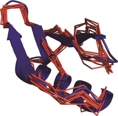
 Two separate groups of scientists have developed methods to uncover proteins’ 3D structure inside living animal cells for the first time.
It is vital to know the structure of a protein to understand its chemical and biological functions. Scientists usually need to purify and crystallise a protein to determine its 3D structure by x-ray crystallography. Not only is this process difficult and lengthy, it can also misrepresent the protein’s structure, as the measuring conditions are vastly different from the conditions inside a living cell.
Now, a researcher team led by Xun-Cheng Su from Nankai University in Tianjin, China,Conggang Li at Chinese Academy of Sciences, and Thomas Huber from the Australian National University, has analysed a protein’s structure using nuclear magnetic resonance (NMR) spectroscopy inside living frog cells.1 The researchers tagged the protein with a beacon, a paramagnetic lanthanide tag, which binds to a cysteine residue on the outside of the protein. ‘The measured effects from the beacons tagged onto the protein give away the positions of the atoms in the protein, in a similar way that a set of satellites can be used to locate the exact position of a GPS receiver,’ Su explains.
Read the full article on Chemistry World >>>


