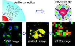
Gold nanoparticles have been widely used for bio-imaging but they need to be coated, commonly in silica, to protect them and stop them aggregating. The conventional method for silica coating is time-consuming but the team say they’ve overcome this tedious process by using an organosilica source. The resulting organosilica shell has –SH and –OH groups inside it, making it easy to functionalise with fluorescent dyes or biomolecules.
By modifying the gold core with Raman reporters and the organosilica shell with a fluorophore, the group produced nanoparticles with three modalities of imaging – Rayleigh scattering, fluorescence and surface enhanced Raman scattering.
Reference:
Au@organosilica multifunctional nanoparticles for the multimodal imaging
Y Cui, X-S Zheng, B Ren, R Wang, J Zhang, N-S Xia and Z-Q Tian, Chem. Sci., 2011, DOI: 10.1039/c1sc00242b


