It’s in the blood

Written by Harriet Brewerton

Scientists have designed a tiny fuel cell that generates electricity from the human body’s blood flow, and for the first time, they have tested the device in a person.

The blood-powered fuel cell connected to a low voltage display

The blood-powered fuel cell connected to a low voltage display

Phones, tablets and other portable electronics are common, but development of equally portable power sources is lagging behind. This is a particular concern for biomedical devices such as pacemakers. Since the 1960s, researchers have made biocompatible fuel cells that generate power inside the body. However, none of these power sources has been successfully demonstrated in a human subject.

To read the full article visit Chemistry World.

Ex vivo electric power generation in human blood using an enzymatic fuel cell in a vein replica
Dmitry Pankratov, Lars Ohlsson, Petri Gudmundsson, Sanela Halak, Lennart Ljunggren, Zoltan Blum and Sergey Shleev
RSC Adv., 2016,6, 70215-70220
DOI: 10.1039/C6RA17122B, Communication

Digg This
Reddit This
Stumble Now!
Share on Facebook
Bookmark this on Delicious
Share on LinkedIn
Bookmark this on Technorati
Post on Twitter
Google Buzz (aka. Google Reader)

A study on spheroid formation in thermosensitive hydrogels

According to an estimate by the World Cancer Report, cancer associated mortality is expected to reach 17 million per year globally by the year 2030. To confront the cancer burden with appropriate clinical interventions, researchers screen cancer-killing drug combinations in monolayer cell cultures. This is a widely used method for the preclinical evaluation of drug efficacy.

A major limitation of monolayer cultures is that they do not, even mildly, recapitulate the complex architecture of a tumor growing in vivo. As an initial step in overcoming this limitation, researchers use scaffolded spheroid cultures – a system wherein cells are grown on hydrogel scaffolds in three dimensions.

Hydrogel scaffolds provide physical and structural support for the formation of a more ‘natural’ setting that better recapitulates cell behavior in vivo. For example, smaller (150um) spheroids have better cell-to-cell contacts and notably different gene expression compared to monolayer cultures; larger (200-500um) ones develop oxygen and nutrient gradients reminiscent of chemical gradients seen in human tumors. However, limitations in design flexibility, handling and interbatch compositional variation have discouraged the routine use of hydrogel scaffolds. In addition, the technical challenge of separating newly formed spheroids from the scaffolding material before drug screening represents a major roadblock.

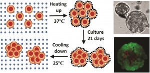
In a study led by Xiaolin Cui and colleagues at the School of Chemical Engineering and the School of Mathematical Sciences, University of Adelaide, Australia, researchers synthesized a thermo-reversible N-Isopropylacrylamide-acrylic acid (NIPAM-AA) hydrogel by free radical emulsion polymerization. In their study using the cervical carcinoma cell line HeLa, the team demonstrates that at 370C the NIPAM-AA hydrogel solidifies and forms a sheath around HeLa cell clusters. As a consequence, these clusters develop into hydrogel scaffolded spheroids over time. At 250C the hydrogel liquefies and releases the newly formed spheroids.

Cell viability assays confirmed that this new state of the art hydrogel is biocompatible. NIPAM-AA derived spheroids were smaller (70-120um),  nearly spherical and showed a narrower size distribution compared to conventional spheroids. The study showed, through time course experiments, that the spheroids remain viable for over 14 days in culture. The study also suggests that spheroids derived via the NIPAM-AA hydrogel method are more viable than those derived from conventional suspension cultures, supporting the notion that hydrogel scaffolding facilitates oxygen and nutrient supply to support cell growth.

The researchers deduced a mathematical model to predict the kinetics of NIPAM-AA derived spheroid growth. Their model accurately recapitulated the growth rate, size and size distribution of the spheroids. The authors propose that hydrogel scaffolding has the potential to evolve into a technology with a wide range of applications including, but not limited to, (1) high throughput screening of anticancer drugs using uniformly sized spheroids; (2) regenerative medicine; and (3) tissue engineering.

Read the full article here:

Xiaolin Cui,   Saber Dini,   Sheng Dai,   Jingxiu Bi,   Benjamin Binder,   Edward Green and   Hu Zhang
Digg This
Reddit This
Stumble Now!
Share on Facebook
Bookmark this on Delicious
Share on LinkedIn
Bookmark this on Technorati
Post on Twitter
Google Buzz (aka. Google Reader)

Top 10 most-downloaded articles: Q2 April–June 2016

Take a look at the most-downloaded RSC Advances articles from the months of April, May and June 2016 and let us know what you think!

Free radicals, natural antioxidants, and their reaction mechanisms
Satish Balasaheb Nimse and Dilipkumar Pal
RSC Adv., 2015,5, 27986-28006
DOI: 10.1039/C4RA13315C

Size-controlled silver nanoparticles synthesized over the range 5–100 nm using the same protocol and their antibacterial efficacy
Shekhar Agnihotri, Soumyo Mukherji and Suparna Mukherji
RSC Adv., 2014,4, 3974-3983
DOI: 10.1039/C3RA44507K

Thermal-runaway experiments on consumer Li-ion batteries with metal-oxide and olivin-type cathodes
Andrey W. Golubkov, David Fuchs, Julian Wagner, Helmar Wiltsche, Christoph Stangl, Gisela Fauler, Gernot Voitic, Alexander Thaler and Viktor Hacker
RSC Adv., 2014,4, 3633-3642
DOI: 10.1039/C3RA45748F

Synthesis and properties of molybdenum disulphide: from bulk to atomic layers
Intek Song, Chibeom Park and Hee Cheul Choi
RSC Adv., 2015,5, 7495-7514
DOI: 10.1039/C4RA11852A

Advancements in the stability of perovskite solar cells: degradation mechanisms and improvement approaches
Bobo Li, Yafang Li, Chaoyue Zheng, Deqing Gao and Wei Huang
RSC Adv., 2016,6, 38079-38091
DOI: 10.1039/C5RA27424A

Dual protection of amino functions involving Boc
Ulf Ragnarsson and Leif Grehn
RSC Adv., 2013,3, 18691-18697
DOI: 10.1039/C3RA42956C

Third-generation solar cells: a review and comparison of polymer:fullerene, hybrid polymer and perovskite solar cells
Junfeng Yan and Brian R. Saunders
RSC Adv., 2014,4, 43286-43314
DOI: 10.1039/C4RA07064J

Colloidal semiconductor nanocrystals: controlled synthesis and surface chemistry in organic media
Jin Chang and Eric R. Waclawik
RSC Adv., 2014,4, 23505-23527
DOI: 10.1039/C4RA02684E

Electrically conductive polymers and composites for biomedical applications
Gagan Kaur, Raju Adhikari, Peter Cass, Mark Bown and Pathiraja Gunatillake
RSC Adv., 2015,5, 37553-37567
DOI: 10.1039/C5RA01851J

Graphene and its nanocomposite material based electrochemical sensor platform for dopamine
Alagarsamy Pandikumar, Gregory Thien Soon How, Teo Peik See, Fatin Saiha Omar, Subramaniam Jayabal, Khosro Zangeneh Kamali, Norazriena Yusoff, Asilah Jamil, Ramasamy Ramaraj, Swamidoss Abraham John, Hong Ngee Lim and Nay Ming Huang
RSC Adv., 2014,4, 63296-63323
DOI: 10.1039/C4RA13777A

Interesting in submitting to RSC Advances? You can submit online today, or email us with your ideas and suggestions.

Digg This
Reddit This
Stumble Now!
Share on Facebook
Bookmark this on Delicious
Share on LinkedIn
Bookmark this on Technorati
Post on Twitter
Google Buzz (aka. Google Reader)

Sweet as sugar, hard as carbon: A hierarchical core-shell 3D graphene network biosensor for glucose detection

A biosensor is a device that uses biological molecules, typically enzymes, to specifically detect the presence of a chemical or a metabolic intermediate (referred to as the analyte) in a diagnostic setting. A biosensor acts as the platform upon which a biochemical reaction, initiated by the analyte, is converted to an electric current that is accurately quantified during a subsequent step. Biosensors have wide clinical applicability. For instance, the detection of blood sugar, which is among the most frequently measured physiological variable, is achieved with biosensors.

Recent years have seen rapid advancements in the use of nanoparticles, nanowires and nanotubes as biosensor platforms. These innovative nanostructures are electrochemically active, chemically stable, have large surface areas and are biocompatible – all of which are desirable attributes for developing biosensors. Of note is the observation that graphene, a substance known for its high electrical conductivity,  lends itself to biosensor development due to its relative ease of manufacture together with its ability to form composites with other electrochemically active nanostructures.

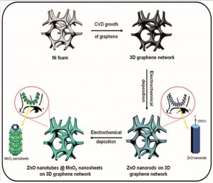
Early prototypes of graphene-based biosensors were inefficient for two main reasons. First, the clumping of graphene sheets reduced the accessible surface area. As a consequence, the biosensor/analyte interface was greatly reduced. Second, the restacking of graphene sheets introduced electrical resistance due to intersheet contacts. To overcome these hurdles, a research group led by Azam Iraji Zad at the Institute for Nanoscience and Nanotechnology (INST), Tehran, Iran developed a freestanding, porous 3D graphene network (3DGN) which was further modified with metal oxide nanostructures as a platform upon which an enzymatic reaction could occur.

This proof-of-concept study uses the glucose oxidase enzyme for the rapid and selective detection of glucose. The 3DGN, a graphene skeleton with multiple pores, is the core of the nanostructure. Atop the 3DGN, the researchers first grew uniformly spaced ZnO nanorods, which served to hold the enzyme in place. In a subsequent step, MnO2, known to be biocompatible and stable, was deposited onto the ZnO nanorods, thus forming a multilayered hierarchical structure with an average diameter of 100nm. The researchers propose that that the complex architecture of the nanostructure serves to facilitate the electron transfer process, which is the fundamental biochemical mechanism driving the enzymatic reaction.

In principle, the inner parts of the ZnO nanotubes increase the accessible surface area of the nanostructure and enhance the biosensor/analyte interface. In theory, the 3DGN biosensor is expected to respond quicker and have improved sensitivity when compared to other enzyme-based glucose detection devices. The study tested the 3DGN biosensor using a method called amperometry which is used routinely in research laboratories to detect ions – the byproduct of enzymatic reactions. The study found that the 3DGN biosensors had a response time of less than 3 seconds; a value indicative of a competitive advantage over other enzyme-based glucose biosensors. Intriguingly, the study also found that the 3DGN was very sensitive and could detect extremely low concentrations (10nM) of glucose.

The study strongly suggests that 3DGN biosensors could be used as an accurate sensing platform for chemicals and biomolecules. The findings further support the argument that composite nanostructures with complex architecture could find applicability in human health and beyond.

Read the full article here:

Elham Asadian, Saeed Shahrokhian and Azam Iraji Zadac
Digg This
Reddit This
Stumble Now!
Share on Facebook
Bookmark this on Delicious
Share on LinkedIn
Bookmark this on Technorati
Post on Twitter
Google Buzz (aka. Google Reader)

Machine-learning accelerates catalytic trend spotting

Written by Anna Meehan

Researchers in Japan have used a machine-learning method to cut the time it takes to predict the catalytic potential of different metals.

Binding between a metal surface and an adsorbate mainly depends on the electronic structure of the metal. More energy at centre of the metal’s d-band creates a stronger bond between its surface and the adsorbate. Based on this theory, scientists have long regarded a value called the d-band centre as a key indicator of a metal’s catalytic activity.

Machine learning helps researchers tackle challenging tasks, such as designing pollution filter catalysts at industrial scale © iStock

Researchers normally compute this value independently for each metal using first-principles calculations. Now, as part of a wider interest in machine-learning applications, Ichigaku Takigawa and his group at Hokkaido University have developed a new method for predicting the d-band centre value. They use readily available data, such as density and electronegativity from other metals or bimetals, to predict the d-band centre for 11 metals and their bimetallic alloys. The results compare favourably with values obtained through density functional theory.

To read the full article please visit Chemistry World.

Machine-learning prediction of the d-band center for metals and bimetals
Ichigaku Takigawa, Ken-ichi Shimizu, Koji Tsuda and Satoru Takakusagi
RSC Adv., 2016,6, 52587-52595
DOI: 10.1039/C6RA04345C, Paper

Digg This
Reddit This
Stumble Now!
Share on Facebook
Bookmark this on Delicious
Share on LinkedIn
Bookmark this on Technorati
Post on Twitter
Google Buzz (aka. Google Reader)

Now you see me: autofluorescent nanoparticles for live cell imaging and biodegradation modeling

Guest Web-writer Anand Devasthanam
Anand has keen interest for storytelling. He has received formal training in the fields of immunology and oncology. As an aspiring science communicator, Anand strives to help scientists take complex scientific data and core messages and turn them into a narrative that is compelling, creative and scientifically accurate.

There is an increasing need for novel technologies to facilitate in vivo tissue visualization and drug delivery. However, this need is largely unmet due to the challenges associated with creating biocompatible materials that meet safety standards. In addition, the potential health risks associated with the accumulation of non-degradable imaging agents and drug carries represents a major obstacle in the innovation pipeline.

The intrinsic autofluorescent, biodegradable and biocompatible properties of Bovine Serum Albumin (BSA) is well appreciated. However, BSA has short excitation and emission wavelengths, which substantially restricts any in vivo biomedical applications.  Motivated by a recent report suggesting that glutaraldehyde (GA)-crosslinking induces autofluorescence in protein-based nanoparticles by modifying a series of C=C and C=N bonds, a team led by Yu Lei at the Department of Biomedical Engineering, University of Connecticut, developed low-cost, non-toxic, BSA-based protein nanoparticles (average size ~40 nm) for live cell imaging and biodegradation analysis.

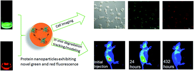
The nanoparticles were generated by adding drops of a prepared BSA solution to glutaraldehyde/n-butanol solution at high-speed, and the resulting product heated at 121°C to ensure sterility. Interestingly, a similar reaction carried out in the absence of the GA crosslinker did not produce autofluorescent BSA nanoparticles, suggesting that GA was indeed playing an important role in chemically transforming BSA. Using UV-visible spectroscopy, the investigators observed that BSA nanoparticles exhibited strong autofluorescence at both green (530 nm) and red (630 nm) wavelengths.

The BSA nanoparticles were not uniform in structure, owing to the random points of crosslinking within BSA, and also due to the ensuing condensation reaction that occurs during the sterilization step. Therefore, a clear mechanistic explanation for the strong autofluorescence warrants further investigation. However, the investigators speculate that GA-crosslinking and heating could result in new C=N bonds, which could synergize with the C=C bonds from tryptophan, tyrosine, phenylalanine and histidine residues with BSA, leading to enhanced green and red fluorescence.

The team went on to demonstrate the utility of the BSA nanoparticles in biomedical applications such as imaging and biodegradation. They used fluorescent microscopy techniques to visualize the entry of BSA nanoparticles into human kidney cells grown in vitro. The study also found that the BSA nanoparticles were completely degraded within 18 days of injection in mice. A mathematical model for the distribution and biodegradation of the nanoparticles was in good agreement with the experimental results. Finally, to add an additional line of evidence supporting the biocompatible nature of the BSA nanoparticles, the investigators looked for signs of tissue damage in the region surrounding the site of injection, together with an analysis of internal organs including the pancreas, liver and kidney, and report that the BSA nanoparticles are biocompatible.

Read the full article here:

Xiaoyu Ma, Derek Hargrove, Qiuchen Dong, Donghui Song, Jun Chen, Shiyao Wang, Xiuling Lu, Yong Ku Cho, Tai-Hsi Fan and  Yu Lei
DOI: 10.1039/c6ra06783b
Digg This
Reddit This
Stumble Now!
Share on Facebook
Bookmark this on Delicious
Share on LinkedIn
Bookmark this on Technorati
Post on Twitter
Google Buzz (aka. Google Reader)

RSC Supramolecular Chemistry Award for Editor-in-Chief Mike Ward

Each year the Royal Society of Chemistry presents prizes and awards to chemical scientists who have made a considerable contribution in their area of research, in industry and academia. This year, we are delighted to announce that RSC Advances Editor-in-Chief, Professor Mike Ward of the University of Sheffield, UK, has been awarded the 2016 RSC Supramolecular Chemistry Award, for his leading contributions to the synthesis, characterisation, host-guest chemistry and functional properties of self-assembled coordination cages.

The Supramolecular Chemistry Award is awarded biennially and recognises studies leading to the design of functionally useful supramolecular species.

In celebration of the 2016 RSC Prizes and Awards, we have collected together some of the research recently published by the winners. This collection showcases articles authored by the winners from across the Royal Society of Chemistry’s journals portfolio, which are free to access for a limited period. A full list of 2016 winners and more information about RSC Prizes and Awards can be found here.

Please join us in congratulating Mike on this achievement!

We would like to highlight the RSC Advances themed collection, Supramolecular chemistry: self-assembly and molecular recognition, Guest Edited by Mike Ward.

The articles in this issue cover many aspects of the formation of, and molecular recognition with, non-covalent self-assembled systems. Systems studied span the range of supramolecular assemblies from MOFs to gels, and potential applications or functional behaviour that are on display here include host/guest chemistry, spin crossover, molecular sensors, and extraction/separation. This collection of articles powerfully illustrates the diversity and increasing importance of supramolecular chemistry.

Digg This
Reddit This
Stumble Now!
Share on Facebook
Bookmark this on Delicious
Share on LinkedIn
Bookmark this on Technorati
Post on Twitter
Google Buzz (aka. Google Reader)

The heat is on: cancer-drug loaded nanoparticles for photothermal therapy

Guest Web-writer Anand Devasthanam
Anand has keen interest for storytelling. He has received formal training in the fields of immunology and oncology. As an aspiring science communicator, Anand strives to help scientists take complex scientific data and core messages and turn them into a narrative that is compelling, creative and scientifically accurate.

Photothermal therapy is an emerging area of cancer treatment. Here, a photothermal agents, often nanoparticles (NPs) with a resonance peak in the 700-1200nm range, are delivered to the tumor site and are subsequently activated by light in the Near Infrared (NIR) range. As a consequence, tumor cells are thermally ablated.

In a study led by Xiaolin Li and colleagues at the Key Laboratory for Thin Film and Microfabrication and Changzheng Hospital in China, scientists used SiO2@Au core-shell NPs chemically conjugated via PEGylation to graphene oxide (GO) in conjunction with a chemotherapeutic agent to target prostate cancer cells in vitro. Using the chemotherapeutic agent Docetaxel (Dtxl),  which is among the leading front line treatments for patients diagnosed with prostate cancer, the team demonstrated that Dtxl-loaded SiO2@Au@GO NPs, when activated with light in the NIR range, significantly curbed the survival of DU145 prostate cancer cells.

While SiO2@Au core-shell NPs have been used previously by other research groups to study their ability to remove tumors, Li’s team fabricated SiO2@Au@GO NPs to take advantage of their relatively low cost, large specific surface area, and efficient loading and delivery of water-soluble aromatic drug molecules. This one-two punch strategy was realized via a double shell, multifunctional approach: the inner core SiO2@Au NPs served as a photothermal inducer to bring about cellular cytotoxicity; the outer GO NPs carried the antitumor drug, Dtxl. The study found that exposing DU145 cells to the NPs alone for 24h did not result in overt cell death, suggesting that the NPs have a good safety profile. Importantly, the study showed that when NP-treated cell cultures were irradiated with a 780nm NIR laser, there was a significant decline in viable cells over a 24h period.

The study demonstrates that Dtxl-loaded SiO2@Au@GO NPs could be manufactured and potentially used an an antitumor agent for the treatment of prostate cancer. Moreover, these findings illuminate the untapped potential of NP-based photothermal agents as adjuvant agents in oncology clinical trials in the near future.

Read the full article here:

Xiaolin Li,   Zhi Yang,   Nantao Hu,   Liying Zhang,   Yafei Zhang and   Lei Yin
DOI: 10.1039/C6RA03886G
Digg This
Reddit This
Stumble Now!
Share on Facebook
Bookmark this on Delicious
Share on LinkedIn
Bookmark this on Technorati
Post on Twitter
Google Buzz (aka. Google Reader)

Top 10 most-downloaded articles: Q1 Jan–Mar 2016

Take a look at the most-downloaded RSC Advances articles from the months of October, November and December 2015 and let us know what you think!

Size-controlled silver nanoparticles synthesized over the range 5–100 nm using the same protocol and their antibacterial efficacy
Shekhar Agnihotri, Soumyo Mukherji and Suparna Mukherji
RSC Adv., 2014,4, 3974-3983
DOI: 10.1039/C3RA44507K

Free radicals, natural antioxidants, and their reaction mechanisms
Satish Balasaheb Nimse and Dilipkumar Pal
RSC Adv., 2015,5, 27986-28006
DOI: 10.1039/C4RA13315C

Thermal-runaway experiments on consumer Li-ion batteries with metal-oxide and olivin-type cathodes
Andrey W. Golubkov, David Fuchs, Julian Wagner, Helmar Wiltsche, Christoph Stangl, Gisela Fauler, Gernot Voitic, Alexander Thaler and Viktor Hacker
RSC Adv., 2014,4, 3633-3642
DOI: 10.1039/C3RA45748F

Synthesis and properties of molybdenum disulphide: from bulk to atomic layers
Intek Song, Chibeom Park and Hee Cheul Choi
RSC Adv., 2015,5, 7495-7514
DOI: 10.1039/C4RA11852A

Orientation dependence of the pseudo-Hall effect in p-type 3C–SiC four-terminal devices under mechanical stress
Hoang-Phuong Phan, Afzaal Qamar, Dzung Viet Dao, Toan Dinh, Li Wang, Jisheng Han, Philip Tanner, Sima Dimitrijev and Nam-Trung Nguyen
RSC Adv., 2015,5, 56377-56381
DOI: 10.1039/C5RA10144A

Formation of organic–inorganic mixed halide perovskite films by thermal evaporation of PbCl<inf>2</inf> and CH<inf>3</inf>NH<inf>3</inf>I compounds
Cheng Gao, Jiang Liu, Cheng Liao, Qinyan Ye, Yongzheng Zhang, Xulin He, Xiaowei Guo, Jun Mei and Woonming Lau
RSC Adv., 2015,5, 26175-26180
DOI: 10.1039/C4RA17316C

Dual protection of amino functions involving Boc
Ulf Ragnarsson and Leif Grehn
RSC Adv., 2013,3, 18691-18697
DOI: 10.1039/C3RA42956C

Third-generation solar cells: a review and comparison of polymer:fullerene, hybrid polymer and perovskite solar cells
Junfeng Yan and Brian R. Saunders
RSC Adv., 2014,4, 43286-43314
DOI: 10.1039/C4RA07064J

Graphene and its nanocomposite material based electrochemical sensor platform for dopamine
Alagarsamy Pandikumar, Gregory Thien Soon How, Teo Peik See, Fatin Saiha Omar, Subramaniam Jayabal, Khosro Zangeneh Kamali, Norazriena Yusoff, Asilah Jamil, Ramasamy Ramaraj, Swamidoss Abraham John, Hong Ngee Lim and Nay Ming Huang
RSC Adv., 2014,4, 63296-63323
DOI: 10.1039/C4RA13777A

Colloidal semiconductor nanocrystals: controlled synthesis and surface chemistry in organic media
Jin Chang and Eric R. Waclawik
RSC Adv., 2014,4, 23505-23527
DOI: 10.1039/C4RA02684E


Interesting in submitting to RSC Advances? You can submit online today, or email us with your ideas and suggestions.


Digg This
Reddit This
Stumble Now!
Share on Facebook
Bookmark this on Delicious
Share on LinkedIn
Bookmark this on Technorati
Post on Twitter
Google Buzz (aka. Google Reader)

Supramolecular cages for chemical weapons

The research of RSC Advances Chief Editor Mike Ward focussing on the development of supramolecular cages that trap chemical weapon stimulants, has been highlighted in Chemistry World.

Mike and his team at Sheffield University have developed new supramolecular cages that exploit the hydrophic effect and bind alkyl phosphonates inside. These phosphonates are very similar to organophosphorous chemical weapons. Cobalt or cadmium dications form the cage vertices and bis(pyrazolyl-pyridine) ligands run along each edge, forming a hydrophobic centre lined with CH groups. So, in water, the phosphonate hydrophobic alkyl tails are attracted to the inside of the cage. Whats more, the cage is luminescent and this luminescence reduces when alkyl phosphonate enters, meaning that the cages can also be used to signal the presence of chemical weapons.

The supramolecular structure

To find out more, read the full Chemistry World article based on this paper:

Binding of chemical warfare agent simulants as guests in a coordination cage: contributions to binding and a fluorescence-based response
Christopher G. P. Taylor, Jerico R. Piper and Michael D. Ward
Chem. Commun., 2016
DOI: 10.1039/C6CC02021F

Digg This
Reddit This
Stumble Now!
Share on Facebook
Bookmark this on Delicious
Share on LinkedIn
Bookmark this on Technorati
Post on Twitter
Google Buzz (aka. Google Reader)