Archive for the ‘Uncategorized’ Category

Photocatalysis – The year in review

Looking back at 2019, we would like to highlight some of the great research that has been published in RSC Advances over the year. We are proud to present a selection of the most popular 2019 articles published on photocatalysis so far.

We hope you enjoy reading these articles – free to read and accessible to everyone.

Happy New Year from the RSC Advances team!

Strain-enhanced properties of van der Waals heterostructure based on blue phosphorus and g-GaN as a visible-light-driven photocatalyst for water splitting
Kai Ren, Sake Wang, Yi Luo, Yujing Xu, Minglei Sun, Jin Yu and Wencheng Tang
RSC Adv., 2019,9, 4816-4823. DOI: 10.1039/c8ra09378d

Photocatalytic dye degradation and biological activities of the Fe2O3/Cu2O nanocomposite
Mavinakere Ramesh Abhilash, Gangadhar Akshatha and Shivanna Srikantaswamy
RSC Adv., 2019,9, 8557-8568. DOI: 10.1039/c8ra09929d

Isolated/interacting Au islands on TiO2 NTs for the switching photocatalytic/photoelectrocatalytic degradation of refractory organic pollutants in wastewater
Dan Zhang, Baohui Wang, Jiaqi Wang, Hongming Wang, Shixu Zhang and Di Gu
RSC Adv., 2019,9, 2784-2791. DOI: 10.1039/c8ra09160a

ZnO decorated Sn3O4 nanosheet nano-heterostructure: a stable photocatalyst for water splitting and dye degradation under natural sunlight
Sagar D. Balgude, Yogesh A. Sethi, Bharat B. Kale, Dinesh P. Amalnerkar and Parag V. Adhyapak
RSC Adv., 2019,9, 10289-10296. DOI: 10.1039/c9ra00788a

Fabrication of interlayer beta-CD/g-C3N4@MoS2 for highly enhanced photodegradation of glyphosate under simulated sunlight irradiation
Xiufang He, Zhansheng Wu, Yongtao Xue, Zhenzhen Gao and Xia Yang
RSC Adv., 2019,9, 4635-4643. DOI: 10.1039/c8ra10190f

Multi-shelled ZnO decorated with nitrogen and phosphorus co-doped carbon quantum dots: synthesis and enhanced photodegradation activity of methylene blue in aqueous solutions
Shaojia Song, Kun Wu, Huadong Wu, Jia Guo and Linfeng Zhang
RSC Adv., 2019,9, 7362-7374. DOI: 10.1039/c9ra00168a

A Bi2WO6/Ag2S/ZnS Z-scheme heterojunction photocatalyst with enhanced visible-light photoactivity towards the degradation of multiple dye pollutants
Soleiman Mosleh, Kheibar Dashtian, Mehrorang Ghaedi and Maryam Amiri
RSC Adv., 2019,9, 30100-30111. DOI: 10.1039/c9ra05372g

Visible-light photocatalytic performance, recovery and degradation mechanism of ternary magnetic Fe3O4/BiOBr/BiOI composite
Jianhui Li, Fan Yang, Quan Zhou, Lijie Wu, Wenying Li, Ruipeng Ren and Yongkang Lv
RSC Adv., 2019,9, 23545-23553. DOI: 10.1039/c9ra04412d

Engineering a CsPbBr3-based nanocomposite for efficient photocatalytic CO2 reduction: improved charge separation concomitant with increased activity sites
Xiao-Xuan Guo, Shang-Feng Tang, Yan-Fei Mu, Li-Yuan Wu, Guang-Xing Dong and Min Zhang
RSC Adv., 2019,9, 34342-34348. DOI: 10.1039/c9ra07236e

TiO2 nanocrystals with the {001} and {101} facets co-exposed with MIL-100(Fe): an egg-like composite nanomaterial for efficient visible light-driven photocatalysis
Wan Wu, Jie Zhu, Yue Hong Deng, Ye Xiang, Ya Wen Tan, Hai Qin Tang, Hao Zou, Yi Feng Xu and Yi Zhou
RSC Adv., 2019,9, 31728-31734. DOI: 10.1039/c9ra06359e

Light-controlled two-dimensional TiO2 plate micromotors
Ying Wang, Zhen Li, Alexander A. Solovev, Gaoshan Huang and Yongfeng Mei
RSC Adv., 2019,9, 29433-29439. DOI: 10.1039/c9ra06426e

Facile synthesis of few-layer MoS2 in MgAl-LDH layers for enhanced visible-light photocatalytic activity
Guoyuan Zheng, Caihong Wu, Jilin Wang, Shuyi Mo, Yanwu Wang, Zhengguang Zou, Bing Zhou and Fei Long
RSC Adv., 2019,9, 24280-24290. DOI: 10.1039/c9ra03858b


RSC Advances Royal Society of Chemistry

Submit to RSC Advances today! Check out our author guidelines for information on our article types or find out more about the advantages of publishing in a Royal Society of Chemistry journal.

Keep up to date with our latest HOT articles, Reviews, Collections & more by following us on Twitter. You can also keep informed by signing up to our E-Alerts.

Digg This
Reddit This
Stumble Now!
Share on Facebook
Bookmark this on Delicious
Share on LinkedIn
Bookmark this on Technorati
Post on Twitter
Google Buzz (aka. Google Reader)

Chemical Biology & Physiology Conference 2019

The organizing committee proudly announces Chemical Biology & Physiology 2019 to be held in Portland, Oregon on December 12-15, 2019. This conference series is a biennial international conference focused on the growing intersection of Chemical Biology and Physiology. The interaction of Chemical Biology and Physiology provides innovative opportunities for drug target discovery and novel therapeutic concepts.

The conference brings together leading scientists from around the world to promote the inspiration and collaboration to stimulate cutting edge research in this exciting research nexus. We are flattered to host some of the leading scientists in the field including Laura Kiessling, Tom Muir, Jennifer Lippincott-Schwartz, Ben Cravatt, and many others.

RSC Advances along with Chemical Science and Organic & Biomolecular Chemistry are pleased to be sponsoring the meeting.

Visit the conference website at www.ohsu.edu/chembiophys2019 for more information.

Digg This
Reddit This
Stumble Now!
Share on Facebook
Bookmark this on Delicious
Share on LinkedIn
Bookmark this on Technorati
Post on Twitter
Google Buzz (aka. Google Reader)

Overwhelmed to death: an anti-cancer gene therapy approach paired with an immune-activating distress signal

Frontline therapies for treating colorectal cancer have shortcomings. These include their inability to impede local tumor recurrence and metastatic spread to distant sites such as the abdomen.  

Researchers have now utilized a gene therapy approach that simultaneously compromises cancer cell survival while activating immune system cells with cancer-killing abilities.

Gene therapy – an advanced technique developed to insert or inject therapeutic genes into human cells – has shown some success in treating the disease. In a previous study, Xiao and co-investigators at State Key Laboratory of Biotherapy, and the Department of Thoracic Oncology Cancer Center, West China Hospital, Sichuan University, had used a gene therapy approach to induce cancer cell death. Their study found that Vesicular Stomatitis Virus Matrix Protein (VSVMP), when inserted into a cancer cell,  compromises the cellular skeletal framework, which is made up of structural proteins. Cell death ensued as a consequence.

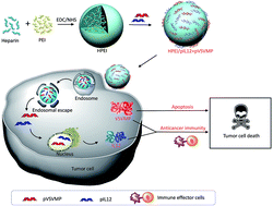
In the current study, the research team further armed with VSVMP gene delivery vessel with Interleukin-12 (IL-12) – a protein known to recruit and switch on the cancer-killing functions of immune cells.

The novel drug particles are based on Heparin-polyethyleneimine (HPEI) nanoparticles. To overcome the high toxicity and non-biocompatible nature of PEI, the team used a method to covalently conjugate this substance with heparin.

Their results, based on lab-grown cancer cells and animal studies, suggest that this novel complexed drug molecule (particle size: 53nm) increases tumor cell death, reduces division frequency, and stimulates the recruitment and activation of two types of cancer-killing cells: T cells and NK cells.

Specifically, the drug inhibited the growth of C-26 colon cancer cells. Animal studies showed that the drug reduced tumor weight. Metastatic spread of tumor cells to the abdomen was also reduced. The team proposes that the drug-derived IL-12 induces a secondary cascade of chemical mediators, which in turn recruit and activate cancer-killing immune cells. Their data supports this proposal. Interestingly, their study also found that the complexed drug molecule did not show adverse side effects within the major organs.

Read the full article here:

Nanoparticles co-delivering pVSVMP and pIL12 for synergistic gene therapy of colon cancer

Yuanyuan Xiao, Yuping Yang, Yujiao Wu, Chunmei Wang, Hao Cheng, Wei Zhao, Yang Li, Beibei Liu, Jianlin Long, Wenhao Guo, Guangping Gaoa and Maling Gou

Digg This
Reddit This
Stumble Now!
Share on Facebook
Bookmark this on Delicious
Share on LinkedIn
Bookmark this on Technorati
Post on Twitter
Google Buzz (aka. Google Reader)

A closer look at clean dishes: detection of domestic detergent residues with LIBS technology

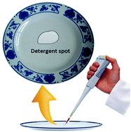
Dish detergents help keep our dishes clean; however, the long term health effects of detergent residues on tableware and cookware is yet to be discussed publicly. Studies suggest that certain household detergents may be linked with disturbances in hormone regulation in humans.

As a first step in uncovering the role of domestic dish detergent in affecting health, a research team comprising scientists from China Agricultural University, China Research Center of Intelligent Equipment for Agriculture, and Beijing Academy  of Agriculture and Forestry Sciences, have developed a method to detect detergent residue rapidly and in real-time, i.e., the process does not involve dissolving, preparing, or conditioning the residue prior to detection.

This method is based on a process called Laser Induced Breakdown Spectroscopy (LIBS). Detergent residues are first vaporized by a high-power laser, leading to the generation of vaporized plasma. At the end of the laser pulse, these atoms and ions spontaneously return from a higher energy state to a lower energy state. This energy decay is associated with the emission of optical radiation of specific wavelengths. The emitted radiation is collected and channeled toward a spectrometer, which converts wavelength information into readable numbers that scientists can record and analyze.

Although the LIBS technology has existed for several years, and is used routinely by researchers in the field, this study let by Zhao an colleagues is the first to use this method to measure household detergents.

Graphical abstract for C7RA04304J

Analogous to how a sensor at a grocery store is programmed to recognize barcodes printed on different items, the scientists used the numbers generated by the spectrometer to generate ‘signatures’ to help them recognize the different detergents used in the study. Using this method, the team found that detergent detection in real-time can be more flexible, used with tableware of different shapes, used to measure trace amounts of detergent, compatible with dry and wet dishes, and safe on tableware.

To demonstrate the utility of the method to real world applications, the team conducted a series of timed dish washes and residue analyses. Their results suggest that a 16-minute rinse removes detergent residues. They also suggest that this information will be useful in designing and programming commercial dishwashers.

This study may someday inspire public health advocates to take a closer look at the prevalence of dish residues in public and household settings. When this day arrives, LIBS technology for residue detection may be pivotal in conducting  studies to better understand the relationship between dish residues and overall wellbeing.

Read the full article here:

Detection of domestic detergent residues on porcelain tableware using laser induced breakdown spectroscopy
Xiande Zhao, Daming Dong, Yang Lic and Chunjiang Zhao
RSC Adv., 2017, 7, 28689-28695 (Open Access)

Digg This
Reddit This
Stumble Now!
Share on Facebook
Bookmark this on Delicious
Share on LinkedIn
Bookmark this on Technorati
Post on Twitter
Google Buzz (aka. Google Reader)

Can this quantum sized double-edged sword help diagnose and treat breast cancer?

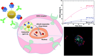
In a study led by Ko and colleagues at the Department of Dental Materials, School of Dentistry, Kyung Hee University, Korea, researchers armed graphene QDs with two therapeutic moieties: a HER-targeting antibody meant to help the therapeutic QDs find HER2-expressing breast cancer cells; and doxorubicin (DOX) – a chemotherapeutic drug used widely in treating breast cancer.

Consistent with previously established criteria (size, shape etc.) for drug carriers, the current study found that the estimated size of 222 nm makes the nanocarriers good candidates for further development toward diagnostic and therapeutic applications. Further, the nanocarriers had excitation and emission wavelengths of 370 nm and 450 nm respectively, making them glow in the ultraviolet range and as a result, optimal for medical imaging applications. The research team showed through chemical binding analysis that anti-HER antibodies were firmly bound to the QDs, and that the QDs were hydrophilic. The team conducted thermal stability studies and showed that the nanocarriers were stable at temperature ranges  much greater than the physiological body temperature range.


The study also analyzed whether the therapeutic nanocarriers were able to specifically target and enter breast cancer cells, release the DOX payload under specific pH and temperature conditions, and subsequently induce breast cancer cell death. Using a HER2-expressing breast cancer cell line, the team showed that the nanocarriers could kill cells in a dose dependent manner. A temperature of 37oC and pH of 5.5 were optimal for DOX release. Results in fluorescent microscopy studies suggested that DOX was released immediately after the nanocarriers entered HER2-expressing cells.


This study proposes that graphene-based QDs, when armed with anti-HER antibodies and DOX, have great potential for translation. In addition, with biomarker-based treatment decisions entering clinical practice in oncology settings, QD-based therapeutic nanocarriers are likely to have a notable impact on cancer therapy.
Read the full article here:

Graphene quantum dot-based theranostic agents for active targeting of breast cancer
N. R. Ko, M. Nafiujjaman, J. S. Lee, H.-N. Lim,a Y.-k. Lee and I. K. Kwon

Digg This
Reddit This
Stumble Now!
Share on Facebook
Bookmark this on Delicious
Share on LinkedIn
Bookmark this on Technorati
Post on Twitter
Google Buzz (aka. Google Reader)

Killing cancer cells with a DNA-based molecular bridge

Monoclonal antibodies (mAbs) are antibodies made by clones of immune cells derived from a common parent cell. These synthesized molecules have achieved widespread clinical utility in the treatment of cancer owing to their high degree of specificity to proteins present on the surface of cancer cells, lower toxicity compared to other classes of targeted therapies, and improved treatment outcomes among patients with advanced stage cancer.

Non-Hodgkin Lymphoma (NHL) is a type of cancer where a subtype of immune cells called B-cells exhibit unrestrained cell division. The abnormal B-cell, now called a malignant B-cell, produces more abnormal cells like it. CD20 is a protein present on the surface of malignant B-cells. Rituximab (RTX) is used to treat patients with NHL because it can bind CD20 and consequently trigger cell death.

To address the growing need for CD20 targeted therapeutics, Cong and colleagues at the Department of Laboratory Diagnosis/Thoracic Surgery, Changhai Hospital Affiliated to The Second Military Medical University, Shanghai, China, developed molecules called aptamers that can bind to CD20 with greater specificity and strength compared to RTX.

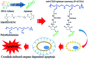
Graphical Abstract

Graphical Abstract

Aptamers are molecules made up of  single stranded DNA that form complex 3D structures and can bind to target proteins, analogous to mAbs. The team used a method called cell-SELEX to retrieve an enriched pool of highly specific CD20-binding aptamers starting with their initial aptamer library. The aptamers used in the study were obtained after 15 rounds of selective refinement.


The study finds that Anti-CD20 DNA Aptamer (ACDA) can bind surface CD20 in NHL cells with greater strength compared to RTX. In the past, experiments have shown that cross-linking surface CD20 with mAbs (i.e. extracellular cross-linking) is a potent method of inducing cell death. A major limitation is that extracellular cross-linking cannot be realized in vivo. Cong et al. develop a method to link two ACDA molecules with polyethyleneimine (PEI) linker’, forming a molecular bridge  – the P-ACDA – capable of spanning the distance between and cross-linking two CD20 molecules. The study finds that P-ACDA led to substantially more cell death compared to ACDA.

Aptamers as a novel class of targeted therapies are expected to outperform mAbs because they do not evoke the body’s endogenous immune response (i.e. less immunogenic) and therefore in good compliance with current FDA recommendations. They are also easier to store since they are stable across a broad temperature range ,less expensive to manufacture, show consistency between production batches and can bind to both protein as well as non-protein targets. For these reasons, the clinical relevance of aptamers in treating HNL and potentially other cancers must be watched closely in the years to come.

Read the full article here:

Cong Wu, Wei Wan, Ji Zhua, Hai Jina, Tiejun Zhao and Huafei Li

Digg This
Reddit This
Stumble Now!
Share on Facebook
Bookmark this on Delicious
Share on LinkedIn
Bookmark this on Technorati
Post on Twitter
Google Buzz (aka. Google Reader)

Serving up anti-cancer cocktails: design, synthesis and evaluation of hybrid inhibitors

Scientists in the area of cancer drug development constantly find ways to target cancer’s Achilles heel. Bcr-Abl is a protein expressed in 95% of all Chronic Myelogenous Leukemia (a type of blood cancer) cases. It remains activated (i.e. switched ON) and instructs cancer cells to divide indefinitely. Bcr-Abl has remained an attractive target for therapy. Yet another attractive therapeutic target is Histone Deacetylase 1 (HDAC1) – a protein known to control cell survival via its ability to influence the turning on and turning off of certain genes.

Previous studies show that the drugs Dasatinib and MS-275 efficiently inhibit the cancer promoting activities of Bcr-Abl and HDAC1 respectively.  Clinical trials also suggest that these drugs, when used independently in separate studies, can be used to treat a variety of solid as well as blood-borne cancers. Bcr-Abl and HDAC1 are components of distinct cellular wiring systems, referred to as signalling pathways, which sustain cell survival and division. Single agent drugs, or drugs that stifle a single cancer-promoting pathway, weed out most cancer cells but also set the stage for drug-resistant cells. Reports suggest that both Dasatinib and MS-275 are associated with cancer drug resistance.

Multi-target inhibitors are a new and evolving class of cancer drugs that can simultaneously inhibit at least two signalling pathways. These compounds  have emerged as a potential solution in circumventing cancer drug resistance. Chen and colleagues at the Department of Organic Chemistry, School of Science, China Pharmaceutical University, China, designed and produced a series of hybrid drug molecules which combine the attributes of the HDAC inhibitor MS-275 with the Bcr-Abl inhibitor Dasatinib.

To determine the effect of the hybrid drugs on cancer cell survival, the research team tested the drug’s ability to halt the growth of three cell types exhibiting features of leukemia, kidney cancer and prostate cancer respectively. They found that all drugs in the series were toxic to cancer cells, with leukemia and kidney cancer cells showing the greatest degree of sensitivity to the hybrid drugs.
To better understand how the hybrid drugs interacted with the Bcr-Abl and HDAC1 active sites (i.e. the ON switch), the team relied on computer-generated three-dimensional models of the hybrid drugs, Bcr-Abl and HDAC1 proteins. Using a method similar to finding the right key for a lock, a computer program found that a hybrid compound termed 6a, which happened to be the most potent compound in the series, fit most snugly into both Bcr-Abl  and HDAC1 active sites. In theory, 6a would prevent both Bcr-Abl and HDAC1 from becoming activated (i.e they remain switched OFF).

On the basis of these observations, this study strengthens the paradigm that chemically melding two cancer drugs to form a novel single molecule may prove to be an effective clinical strategy for anticancer treatment. On a broader scale, this is one among many studies advocating for the use of multi-target agents in cancer treatment, highlighting an imminent upsurge in single molecule combination therapies.

Read the full article here:

Digg This
Reddit This
Stumble Now!
Share on Facebook
Bookmark this on Delicious
Share on LinkedIn
Bookmark this on Technorati
Post on Twitter
Google Buzz (aka. Google Reader)

Micelles meet transplantation medicine: How a novel nanoparticle based immune cell blocker might benefit human organ transplantation

Organ transplantation saves lives. According to the Organ Procurement and Transplantation Network, U.S Department of Health and Human Services, over 22,000 organs transplantation surgeries have been conducted between January and September 2016.

Ischemia Reperfusion Injury (IRI) is a well characterized cardiac transplantation-related complication wherein the host tissue (graft), deprived of blood supply for prolonged periods, undergoes damage when blood supply is restored post-implantation. Immune cells at the interface of the graft and recipient tissue mediate damage by releasing inflammation-promoting chemicals and free radicals.

In a study led by Nadig and colleagues at the Department of Surgery, Division of Transplant, Medical University of South Carolina, USA, researchers first acknowledge the central role played by endothelial cells (EC) in promoting IRI-associated tissue damage and subsequently developed a novel pH-sensitive, immunosuppressive drug-loaded, targeted micelle nanoparticle to curb the damaging effects of ECs. The team choose rapamycin as their immunosuppressive drug of choice given its dual roles in limiting cytotoxic immune cell functions and in protecting tissues that make up blood vessels.

While treating patients with immunosuppressive drugs prior to surgery is currently a standard practice, a major drawback of this approach is that these drugs prevent immune system activity throughout the body, placing patients at risk for diseases including diabetes and cancer. As an initial step in addressing this limitation, Nadig et al. coated the micelles with cyclic arginine-glycine-aspartate moieties, which specifically bind to and integrin protein (the alpha v beta 3 receptor protein) present almost exclusively on ECs. As a finishing touch, the team attached fluorescent chemical compounds to allow for tracking and visualization in their studies.

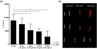
Graphical abstract of "Immunosuppressive nano-therapeutic micelles downregulate endothelial cell inflammation and immunogenicity"Their studies showed that the rapamycin-loaded nanoparticles were stable and biocompatible when tested in human endothelial cells. Further, the rapamycin release could be controlled by adjusting the pH values lower than 7 or higher than 8. The study found that the micelles were being taken up by cells within 6h after incubation. The study also demonstrates the specificity of the micelles by showing that what the cells were pre-treated with an integrin inhibitor,  they were  less likely to take up the micelles.
To demonstrate the clinical utility of their idea, the researchers exposed human endothelial cell cultures to hydrogen peroxide to mimic IRI-like conditions. The cells responded by increasing their production of inflammation-promoting chemicals. Importantly, the rapamycin-loaded nanoparticle micelles significantly curbed this response. Nadig et al. propose that the ultimate goal is to incorporate this technology into organ storage media to minimize the harmful effects of IRI.

Read the full article here:

Satish N. Nadig, Suraj K. Dixit, Natalie Levey, Scott Esckilsen, Kayla Miller, William Dennis, Carl Atkinson and Ann-Marie Broome

RSC Adv., 2015, 5, 43552-43562

DOI: 10.1039/C5RA04057D

Digg This
Reddit This
Stumble Now!
Share on Facebook
Bookmark this on Delicious
Share on LinkedIn
Bookmark this on Technorati
Post on Twitter
Google Buzz (aka. Google Reader)

Now you see me: autofluorescent nanoparticles for live cell imaging and biodegradation modeling

There is an increasing need for novel technologies to facilitate in vivo tissue visualization and drug delivery. However, this need is largely unmet due to the challenges associated with creating biocompatible materials that meet safety standards. In addition, the potential health risks associated with the accumulation of non-degradable imaging agents and drug carries represents a major obstacle in the innovation pipeline.

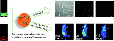
The intrinsic autofluorescent, biodegradable and biocompatible properties of Bovine Serum Albumin (BSA) is well appreciated. However, BSA has short excitation and emission wavelengths, which substantially restricts any in vivo biomedical applications.  Motivated by a recent report suggesting that glutaraldehyde (GA)-crosslinking induces autofluorescence in protein-based nanoparticles by modifying a series of C=C and C=N bonds, a team led by Yu Lei at the Department of Biomedical Engineering, University of Connecticut, developed low-cost, non-toxic, BSA-based protein nanoparticles (average size ~40 nm) for live cell imaging and biodegradation analysis.

The nanoparticles were generated by adding drops of a prepared BSA solution to glutaraldehyde/n-butanol solution at high-speed, and the resulting product heated at 121°C to ensure sterility. Interestingly, a similar reaction carried out in the absence of the GA crosslinker did not produce autofluorescent BSA nanoparticles, suggesting that GA was indeed playing an important role in chemically transforming BSA. Using UV-visible spectroscopy, the investigators observed that BSA nanoparticles exhibited strong autofluorescence at both green (530 nm) and red (630 nm) wavelengths.

The BSA nanoparticles were not uniform in structure, owing to the random points of crosslinking within BSA, and also due to the ensuing condensation reaction that occurs during the sterilization step. Therefore, a clear mechanistic explanation for the strong autofluorescence warrants further investigation. However, the investigators speculate that GA-crosslinking and heating could result in new C=N bonds, which could synergize with the C=C bonds from tryptophan, tyrosine, phenylalanine and histidine residues with BSA, leading to enhanced green and red fluorescence.

The team went on to demonstrate the utility of the BSA nanoparticles in biomedical applications such as imaging and biodegradation. They used fluorescent microscopy techniques to visualize the entry of BSA nanoparticles into human kidney cells grown in vitro. The study also found that the BSA nanoparticles were completely degraded within 18 days of injection in mice. A mathematical model for the distribution and biodegradation of the nanoparticles was in good agreement with the experimental results. Finally, to add an additional line of evidence supporting the biocompatible nature of the BSA nanoparticles, the investigators looked for signs of tissue damage in the region surrounding the site of injection, together with an analysis of internal organs including the pancreas, liver and kidney, and report that the BSA nanoparticles are biocompatible.

Read the full article here:

Xiaoyu Ma, Derek Hargrove, Qiuchen Dong, Donghui Song, Jun Chen, Shiyao Wang, Xiuling Lu, Yong Ku Cho, Tai-Hsi Fan and  Yu Lei
DOI: 10.1039/c6ra06783b
Digg This
Reddit This
Stumble Now!
Share on Facebook
Bookmark this on Delicious
Share on LinkedIn
Bookmark this on Technorati
Post on Twitter
Google Buzz (aka. Google Reader)

Modelling lung cancer: tumor cells on collagen scaffolds

Non-small-cell lung cancer (NSCLC) is among the leading causes of cancer-related deaths globally. Our understanding of the way tumors grow, spread and respond to therapy is driven largely by studies conducted on tumor cells growing as monolayers in plastic cell culture flasks in laboratories across the world. The ability to develop novel and more effective cancer-fighting drugs is dependent, in part, on developing cell culture systems that allow scientists to better observe how tumor cells grow in a three dimensional, physiologically relevant environment.

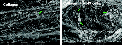
SEM images of the collagen meshwork and A549 cell aggregates (noted by the arrow head) formed during the
3D cultivation in vitro.

The tumor microenvironment (TM) is the area that immediately surrounds a tumor and includes non-cancer cells together with secreted proteins called the extracellular matrix (ECM), which supports tumor growth. Monolayer cell cultures, although utilized widely, cannot accurately mimic the TM. For instance, cell-cell and cell-ECM interactions that influence tumor growth cannot be observed in great detail with conventional monolayer cultures. Inspired by the up-and-coming field of tumor engineering, which aims to construct culture models that recapitulate aspects of the TM, a team of researchers led by Dr. Dan-Dan Wang at the Chinese Academy of Sciences developed a 3D culture system wherein A549 cells (immortal lung cancer cells of human origin) grow on a collagen hydrogel scaffold.

To demonstrate the utility of the 3D culture system, the study measured cell viability and showed that cells in the collagen hydrogel scaffold were alive for extended periods (>12 days) in vitro. The study also assessed the appearance of artificial A549 tumors growing on the hydrogel to demonstrate that 3D cultures more closely recapitulate the morphology of tumors growing within human tissues.

The proliferation of A549 cells is driven by the activation of a cell surface protein called Epidermal Growth Factor Receptor (EGFR), which in turn switches on genes that sustain cell growth and cell division. The team observed that Gefitinib, a drug known to disrupt growth-promoting signals arising at EGFR, was able to significantly constrain A549 cell proliferation in 3D cultures. Interestingly, the team reports that a higher concentration of Gefitinib was required to curb cell growth in 3D cultures compared to monolayers due to the complex architecture of the artificial tumors in 3D cultures.

Collectively, this study demonstrates an improved culture model of human lung cancer. Since collagen is an important component of the ECM, the study sets the stage for future efforts to better recapitulate the TM in vitro. The collagen hydrogel scaffold system could serve as in important tool in the discovery of targeted therapies for lung cancer.

Read the full article here:

Dan-Dan Wang,   Wei Liu,   Jing-Jie Chang,   Xu Cheng,   Xiu-Zhen Zhang,   Hong Xu,   Di Feng,   Li-Jun Yu and   Xiu-Li Wang
RSC Adv., 2016, 6, 24083-24090
DOI: 10.1039/C6RA00229C
Digg This
Reddit This
Stumble Now!
Share on Facebook
Bookmark this on Delicious
Share on LinkedIn
Bookmark this on Technorati
Post on Twitter
Google Buzz (aka. Google Reader)