Archive for the ‘RSC Advances’ Category

RSC Advances Reviewer Panel: 2017 Outstanding Reviewers

We are delighted to highlight the Outstanding Reviewers for RSC Advances in 2017, as selected by the editorial team, for their significant contribution to the journal. The reviewers have been chosen from the reviewer panel based on the quantity, quality and timeliness of the reports completed over the last 12 months.

A big thank you to those individuals listed here as well as to all of the reviewers on the RSC Advances reviewer panel that have supported the journal.

Each Outstanding Reviewer will receive a certificate to give recognition for their significant contribution.

Mr Rok Borstnar, Laboratory for genotoxicity
Dr Nghia Truong, Phuoc Monash University, ORCID: 0000-0001-9900-2644
Dr Wujun Fu, Oak Ridge National Laboratory
Dr S. Girish Kumar, CMR University, ORCID: 0000-0001-9132-1202
Dr Nicholas Geitner, Duke University, ORCID: 0000-0003-4313-372X
Dr Emanuele Curotto, University of Arcadia, ORCID: 0000-0001-9119-3263
Dr Yoong Ahm Kim, Chonnam National University, ORCID: 0000-0003-4074-7515
Dr Paul Trippier, Texas Tech University
Dr Michele Ceotto, Universita’ degli Studi di Milano, ORCID: 0000-0002-8270-3409
Dr Chunping Yang, Hunan University, ORCID: 0000-0003-3987-2722
Dr Wei Li, Utah State University, ORCID: 0000-0003-2802-7443
Dr Mark Waterland, Massey University, ORCID: 0000-0002-8493-9407
Dr Leo Small, Sandia National Laboratories, ORCID: 0000-0003-0404-6287
Dr Marija Gizdavic-Nikolaidis, The University of Auckland, ORCID: 0000-0002-8076-8508
Dr Xin Liu, State Key Laboratory of Fine Chemicals, ORCID: 0000-0002-4422-4108
Dr Zhijie Ma, University of Colorado Boulder, ORCID: 0000-0002-0734-1903
Dr Juliano Bonacin, University of Campinas, ORCID: 0000-0001-9399-1031
Dr Daniela Giacomazza, Istituto di Biofisica, ORCID: 0000-0002-6667-0205
Dr Ekkehard Lindner, Universitat Tubingen
Professor Zhenghua Tang, South China University of Technology, ORCID: 0000-0003-0718-3164
Dr Weixia Zhang, Harvard University, ORCID: 0000-0002-5835-2020
Dr Sreekuttan Unni, Central Electrochemical Research Institute, ORCID: 0000-0002-0403-9186
Professor Christian Robl, Friedrich-Schiller-Universität Jena
Professor Stanislaw Slomkowski, Center of Molecular and Macromolecular Studies, ORCID: 0000-0003-1543-535X
Dr Rui Oliveira, Universidade do Minho, ORCID: 0000-0002-3989-8925
Dr Wan Basirun, University of Malaya, ORCID: 0000-0001-8050-6113
Dr Yang Zhang, Arizona State University
Dr Maria Timofeeva, Novosibirsk State Technichal University
Dr Luis Simon, University of Salamanca, ORCID: 0000-0002-3781-0803
Dr Tsinghai Wang, National Tsing Hua University, ORCID: 0000-0003-4629-2005
Dr Thomas Mayer-Gall, Deutsches Textilforschungszentrum Nord-West, ORCID: 0000-0002-2822-6461
Dr Guowei Zhou Qilu, University of Technology
Dr Xiehong Cao, Nanyang Technological University, ORCID: 0000-0002-3004-7518
Dr Quanjun Xiang, University of Electronic Science and Technology of China, ORCID: 0000-0002-4486-7429
Dr Miklós Kubinyi, Budapest University of Technology and Economics, ORCID: 0000-0002-6343-0820
Dr Hu Li, Guizhou University, ORCID: 0000-0003-3604-9271
Dr Xuefeng Guo, Nanjing University, ORCID: 0000-0002-5492-5899
Dr Ahmad Zoolfakar, Universiti Teknologi MARA
Dr Bogdan-Marian Tofanica, Technical University of Iasi, ORCID: 0000-0002-4975-4650
Dr Zhiwei Xu, Tianjin Polytechnic University, ORCID: 0000-0003-1308-8884
Dr Tamás Vidóczy, Institute of Structuraél Chemistry
Dr Marinos Pitsikalis, University of Athens, ORCID: 0000-0002-7836-4862
Dr Haibo Shu, China Jiliang University, ORCID: 0000-0003-1728-2190
Dr Lin Zhang, Auburn University
Dr Ignacio Alfonso, Instituto de Química Avanzada de Cataluña, ORCID: 0000-0003-0678-0362
Dr Igor Komarov, Taras Shevchenko National University of Kyiv, ORCID: 0000-0002-7908-9145
Dr Xiao-Yu Hu, Nanjing University, ORCID: 0000-0002-9634-315X
Dr Zhe Wang, National Institutes of Health
Dr Muhammad Hossain, Yeungnam University, ORCID: 0000-0002-3428-8271
Dr Vaibhav Mehta, Marwadi University, ORCID: 0000-0003-4426-3374

Thank you to the RSC Advances board and our community for their continued support of the journal, as authors, reviewers and readers.

If you would like to become a reviewer for our journal, just email us with details of your research interests and an up-to-date CV or résumé.  You can find more details in our author and reviewer resource centre

Follow us on Twitter to keep informed!

Digg This
Reddit This
Stumble Now!
Share on Facebook
Bookmark this on Delicious
Share on LinkedIn
Bookmark this on Technorati
Post on Twitter
Google Buzz (aka. Google Reader)

Read our most downloaded RSC Advances articles of 2017

We are delighted to present a collection which showcases some of the most accessed RSC Advances articles published in 2017. This provides an easy way to access the most important papers published in RSC Advances in this year in your area of research.

RSC Advances is the largest open access chemistry journal, bringing you the latest research from right across the chemical sciences. For enhanced browsing and discoverability, topic-modelling technology automatically categorises articles into one or more of the 12 main subject categories and over 100 further subcategories.

The articles in the collection highlight the most exciting and important research published across analytical chemistry, biological chemistry, catalysis, chemical biology and medicinal chemistry, energy, environmental chemistry, inorganic chemistry, materials chemistry, nanoscience, organic chemistry and physical chemistry.

Follow the link to find our most downloaded articles in your research area.

We hope you enjoy reading these articles!

Digg This
Reddit This
Stumble Now!
Share on Facebook
Bookmark this on Delicious
Share on LinkedIn
Bookmark this on Technorati
Post on Twitter
Google Buzz (aka. Google Reader)

RSC Advances celebrates Open Access week 2017!

As we prepare to celebrate the 8th International Open Access week from 23-29 October 2017, it is an opportunity to highlight the benefits of Open Access and what it means for the RSC Advances research community…

We have published over 5600 open access articles so far in 2017, all of which are freely available to read on our website!

Open access refers to the free and permanent unrestricted online access to scholarly research and aims to maximise the visibility of research.

RSC Advances provides a high quality, open access option that helps get our authors’ work the attention that it deserves. Community-led, with an international team of associate editors, a dedicated reviewer panel and features such as article-based publishing, RSC Advances has been gold open access since January 2017, with one of the lowest article processing charges in the industry.

We deliberately push the boundaries with RSC Advances, always looking for new and unique ways to make the scientific developments we publish accessible to the widest possible audience.

See below for a sample of some of these articles that can be read for free, starting with our 5000th published article this year!

Fabrication of nanoporous copper with tunable ligaments and promising sonocatalytic performance by dealloying Cu–Y metallic glasses

Ning Wang, Ye Pan, Shikai Wu, Enming Zhang and Weiji Dai

RSC Adv., 2017,7, 43255-43265

DOI: 10.1039/C7RA08390D

High-quality CsPbBr3 perovskite nanocrystals for quantum dot light-emitting diodes

Xiafang Du, Guan Wu, Jian Cheng, Hui Dang, Kangzhe Ma, Ya-Wen Zhang, Peng-Feng Tan and Su Chen

RSC Adv., 2017,7, 10391-10396

DOI: 10.1039/C6RA27665b

Auxetic mechanical metamaterials

H. M. A. Kolken and A. A. Zadpoor

RSC Adv., 2017,7, 5111-5129

DOI: 10.1039/C7RA27333e

Enhanced seed germination and plant growth by atmospheric pressure cold air plasma: combined effect of seed and water treatment

L. Sivachandiran and A. Khacef

RSC Adv., 2017,7, 1822-1832

DOI: 10.1039/C6RA24762H


Digg This
Reddit This
Stumble Now!
Share on Facebook
Bookmark this on Delicious
Share on LinkedIn
Bookmark this on Technorati
Post on Twitter
Google Buzz (aka. Google Reader)

Overwhelmed to death: an anti-cancer gene therapy approach paired with an immune-activating distress signal

Frontline therapies for treating colorectal cancer have shortcomings. These include their inability to impede local tumor recurrence and metastatic spread to distant sites such as the abdomen.  

Researchers have now utilized a gene therapy approach that simultaneously compromises cancer cell survival while activating immune system cells with cancer-killing abilities.

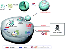
Gene therapy – an advanced technique developed to insert or inject therapeutic genes into human cells – has shown some success in treating the disease. In a previous study, Xiao and co-investigators at State Key Laboratory of Biotherapy, and the Department of Thoracic Oncology Cancer Center, West China Hospital, Sichuan University, had used a gene therapy approach to induce cancer cell death. Their study found that Vesicular Stomatitis Virus Matrix Protein (VSVMP), when inserted into a cancer cell,  compromises the cellular skeletal framework, which is made up of structural proteins. Cell death ensued as a consequence.

In the current study, the research team further armed with VSVMP gene delivery vessel with Interleukin-12 (IL-12) – a protein known to recruit and switch on the cancer-killing functions of immune cells.

The novel drug particles are based on Heparin-polyethyleneimine (HPEI) nanoparticles. To overcome the high toxicity and non-biocompatible nature of PEI, the team used a method to covalently conjugate this substance with heparin.

Their results, based on lab-grown cancer cells and animal studies, suggest that this novel complexed drug molecule (particle size: 53nm) increases tumor cell death, reduces division frequency, and stimulates the recruitment and activation of two types of cancer-killing cells: T cells and NK cells.

Specifically, the drug inhibited the growth of C-26 colon cancer cells. Animal studies showed that the drug reduced tumor weight. Metastatic spread of tumor cells to the abdomen was also reduced. The team proposes that the drug-derived IL-12 induces a secondary cascade of chemical mediators, which in turn recruit and activate cancer-killing immune cells. Their data supports this proposal. Interestingly, their study also found that the complexed drug molecule did not show adverse side effects within the major organs.

Read the full article here:

Nanoparticles co-delivering pVSVMP and pIL12 for synergistic gene therapy of colon cancer

Yuanyuan Xiao, Yuping Yang, Yujiao Wu, Chunmei Wang, Hao Cheng, Wei Zhao, Yang Li, Beibei Liu, Jianlin Long, Wenhao Guo, Guangping Gaoa and Maling Gou

Digg This
Reddit This
Stumble Now!
Share on Facebook
Bookmark this on Delicious
Share on LinkedIn
Bookmark this on Technorati
Post on Twitter
Google Buzz (aka. Google Reader)

Killing cancer cells with a DNA-based molecular bridge

Monoclonal antibodies (mAbs) are antibodies made by clones of immune cells derived from a common parent cell. These synthesized molecules have achieved widespread clinical utility in the treatment of cancer owing to their high degree of specificity to proteins present on the surface of cancer cells, lower toxicity compared to other classes of targeted therapies, and improved treatment outcomes among patients with advanced stage cancer.

Non-Hodgkin Lymphoma (NHL) is a type of cancer where a subtype of immune cells called B-cells exhibit unrestrained cell division. The abnormal B-cell, now called a malignant B-cell, produces more abnormal cells like it. CD20 is a protein present on the surface of malignant B-cells. Rituximab (RTX) is used to treat patients with NHL because it can bind CD20 and consequently trigger cell death.

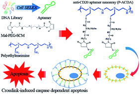
To address the growing need for CD20 targeted therapeutics, Cong and colleagues at the Department of Laboratory Diagnosis/Thoracic Surgery, Changhai Hospital Affiliated to The Second Military Medical University, Shanghai, China, developed molecules called aptamers that can bind to CD20 with greater specificity and strength compared to RTX.

Graphical Abstract

Graphical Abstract

Aptamers are molecules made up of  single stranded DNA that form complex 3D structures and can bind to target proteins, analogous to mAbs. The team used a method called cell-SELEX to retrieve an enriched pool of highly specific CD20-binding aptamers starting with their initial aptamer library. The aptamers used in the study were obtained after 15 rounds of selective refinement.


The study finds that Anti-CD20 DNA Aptamer (ACDA) can bind surface CD20 in NHL cells with greater strength compared to RTX. In the past, experiments have shown that cross-linking surface CD20 with mAbs (i.e. extracellular cross-linking) is a potent method of inducing cell death. A major limitation is that extracellular cross-linking cannot be realized in vivo. Cong et al. develop a method to link two ACDA molecules with polyethyleneimine (PEI) linker’, forming a molecular bridge  – the P-ACDA – capable of spanning the distance between and cross-linking two CD20 molecules. The study finds that P-ACDA led to substantially more cell death compared to ACDA.

Aptamers as a novel class of targeted therapies are expected to outperform mAbs because they do not evoke the body’s endogenous immune response (i.e. less immunogenic) and therefore in good compliance with current FDA recommendations. They are also easier to store since they are stable across a broad temperature range ,less expensive to manufacture, show consistency between production batches and can bind to both protein as well as non-protein targets. For these reasons, the clinical relevance of aptamers in treating HNL and potentially other cancers must be watched closely in the years to come.

Read the full article here:

Cong Wu, Wei Wan, Ji Zhua, Hai Jina, Tiejun Zhao and Huafei Li

Digg This
Reddit This
Stumble Now!
Share on Facebook
Bookmark this on Delicious
Share on LinkedIn
Bookmark this on Technorati
Post on Twitter
Google Buzz (aka. Google Reader)

A study on spheroid formation in thermosensitive hydrogels

According to an estimate by the World Cancer Report, cancer associated mortality is expected to reach 17 million per year globally by the year 2030. To confront the cancer burden with appropriate clinical interventions, researchers screen cancer-killing drug combinations in monolayer cell cultures. This is a widely used method for the preclinical evaluation of drug efficacy.

A major limitation of monolayer cultures is that they do not, even mildly, recapitulate the complex architecture of a tumor growing in vivo. As an initial step in overcoming this limitation, researchers use scaffolded spheroid cultures – a system wherein cells are grown on hydrogel scaffolds in three dimensions.

Hydrogel scaffolds provide physical and structural support for the formation of a more ‘natural’ setting that better recapitulates cell behavior in vivo. For example, smaller (150um) spheroids have better cell-to-cell contacts and notably different gene expression compared to monolayer cultures; larger (200-500um) ones develop oxygen and nutrient gradients reminiscent of chemical gradients seen in human tumors. However, limitations in design flexibility, handling and interbatch compositional variation have discouraged the routine use of hydrogel scaffolds. In addition, the technical challenge of separating newly formed spheroids from the scaffolding material before drug screening represents a major roadblock.

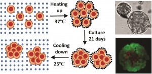
In a study led by Xiaolin Cui and colleagues at the School of Chemical Engineering and the School of Mathematical Sciences, University of Adelaide, Australia, researchers synthesized a thermo-reversible N-Isopropylacrylamide-acrylic acid (NIPAM-AA) hydrogel by free radical emulsion polymerization. In their study using the cervical carcinoma cell line HeLa, the team demonstrates that at 370C the NIPAM-AA hydrogel solidifies and forms a sheath around HeLa cell clusters. As a consequence, these clusters develop into hydrogel scaffolded spheroids over time. At 250C the hydrogel liquefies and releases the newly formed spheroids.

Cell viability assays confirmed that this new state of the art hydrogel is biocompatible. NIPAM-AA derived spheroids were smaller (70-120um),  nearly spherical and showed a narrower size distribution compared to conventional spheroids. The study showed, through time course experiments, that the spheroids remain viable for over 14 days in culture. The study also suggests that spheroids derived via the NIPAM-AA hydrogel method are more viable than those derived from conventional suspension cultures, supporting the notion that hydrogel scaffolding facilitates oxygen and nutrient supply to support cell growth.

The researchers deduced a mathematical model to predict the kinetics of NIPAM-AA derived spheroid growth. Their model accurately recapitulated the growth rate, size and size distribution of the spheroids. The authors propose that hydrogel scaffolding has the potential to evolve into a technology with a wide range of applications including, but not limited to, (1) high throughput screening of anticancer drugs using uniformly sized spheroids; (2) regenerative medicine; and (3) tissue engineering.

Read the full article here:

Xiaolin Cui,   Saber Dini,   Sheng Dai,   Jingxiu Bi,   Benjamin Binder,   Edward Green and   Hu Zhang
Digg This
Reddit This
Stumble Now!
Share on Facebook
Bookmark this on Delicious
Share on LinkedIn
Bookmark this on Technorati
Post on Twitter
Google Buzz (aka. Google Reader)

Sweet as sugar, hard as carbon: A hierarchical core-shell 3D graphene network biosensor for glucose detection

A biosensor is a device that uses biological molecules, typically enzymes, to specifically detect the presence of a chemical or a metabolic intermediate (referred to as the analyte) in a diagnostic setting. A biosensor acts as the platform upon which a biochemical reaction, initiated by the analyte, is converted to an electric current that is accurately quantified during a subsequent step. Biosensors have wide clinical applicability. For instance, the detection of blood sugar, which is among the most frequently measured physiological variable, is achieved with biosensors.

Recent years have seen rapid advancements in the use of nanoparticles, nanowires and nanotubes as biosensor platforms. These innovative nanostructures are electrochemically active, chemically stable, have large surface areas and are biocompatible – all of which are desirable attributes for developing biosensors. Of note is the observation that graphene, a substance known for its high electrical conductivity,  lends itself to biosensor development due to its relative ease of manufacture together with its ability to form composites with other electrochemically active nanostructures.

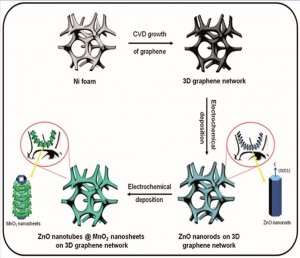
Early prototypes of graphene-based biosensors were inefficient for two main reasons. First, the clumping of graphene sheets reduced the accessible surface area. As a consequence, the biosensor/analyte interface was greatly reduced. Second, the restacking of graphene sheets introduced electrical resistance due to intersheet contacts. To overcome these hurdles, a research group led by Azam Iraji Zad at the Institute for Nanoscience and Nanotechnology (INST), Tehran, Iran developed a freestanding, porous 3D graphene network (3DGN) which was further modified with metal oxide nanostructures as a platform upon which an enzymatic reaction could occur.

This proof-of-concept study uses the glucose oxidase enzyme for the rapid and selective detection of glucose. The 3DGN, a graphene skeleton with multiple pores, is the core of the nanostructure. Atop the 3DGN, the researchers first grew uniformly spaced ZnO nanorods, which served to hold the enzyme in place. In a subsequent step, MnO2, known to be biocompatible and stable, was deposited onto the ZnO nanorods, thus forming a multilayered hierarchical structure with an average diameter of 100nm. The researchers propose that that the complex architecture of the nanostructure serves to facilitate the electron transfer process, which is the fundamental biochemical mechanism driving the enzymatic reaction.

In principle, the inner parts of the ZnO nanotubes increase the accessible surface area of the nanostructure and enhance the biosensor/analyte interface. In theory, the 3DGN biosensor is expected to respond quicker and have improved sensitivity when compared to other enzyme-based glucose detection devices. The study tested the 3DGN biosensor using a method called amperometry which is used routinely in research laboratories to detect ions – the byproduct of enzymatic reactions. The study found that the 3DGN biosensors had a response time of less than 3 seconds; a value indicative of a competitive advantage over other enzyme-based glucose biosensors. Intriguingly, the study also found that the 3DGN was very sensitive and could detect extremely low concentrations (10nM) of glucose.

The study strongly suggests that 3DGN biosensors could be used as an accurate sensing platform for chemicals and biomolecules. The findings further support the argument that composite nanostructures with complex architecture could find applicability in human health and beyond.

Read the full article here:

Elham Asadian, Saeed Shahrokhian and Azam Iraji Zadac
Digg This
Reddit This
Stumble Now!
Share on Facebook
Bookmark this on Delicious
Share on LinkedIn
Bookmark this on Technorati
Post on Twitter
Google Buzz (aka. Google Reader)

RSC Supramolecular Chemistry Award for Editor-in-Chief Mike Ward

Each year the Royal Society of Chemistry presents prizes and awards to chemical scientists who have made a considerable contribution in their area of research, in industry and academia. This year, we are delighted to announce that RSC Advances Editor-in-Chief, Professor Mike Ward of the University of Sheffield, UK, has been awarded the 2016 RSC Supramolecular Chemistry Award, for his leading contributions to the synthesis, characterisation, host-guest chemistry and functional properties of self-assembled coordination cages.

The Supramolecular Chemistry Award is awarded biennially and recognises studies leading to the design of functionally useful supramolecular species.

In celebration of the 2016 RSC Prizes and Awards, we have collected together some of the research recently published by the winners. This collection showcases articles authored by the winners from across the Royal Society of Chemistry’s journals portfolio, which are free to access for a limited period. A full list of 2016 winners and more information about RSC Prizes and Awards can be found here.

Please join us in congratulating Mike on this achievement!

We would like to highlight the RSC Advances themed collection, Supramolecular chemistry: self-assembly and molecular recognition, Guest Edited by Mike Ward.

The articles in this issue cover many aspects of the formation of, and molecular recognition with, non-covalent self-assembled systems. Systems studied span the range of supramolecular assemblies from MOFs to gels, and potential applications or functional behaviour that are on display here include host/guest chemistry, spin crossover, molecular sensors, and extraction/separation. This collection of articles powerfully illustrates the diversity and increasing importance of supramolecular chemistry.

Digg This
Reddit This
Stumble Now!
Share on Facebook
Bookmark this on Delicious
Share on LinkedIn
Bookmark this on Technorati
Post on Twitter
Google Buzz (aka. Google Reader)

The heat is on: cancer-drug loaded nanoparticles for photothermal therapy

Photothermal therapy is an emerging area of cancer treatment. Here, a photothermal agents, often nanoparticles (NPs) with a resonance peak in the 700-1200nm range, are delivered to the tumor site and are subsequently activated by light in the Near Infrared (NIR) range. As a consequence, tumor cells are thermally ablated.

In a study led by Xiaolin Li and colleagues at the Key Laboratory for Thin Film and Microfabrication and Changzheng Hospital in China, scientists used SiO2@Au core-shell NPs chemically conjugated via PEGylation to graphene oxide (GO) in conjunction with a chemotherapeutic agent to target prostate cancer cells in vitro. Using the chemotherapeutic agent Docetaxel (Dtxl),  which is among the leading front line treatments for patients diagnosed with prostate cancer, the team demonstrated that Dtxl-loaded SiO2@Au@GO NPs, when activated with light in the NIR range, significantly curbed the survival of DU145 prostate cancer cells.

While SiO2@Au core-shell NPs have been used previously by other research groups to study their ability to remove tumors, Li’s team fabricated SiO2@Au@GO NPs to take advantage of their relatively low cost, large specific surface area, and efficient loading and delivery of water-soluble aromatic drug molecules. This one-two punch strategy was realized via a double shell, multifunctional approach: the inner core SiO2@Au NPs served as a photothermal inducer to bring about cellular cytotoxicity; the outer GO NPs carried the antitumor drug, Dtxl. The study found that exposing DU145 cells to the NPs alone for 24h did not result in overt cell death, suggesting that the NPs have a good safety profile. Importantly, the study showed that when NP-treated cell cultures were irradiated with a 780nm NIR laser, there was a significant decline in viable cells over a 24h period.

The study demonstrates that Dtxl-loaded SiO2@Au@GO NPs could be manufactured and potentially used an an antitumor agent for the treatment of prostate cancer. Moreover, these findings illuminate the untapped potential of NP-based photothermal agents as adjuvant agents in oncology clinical trials in the near future.

Read the full article here:

Xiaolin Li,   Zhi Yang,   Nantao Hu,   Liying Zhang,   Yafei Zhang and   Lei Yin
DOI: 10.1039/C6RA03886G
Digg This
Reddit This
Stumble Now!
Share on Facebook
Bookmark this on Delicious
Share on LinkedIn
Bookmark this on Technorati
Post on Twitter
Google Buzz (aka. Google Reader)

Supramolecular cages for chemical weapons

The research of RSC Advances Chief Editor Mike Ward focussing on the development of supramolecular cages that trap chemical weapon stimulants, has been highlighted in Chemistry World.

Mike and his team at Sheffield University have developed new supramolecular cages that exploit the hydrophic effect and bind alkyl phosphonates inside. These phosphonates are very similar to organophosphorous chemical weapons. Cobalt or cadmium dications form the cage vertices and bis(pyrazolyl-pyridine) ligands run along each edge, forming a hydrophobic centre lined with CH groups. So, in water, the phosphonate hydrophobic alkyl tails are attracted to the inside of the cage. Whats more, the cage is luminescent and this luminescence reduces when alkyl phosphonate enters, meaning that the cages can also be used to signal the presence of chemical weapons.

The supramolecular structure

To find out more, read the full Chemistry World article based on this paper:

Binding of chemical warfare agent simulants as guests in a coordination cage: contributions to binding and a fluorescence-based response
Christopher G. P. Taylor, Jerico R. Piper and Michael D. Ward
Chem. Commun., 2016
DOI: 10.1039/C6CC02021F

Digg This
Reddit This
Stumble Now!
Share on Facebook
Bookmark this on Delicious
Share on LinkedIn
Bookmark this on Technorati
Post on Twitter
Google Buzz (aka. Google Reader)