Archive for April, 2016

Top 10 most-downloaded articles: Q1 Jan–Mar 2016

Take a look at the most-downloaded RSC Advances articles from the months of October, November and December 2015 and let us know what you think!

Size-controlled silver nanoparticles synthesized over the range 5–100 nm using the same protocol and their antibacterial efficacy
Shekhar Agnihotri, Soumyo Mukherji and Suparna Mukherji
RSC Adv., 2014,4, 3974-3983
DOI: 10.1039/C3RA44507K

Free radicals, natural antioxidants, and their reaction mechanisms
Satish Balasaheb Nimse and Dilipkumar Pal
RSC Adv., 2015,5, 27986-28006
DOI: 10.1039/C4RA13315C

Thermal-runaway experiments on consumer Li-ion batteries with metal-oxide and olivin-type cathodes
Andrey W. Golubkov, David Fuchs, Julian Wagner, Helmar Wiltsche, Christoph Stangl, Gisela Fauler, Gernot Voitic, Alexander Thaler and Viktor Hacker
RSC Adv., 2014,4, 3633-3642
DOI: 10.1039/C3RA45748F

Synthesis and properties of molybdenum disulphide: from bulk to atomic layers
Intek Song, Chibeom Park and Hee Cheul Choi
RSC Adv., 2015,5, 7495-7514
DOI: 10.1039/C4RA11852A

Orientation dependence of the pseudo-Hall effect in p-type 3C–SiC four-terminal devices under mechanical stress
Hoang-Phuong Phan, Afzaal Qamar, Dzung Viet Dao, Toan Dinh, Li Wang, Jisheng Han, Philip Tanner, Sima Dimitrijev and Nam-Trung Nguyen
RSC Adv., 2015,5, 56377-56381
DOI: 10.1039/C5RA10144A

Formation of organic–inorganic mixed halide perovskite films by thermal evaporation of PbCl<inf>2</inf> and CH<inf>3</inf>NH<inf>3</inf>I compounds
Cheng Gao, Jiang Liu, Cheng Liao, Qinyan Ye, Yongzheng Zhang, Xulin He, Xiaowei Guo, Jun Mei and Woonming Lau
RSC Adv., 2015,5, 26175-26180
DOI: 10.1039/C4RA17316C

Dual protection of amino functions involving Boc
Ulf Ragnarsson and Leif Grehn
RSC Adv., 2013,3, 18691-18697
DOI: 10.1039/C3RA42956C

Third-generation solar cells: a review and comparison of polymer:fullerene, hybrid polymer and perovskite solar cells
Junfeng Yan and Brian R. Saunders
RSC Adv., 2014,4, 43286-43314
DOI: 10.1039/C4RA07064J

Graphene and its nanocomposite material based electrochemical sensor platform for dopamine
Alagarsamy Pandikumar, Gregory Thien Soon How, Teo Peik See, Fatin Saiha Omar, Subramaniam Jayabal, Khosro Zangeneh Kamali, Norazriena Yusoff, Asilah Jamil, Ramasamy Ramaraj, Swamidoss Abraham John, Hong Ngee Lim and Nay Ming Huang
RSC Adv., 2014,4, 63296-63323
DOI: 10.1039/C4RA13777A

Colloidal semiconductor nanocrystals: controlled synthesis and surface chemistry in organic media
Jin Chang and Eric R. Waclawik
RSC Adv., 2014,4, 23505-23527
DOI: 10.1039/C4RA02684E


Interesting in submitting to RSC Advances? You can submit online today, or email us with your ideas and suggestions.


Digg This
Reddit This
Stumble Now!
Share on Facebook
Bookmark this on Delicious
Share on LinkedIn
Bookmark this on Technorati
Post on Twitter
Google Buzz (aka. Google Reader)

Supramolecular cages for chemical weapons

The research of RSC Advances Chief Editor Mike Ward focussing on the development of supramolecular cages that trap chemical weapon stimulants, has been highlighted in Chemistry World.

Mike and his team at Sheffield University have developed new supramolecular cages that exploit the hydrophic effect and bind alkyl phosphonates inside. These phosphonates are very similar to organophosphorous chemical weapons. Cobalt or cadmium dications form the cage vertices and bis(pyrazolyl-pyridine) ligands run along each edge, forming a hydrophobic centre lined with CH groups. So, in water, the phosphonate hydrophobic alkyl tails are attracted to the inside of the cage. Whats more, the cage is luminescent and this luminescence reduces when alkyl phosphonate enters, meaning that the cages can also be used to signal the presence of chemical weapons.

The supramolecular structure

To find out more, read the full Chemistry World article based on this paper:

Binding of chemical warfare agent simulants as guests in a coordination cage: contributions to binding and a fluorescence-based response
Christopher G. P. Taylor, Jerico R. Piper and Michael D. Ward
Chem. Commun., 2016
DOI: 10.1039/C6CC02021F

Digg This
Reddit This
Stumble Now!
Share on Facebook
Bookmark this on Delicious
Share on LinkedIn
Bookmark this on Technorati
Post on Twitter
Google Buzz (aka. Google Reader)

Modelling lung cancer: tumor cells on collagen scaffolds

Non-small-cell lung cancer (NSCLC) is among the leading causes of cancer-related deaths globally. Our understanding of the way tumors grow, spread and respond to therapy is driven largely by studies conducted on tumor cells growing as monolayers in plastic cell culture flasks in laboratories across the world. The ability to develop novel and more effective cancer-fighting drugs is dependent, in part, on developing cell culture systems that allow scientists to better observe how tumor cells grow in a three dimensional, physiologically relevant environment.

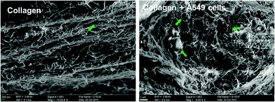
SEM images of the collagen meshwork and A549 cell aggregates (noted by the arrow head) formed during the
3D cultivation in vitro.

The tumor microenvironment (TM) is the area that immediately surrounds a tumor and includes non-cancer cells together with secreted proteins called the extracellular matrix (ECM), which supports tumor growth. Monolayer cell cultures, although utilized widely, cannot accurately mimic the TM. For instance, cell-cell and cell-ECM interactions that influence tumor growth cannot be observed in great detail with conventional monolayer cultures. Inspired by the up-and-coming field of tumor engineering, which aims to construct culture models that recapitulate aspects of the TM, a team of researchers led by Dr. Dan-Dan Wang at the Chinese Academy of Sciences developed a 3D culture system wherein A549 cells (immortal lung cancer cells of human origin) grow on a collagen hydrogel scaffold.

To demonstrate the utility of the 3D culture system, the study measured cell viability and showed that cells in the collagen hydrogel scaffold were alive for extended periods (>12 days) in vitro. The study also assessed the appearance of artificial A549 tumors growing on the hydrogel to demonstrate that 3D cultures more closely recapitulate the morphology of tumors growing within human tissues.

The proliferation of A549 cells is driven by the activation of a cell surface protein called Epidermal Growth Factor Receptor (EGFR), which in turn switches on genes that sustain cell growth and cell division. The team observed that Gefitinib, a drug known to disrupt growth-promoting signals arising at EGFR, was able to significantly constrain A549 cell proliferation in 3D cultures. Interestingly, the team reports that a higher concentration of Gefitinib was required to curb cell growth in 3D cultures compared to monolayers due to the complex architecture of the artificial tumors in 3D cultures.

Collectively, this study demonstrates an improved culture model of human lung cancer. Since collagen is an important component of the ECM, the study sets the stage for future efforts to better recapitulate the TM in vitro. The collagen hydrogel scaffold system could serve as in important tool in the discovery of targeted therapies for lung cancer.

Read the full article here:

Dan-Dan Wang,   Wei Liu,   Jing-Jie Chang,   Xu Cheng,   Xiu-Zhen Zhang,   Hong Xu,   Di Feng,   Li-Jun Yu and   Xiu-Li Wang
RSC Adv., 2016, 6, 24083-24090
DOI: 10.1039/C6RA00229C
Digg This
Reddit This
Stumble Now!
Share on Facebook
Bookmark this on Delicious
Share on LinkedIn
Bookmark this on Technorati
Post on Twitter
Google Buzz (aka. Google Reader)