Imagine a scenario where one morning, a close friend of yours calls you and painfully conveys the news of him being diagnosed with cancer and you, instead of sitting horrified and helpless, casually say “Hey don’t worry man, we have PDT!” That sounds fascinating right?! Yes, Photodynamic therapy has shown potential to do that. With the same fascination towards the idea of photodynamic therapy, inventors of PDT pursued research on this therapy and shaped an unconventional out of box method of treating cancer. The simple mechanism of working of this technique is widely known. Drugs used in this technique are light sensitive. In response to specific light irradiated on the drug molecule, it converts surrounding molecular oxygen into form of oxygen which kills nearby cancer cells. The reasons this therapy called as out of box here are multifold. First, there are many photosensitizers easily available approved by FDA which can easily respond to specific light and produced the effect explained above. Second it makes use of naturally available oxygen molecules surrounding cancer cells. Last and importantly all the conventional drugs/ therapies for the cancer are immunosuppressive meaning they suppress our immune system after treatment unlike PDT, which is immunostimulative which stimulates immune system of the patient after treatment.
Can you treat cancer this way too?! Really?!!
Writers Choice: Amphiphilic chitosan modified upconversion nanoparticles for in vivo photodynamic therapy induced by near-infrared light
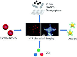
NIR Luminescent Nanomaterials for Biomedical Imaging
Commonly in my household the phrase “you make a better door than you do a window” is often fired at whichever thoughtless member is blocking the latest episode of whatever intelligence diluting programme is being watched at the time. However, this same, seemingly mundane problem, of human solidity is also being suffered by scientists developing new techniques for biomedical imaging.
Luminescent labels have been widely used for biological applications, primarily bioimaging and assays. They offer advantages over tomographic imaging techniques (e.g. CT, PET and MRI) including fast feedback and high selectivity and resolution. Unfortunately, adsorption and scattering of the photos emitted by these labels caused by biological tissue and water inside the body create problems such as weak signals and limited depth of detection.
Luckily, there are some wavelengths of light that are not adsorbed by the body and fall into what is known as the “biological transparency window”. There are two ranges: NIR I 650 – 900 nm and NIR II 1000 – 1450 nm. Since the discovery of these NIR regions research has increased with the focus of developing nanomaterials that can be excited or emitted within these wavelengths. The main content of this review written by Wang and Zhang is an overview of these novel nanomaterials, divided into four main species: lanthanide based nanomaterials, carbon based nanomaterials, quantum dots and noble metal nanoparticles. Covering their fabrication and application and also their shortcomings and what challenges and opportunities there are in the future.
NIR Luminescent Nanomaterials for Biomedical Imaging
Rui Wang and Fan Zhang
J. Mater Chem. B, 2014, 2, 2422-2443. C3TB21447H
H. L. Parker is a guest web writer for the Journal of Materials Chemistry blog. She currently works at the Green Chemistry Centre of Excellence, the University of York.
To keep up-to-date with all the latest research, sign-up to our RSS feed or Table of contents alert.
A good hair day for glowing nanoparticles
Written by Jennifer Newton for Chemistry World.
By raiding their local barber’s shop, scientists in China have found the ideal raw material for an emerging class of fluorescent nanoparticles.
The desirable optical properties, chemical inertness and biocompatibility of carbon dots has led researchers to explore their application in anti-counterfeiting fields and flat panel displays…
Interested? Read the full article at Chemistry World.

Photographs of carbon dot ink patterns under UV light
The original article can be read below:
Hair-Derived Carbon Dots toward Versatile Multidimensional Fluorescent Hybrid Materials
Si-Si Liu, Cai-Feng Wang, Chen-Xiong Li, Jing Wang, Li-Hua Mao and Su Chen
J. Mater. Chem. C, 2014, Accepted Manuscript
DOI: 10.1039/C4TC00636D
Henry Snaith wins the 2014 Journal of Materials Chemistry Lectureship
On behalf of the Journal of Materials Chemistry Executive Editorial Board we are delighted to announce Professor Henry Snaith is the winner of the Journal of Materials Chemistry Lectureship 2014.
Professor Snaith is the fifth winner of the Journal of Materials Chemistry Lectureship. The Journal of Materials Chemistry Executive Editorial Board chose Professor Snaith in recognition of the remarkable contribution he has made to the materials chemistry field.
This annual lectureship honours a younger scientist who has made a significant contribution to the field of materials chemistry. Further details of the Lectureship, including the lecture location, will be announced in due course.
Also of interest: You can now read all three of the 2014 Emerging Investigators Issues of Journal of Materials Chemistry A, B and C, which highlight the work of emerging investigators across the field of materials chemistry. There are a mix of review-type articles, Communications and Full Papers, as well as Profile articles showcasing the authors.
Follow the latest journal news on Twitter @JMaterChem or go to our Facebook page.
Hot Article: A simple, low-cost CVD route to thin films of BiFeO3 for efficient water photo-oxidation
Hydrogen holds immeasurable promise in our search for alternative, sustainable, cleaner fuels. However, the simple, cheap production of hydrogen is still proving a problem. Water photolysis is a great way to achieve pure H2 and as O2 is the only side product it does not result in the harmful greenhouse gas emissions that arise from using hydrocarbons to produce H2. Unfortunately, the generation of H2 by water photolysis is challenging as the reaction that forms O2 is much slower than the H2 forming reaction. The use of an efficient photocatalyst can significantly improve the success of this process.
This paper by Moniz et al. details the development of just such a photocatalyst. In this work a bimetallic BiFeO3 catalyst is prepared using a novel method of Aerosol Assisted Chemical Vapour Deposition (AA CVD). This is the first time that this method has been used to prepare a photocatalyst of this type. The team go on to test this photocatalyst for the electrolysis of water using both UV and solar irridation and encouragingly, activity is confirmed for the BiFeO3 catalyst. Even more impressively the catalyst greatly outperforms both a commercially available photocatalyst (TiO2 Activ® glass) and another recently published photocatalyst (B-doped TiO2 films).
The novel synthetic methodology presented in this paper enables large area thin film deposition and as a result has potential for high volume applications in the future.
A simple, low-cost CVD route to thin films of BiFeO3 for efficient water photo-oxidation
Savio J. A. Moniz, Raul Quessada-Cabrera, Christopher S. Blackman, Junwang Tang, Paul Southern, Paul M. Weaver and Claire J. Carmalt,
J. Mater. Chem. A, 2014, 2, 2922-2927 C3TA14824F
H. L. Parker is a guest web writer for the Journal of Materials Chemistry blog. She currently works at the Green Chemistry Centre of Excellence, the University of York.
To keep up-to-date with all the latest research, sign-up to our
Hot Article: A highly luminescent chameleon: fine-tuned emission trajectory and controllable energy transfer
Luminescent materials are a part of our everyday life featuring in lighting, television screens, etc. The recent emergence of lanthanide-based metal-organic frameworks (Ln-MOFs) has illuminated the future development of new functional luminescent materials. Research into Ln-MOFs is still at its early stages but they have shown promise in the development of effective novel compounds.
This paper by Zhang et al. takes Ln-MOFs to the next level and presents the first example of mixed-lanthanide MOFs. The work combines Eu3+, Gd3+ and Tb3+ as co-doped ions on to one MOF framework. The co-doped Ln-MOF is capable of excitation-dependent mutual conversion between blue, white and yellow emission chromaticity…I am guessing this is where the rather whimsical title has come from.
This succinctly written communication gives a first look at the synthesis and testing of this exciting new Ln-MOF and gives an idea of where the research into Ln-MOFs might be heading in the future.
A highly luminescent chameleon: fine tuned emission trajectory and controllable energy transfer
Huabin Zhang, Xiaochen Shan, Zuju Ma, Liujiang Zhou. Mingjian Zhang, Ping Lin, Shengmin Hu, En Ma, Renfu Li and Shaowu Du
J. Mater Chem. C, 2014, 2, 1367-1371. C3TC31624F
H. L. Parker is a guest web writer for the Journal of Materials Chemistry blog. She currently works at the Green Chemistry Centre of Excellence, the University of York.
To keep up-to-date with all the latest research, sign-up to our RSS feed or Table of contents alert.
Is it a bird? Is it a plane? Possibly, its Graphene the Wonder Material!
Hot Article: Graphene–organic composites for electronics: optical and electronic interactions in vacuum, liquids and thin solid films
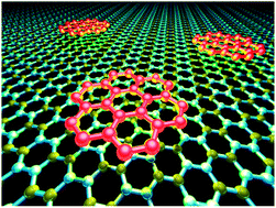
“Wonder material” they called it when it was discovered last decade and it started to be center of attention from 2010. To understand the reasoning behind calling graphene a wonder material, one need not be a rocket scientist. The beauty of this material is so conspicuous that it can fascinate anybody on the globe. Graphene is one the few materials in existence which is very thin, conductive, transparent and flexible at the same time. The wonder of this material’s thinness is so intriguing, that according to scientists, just ounce of graphene could cover 28 football fields. Only a single atom thick, Graphene could take electronics to the next level with much thinner, faster and cheaper components compared to the current silicon based electronics.
We all know the saying nothing is perfect, however researchers all over the globe are claiming that Graphene may not be perfect but it is close to perfect. The major challenges for making Graphene a game changer in electronics are control over chemical and physical properties by chemical functionalization and processing them upon up-scalable approaches.
Graphene-organic composites for electronics: optical and electronic interactions in vacuum, liquids and thin solid films
A. Schlierf, P. Samori and V. Palermo
J. Mater. Chem. C, 2014, 2, 3129-3143. DOI:10.1039/C3TC32153C
Padmanabh Joshi is a guest web writer for the Journal of Materials Chemistry blog. He currently works at the Department of Chemistry, University of Cincinnati.
To keep up-to-date with all the latest research, sign-up to our RSS feed or Table of contents alert.
Endothelial cell thrombogenicity is reduced by ATRP-mediated grafting of gelatin onto PCL surfaces
One of the most promising areas of materials chemistry research is the creation of synthetic analogues of biological tissue. The development of such materials would reduce the need for transplants and their associated problems. For example, polymer-based, artificial vascular grafts are a promising candidate for use in bypass operations. Unfortunately, they increase the risk of increasing thrombus (blood clot) formation. This in turn can lead to risk of serious medical problems such as stroke, heart attack and pulmonary embolism.
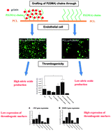
Researchers at Nanyang Technological University in Singapore have found that using functionalised polycaprolactone (PCL) can lead to reduced thrombogenicity. PCL itself is already widely used for in vivo applications although its hydrophobicity reduces its usefulness as an artificial blood vessel. Hydrophobic materials are also thought to be unsuitable because of their weak interactions with endothelial cells (ECs) – something commonly implicated in thrombus formation. To overcome these limitations, poly(glycidyl methacrylate) (PGMA) was grown from PCL surfaces via a graft-from living polymerisation. The side chains of the PGMA were then used to attach gelatine molecules. It was found that the gelatin coat decreased the hydrophobicity of the surface leading to improved EC adhesion and a corresponding reduction in thrombogenicity.
Endothelial cell thrombogenicity is reduced by ATRP-mediated grafting of gelatin onto PCL surfaces
Gordon Minru Xiong, Shaojun Yuan, Chek Kun Tan, Jun Kit Wang, Yang Liu, Timothy Thatt Yang Tan, Nguan Soon Tan and Cleo Choong
J. Mater. Chem. B, 2014, 2, 485-493. DOI:10.1039/C3TB20760a
James Serginson is a guest web writer for the Journal of Materials Chemistry blog. He currently works at Imperial College London carrying out research into nanocomposites.
To keep up-to-date with all the latest research, sign-up to our RSS feed or Table of contents alert.
Slow-setting bone glue for easier post-surgery access
A patient is rushed into hospital. Doctors crack the patient’s chest to operate. A life is saved, but what comes next? In order to access the heart and lungs, the sternum had to be opened, but closing it again and healing the wound is often more problematic than the surgery itself.
Researchers in Ireland and Germany have developed an adhesive to address this issue. Standard practise for sternal closure winds steel wires around the bones to immobilise them as they heal. However, displacement can occur in active patients and older bones can be weak, resulting in the wires cutting through. If the bones part again, a patient can be in agony.
Interested? Read the full article at Chemistry World.

Cohesive coupling, complexation and redox radical polymerisation work together to resist displacement
The original article can be read below:
Biomimetic Hyperbranched Poly(amino ester)-based Nanocomposite as Tunable Bone Adhesive for Sternal Closure
Wenxin Wang, Hong Zhang, Ligia Bre, Tianyu Zhao, Ben Newland and Mark Dacosta
J. Mater. Chem. B, 2014, Accepted Manuscript
DOI: 10.1039/C4TB00155A
Hot Article: Chemical modification of inorganic nanostructures for targeted and controlled drug delivery in cancer treatment
Cancer nanotechnology, small but mighty!
The use of engineered nanostructures in biomedical applications and optimized therapy is revolutionising medicine and the way we treat disease. It probably doesn’t come as a surprise that cancer is one of the biggest driving forces responsible for development of therapeutic nanotechnologies. The potential for earlier detection and targeted treatment of tumours using nanotechnologies will act not only to reduce the number of cancer deaths but also reduce the side effects and increase the efficacy of treatments.
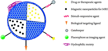
This review by Zhang et al. examines the recent advances in nanotechnology for targeted drug delivery and controlled drug release in cancer treatment. The focus of the introduction is on inorganic nanostructures, highlighting the advantages of these materials over bioorganic nanomaterials, namely the ease of synthesis, modification and the control of size, shape and surface functionalization can be carried out. All of which allow for the design of materials for specific tissue or cell type targeting, controlled drug delivery and in vivo diagnostic imaging.
The review also covers the mechanisms of systematic targeted drug delivery, stimuli-responsive drug release and biocompatibility of these inorganic nanostructures. Overall, this review gives a clear and varied look at the different technologies under development that I would recommend to many scientists, not just those working in this field.
Chemical modification of inorganic nanostructures for targeted and controlled drug delivery in cancer treatment
Lei Zhang, Yecheng Li and Jimmy C. Yu
J. Mater Chem. B, 2014, 2, 452-470. C3TB21196G
H. L. Parker is a guest web writer for the Journal of Materials Chemistry blog. She currently works at the Green Chemistry Centre of Excellence, the University of York.
To keep up-to-date with all the latest research, sign-up to our RSS feed or Table of contents alert.


