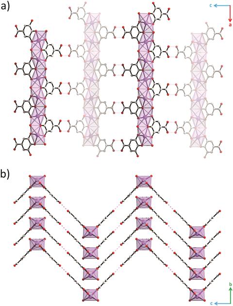
Crystal structure of bismuth subgallate viewed along (a) [010] and (b) [100]. Bismuth, carbon and oxygen atoms are coloured purple, grey and red, respectively. Hydrogen atoms and water molecules in the pores have been omitted for clarity.
Now, Andrew Kentaro Inge from Stockholm University and his team have overcome these issues. By combining continuous rotational data collection with a cooling technique, they avoided the electron beam damage, poor resolution and diffuse scattering holding them and others back. ‘Continuous rotation electron diffraction is a promising way to elucidate the structures of hard to obtain, or very hard to crystallise, pharmaceutical forms. For this purpose, it’s an up-and-coming method,’ says Tomislav Friŝĉić, an expert in materials chemistry at McGill University in Canada.
Read the full story by Tabitha Watson on Chemistry World.


")