There is an increasing need for novel technologies to facilitate in vivo tissue visualization and drug delivery. However, this need is largely unmet due to the challenges associated with creating biocompatible materials that meet safety standards. In addition, the potential health risks associated with the accumulation of non-degradable imaging agents and drug carries represents a major obstacle in the innovation pipeline.
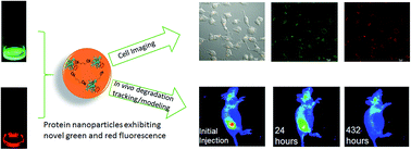
The intrinsic autofluorescent, biodegradable and biocompatible properties of Bovine Serum Albumin (BSA) is well appreciated. However, BSA has short excitation and emission wavelengths, which substantially restricts any in vivo biomedical applications. Motivated by a recent report suggesting that glutaraldehyde (GA)-crosslinking induces autofluorescence in protein-based nanoparticles by modifying a series of C=C and C=N bonds, a team led by Yu Lei at the Department of Biomedical Engineering, University of Connecticut, developed low-cost, non-toxic, BSA-based protein nanoparticles (average size ~40 nm) for live cell imaging and biodegradation analysis.
|

|
|
|
|
The nanoparticles were generated by adding drops of a prepared BSA solution to glutaraldehyde/n-butanol solution at high-speed, and the resulting product heated at 121°C to ensure sterility. Interestingly, a similar reaction carried out in the absence of the GA crosslinker did not produce autofluorescent BSA nanoparticles, suggesting that GA was indeed playing an important role in chemically transforming BSA. Using UV-visible spectroscopy, the investigators observed that BSA nanoparticles exhibited strong autofluorescence at both green (530 nm) and red (630 nm) wavelengths.
The BSA nanoparticles were not uniform in structure, owing to the random points of crosslinking within BSA, and also due to the ensuing condensation reaction that occurs during the sterilization step. Therefore, a clear mechanistic explanation for the strong autofluorescence warrants further investigation. However, the investigators speculate that GA-crosslinking and heating could result in new C=N bonds, which could synergize with the C=C bonds from tryptophan, tyrosine, phenylalanine and histidine residues with BSA, leading to enhanced green and red fluorescence.
The team went on to demonstrate the utility of the BSA nanoparticles in biomedical applications such as imaging and biodegradation. They used fluorescent microscopy techniques to visualize the entry of BSA nanoparticles into human kidney cells grown in vitro. The study also found that the BSA nanoparticles were completely degraded within 18 days of injection in mice. A mathematical model for the distribution and biodegradation of the nanoparticles was in good agreement with the experimental results. Finally, to add an additional line of evidence supporting the biocompatible nature of the BSA nanoparticles, the investigators looked for signs of tissue damage in the region surrounding the site of injection, together with an analysis of internal organs including the pancreas, liver and kidney, and report that the BSA nanoparticles are biocompatible.
Read the full article here:
Xiaoyu Ma, Derek Hargrove, Qiuchen Dong, Donghui Song, Jun Chen, Shiyao Wang, Xiuling Lu, Yong Ku Cho, Tai-Hsi Fan and Yu Lei
DOI: 10.1039/c6ra06783b


