Commonly in my household the phrase “you make a better door than you do a window” is often fired at whichever thoughtless member is blocking the latest episode of whatever intelligence diluting programme is being watched at the time. However, this same, seemingly mundane problem, of human solidity is also being suffered by scientists developing new techniques for biomedical imaging.
Luminescent labels have been widely used for biological applications, primarily bioimaging and assays. They offer advantages over tomographic imaging techniques (e.g. CT, PET and MRI) including fast feedback and high selectivity and resolution. Unfortunately, adsorption and scattering of the photos emitted by these labels caused by biological tissue and water inside the body create problems such as weak signals and limited depth of detection.
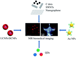
Luckily, there are some wavelengths of light that are not adsorbed by the body and fall into what is known as the “biological transparency window”. There are two ranges: NIR I 650 – 900 nm and NIR II 1000 – 1450 nm. Since the discovery of these NIR regions research has increased with the focus of developing nanomaterials that can be excited or emitted within these wavelengths. The main content of this review written by Wang and Zhang is an overview of these novel nanomaterials, divided into four main species: lanthanide based nanomaterials, carbon based nanomaterials, quantum dots and noble metal nanoparticles. Covering their fabrication and application and also their shortcomings and what challenges and opportunities there are in the future.
NIR Luminescent Nanomaterials for Biomedical Imaging
Rui Wang and Fan Zhang
J. Mater Chem. B, 2014, 2, 2422-2443. C3TB21447H
H. L. Parker is a guest web writer for the Journal of Materials Chemistry blog. She currently works at the Green Chemistry Centre of Excellence, the University of York.
To keep up-to-date with all the latest research, sign-up to our RSS feed or Table of contents alert.


