Author Archive

Making a splash

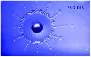
When a droplet impacts on a solid surface it deforms. Depending on the properties of the drop and the surface (velocity, viscosity, surface tension, hydrophobicity etc.) this deformation can be temporary, the droplet spreads out before retracting and bouncing back, or permanent, the drop breaks apart on impact making a splash.

Air also plays an important role in determining the behaviour of the impacting drop. Detlef Lohse, University of Twente, is interested in understanding how and why the air layer profile under a drop influences its deformation as it falls and subsequently hits a surface. His group has developed an ultra-high speed colour interferometry imaging method, allowing them to resolve the dynamics of an impacting drop on short timescales. Using this method, Lohse has been able to measure the time evolution of the drop before and during impact on a solid surface. As the drop falls the air between the drop and surface is strongly squeezed. A region of high pressure builds up, which leads to the formation of a dimple on the underside of the drop. At high velocities this can result in splashing as the air is compressed on impact. The presence of an air bubble can also stop the droplet from touching the surface at all leading to some interesting effects.

When a liquid droplet impacts a surface heated above the liquid’s boiling point three impact behaviour regimes are observed. In the first, ‘contact boiling’, the droplet immediately boils as it comes into contact with the surface. The droplet contacts the surface and spreads out. Bubbles then form and the liquid evaporates. For ‘gentle film boiling’ the droplet appears to hit the surface before bouncing back. In this regime a Leidenfrost vapour layer forms under the drop before it hits, preventing it from coming into direct contact with the surface. The final behavioural regime is ‘spray film boiling’. In this case, although the droplet does not contact the surface, breakup does occur. A Leidenfrost vapour layer forms below the drop. As the vapour tries to escape it drags fluid out with it and the droplet forms a thinner pancake shape. Tiny drops are the ejected upwards in what can be quite a violent spray event. The impact conditions under which each regime can be observed were recently published in Physical Review Letters.

Lohse has also looked at how the structure of the surface can influence the splashing dynamics for high velocity impacting drops. In this case the surfaces are all at room temperature. This is discussed in detail for Newtonian and non-Newtonian liquids in two recent Soft Matter papers. Directional splashing can be tuned and suppressed by varying the periodicity of the lattice and, or the air pressure. A number of videos of the impacting drops can be found in the supplementary information accompanying the Soft Matter article and are well worth a look.

Digg This
Reddit This
Stumble Now!
Share on Facebook
Bookmark this on Delicious
Share on LinkedIn
Bookmark this on Technorati
Post on Twitter
Google Buzz (aka. Google Reader)

Micron Microfluidics

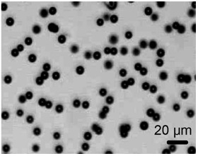
8 micrometer bubbles produced by Kumacheva via bubble dissolution.

Microfluidics is a very successful technology widely used to produce droplets with sizes in the range of 10-200 µm. However, the production and behaviour of droplets smaller than this, around 1 µm, has not really been explored. Droplets of this size are highly desirable due, amongst other things, to their biomedical applications.

There are a number of possible methods, which could be used to generate micron sized droplets. Kumacheva et al. formed bubbles of < 10 µm using a dissolution technique. Micro-bubbles of 50-100 µm were formed initially and allowed to carefully dissolve until they reached the desired size. Anna et al., on the other hand, used tip streaming through a flow focusing device to form micron sized liquid drops. While both of these methods have successfully formed droplets smaller than 10 µm, droplet formation using these techniques can be hard to control as both methods rely on specific physiochemical conditions and have relatively low throughputs.

In a recent talk, Patrick Tabeling discussed a brute force method, which he used to form simple droplets, multiple droplets, particles and Janus particles with diameters of 900 nm – 3 µm. The microfluidic devices used by Tabeling et al. contain a submicrometric channel, with a cross junction (where the dispersed and continuous phases meet), followed by a terrace. The interface between the two fluids forms a tongue as it flows down the nanofluidic terrace. The terrace empties into a reservoir, who’s depth is much greater than that of the nanochannel. As the tongue tip crosses into the reservoir it becomes unstable and droplets are formed. Highly monodisperse droplets can be generated using this technique at a rate of 5-15 kHz.

In his talk, Tabeling demonstrated the use of these micron sized droplets for targeted drug delivery in rats. Fluorescence containing droplets were injected into rats, targeted disruption of the droplets and delivery of the fluorescence was achieved using acoustic waves.

Digg This
Reddit This
Stumble Now!
Share on Facebook
Bookmark this on Delicious
Share on LinkedIn
Bookmark this on Technorati
Post on Twitter
Google Buzz (aka. Google Reader)

Active soft matter

What does softness do for life? According to Zhigang Suo, Havard University, for life the important feature of soft materials is that they are easily deformable. In the human body the deformation of soft materials enables the heart to beat, the vocal folds to produce sound and the eye to focus. In all these examples, a stimulus is applied to the soft material and a large reversible deformation occurs. This deformation in turns provides a function.

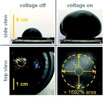
Soft active materials are not only important in life, but also have technological relevance for example in adaptive optics, self-regulated fluidics and soft robotics. Suo is interested in how mechanics, chemistry and electrostatics all work together to generate large deformations in soft materials. In soft dielectrics actuation can be readily observed by applying a voltage across the membrane, causing the thickness to reduce and the area to expand. In polymers, strains of up to 30% are easily achieved using this method.

To get higher deformations two limitations need to be overcome; electrical breakdown and electromechanical instability. An electromechanical instability is observed in compliant dielectrics such as elastomers, when the applied voltage causes the material to thin down excessively, amplifying the electric field. This is known as a snap-though instability and often occurs prior to electrical breakdown. Suo has shown, however, that if this electromechanical instability can be overcome, and the elastomer reaches a stable state without breakdown occurring, giant voltage-induced deformations of over 1000% are achievable.

Suo’s theory shows that the instability can be eliminated when pre-stretched or short-chain polymers, where the elastic strain is sufficiently low, are used. The elastomer is compliant at low deformations and stiffens rapidly as it is slowly stretched. This stiffening averts the rapid excessive deformation that would otherwise cause the material to fail. The elastomer survives the instability and reaches a steady state without breakdown occurring. Suo and his collaborators have used these ideas to carefully design and tailor the properties of soft materials realising area expansions of 1692%.

For more information see:

Keplinger et al., Harnessing snap-through instability in soft dielectrics to achieve giant voltage-triggered deformation, Soft Matter, 8,  285-288, 2012.

Lu et al., Dielectric elastomer actuators under equal-biaxial forces, uniaxial forces, and uniaxial constraint of stiff fibers, Soft Matter, 8, 6167-6173, 2012.

Zhao and Suo, Theory of Dielectric Elastomers Capable of Giant Deformation of Actuation, Phys. Rev. Lett., 104, 178302, 2010.

Digg This
Reddit This
Stumble Now!
Share on Facebook
Bookmark this on Delicious
Share on LinkedIn
Bookmark this on Technorati
Post on Twitter
Google Buzz (aka. Google Reader)

Using DNA to detect DNA

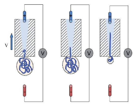
Schematic of DNA translocation in a glass capillary. Image taken from Soft Matter, 2012, doi:10.1039/C2SM25346A

Detecting single strands of DNA is a tricky business. One way in which it can be done is by creating a pore and sensing the DNA strand as it passes through the pore. This is what Ullrich Keyser from the University of Cambridge, UK and his group have been doing.

Keyser forms nanopores with diameters of less than 100nm from glass capillary tubes (the diameters can be as small as 20nm). These nanocapillaries act as single molecule sensors. Using electrophoresis, the negatively charged DNA is pulled towards a positively charged electrode inside the capillary, in a process known as DNA translocation. As the strand enters the capillary the resistance across the capillary pore changes, allowing the DNA to be detected. The change in ionic current is dependent not only on the presence of a DNA strand, but also on its folded state. This method offers a simple and cost-effective method for the detection of single molecules and for DNA sequencing.

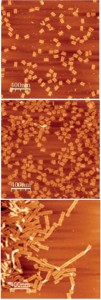
Examples of DNA origami. Image taken from Soft Matter, 2011, 7, 4636.

Full control over DNA translocation can be achieved using optical trapping, where the DNA is attached to a colloidal particle, held in place by an optical trap. The DNA can then be moved in and out of the capillary pore at will. Using this method, Keyser and his group have measured the capture force due to the electric field acting on the DNA. Their results show that the DNA capture force is linearly dependent on the number of strands captured in the capillary.

Whether using glass capillaries, or pores formed in silicon nitride membranes via focussed ion beam milling, control over the exact shape and functionality of the nanocavity can be problematic. Keyser has taken DNA detection yet another step further by using the DNA itself to create the nanopore. The shape into which a DNA strand folds can be controlled in a a process known as DNA origami; the DNA is synthesised such that it will self-assemble into a pre-designed three-dimensional shape. Using this origami, it is possible to design and fabricate virtually any nanosized shape that you want.

Keyser designed the DNA so that it folded into a funnel like shape with a long tail. This structure was then pulled through a pore in a  silicon nitride membrane to form a hybrid nanopore with a diameter of 7.5nm. The assembly of the hybrid pore is robust and easily reversible. These DNA/silicon nitride pores have been successfully used to detect single strands of DNA. The hybrid nanopores offer a novel way to change the size, shape and functionality of pores.

Relevant papers in SoftMatter:

Chen, Q. et al., How does a supercoiled DNA chain pass through a small conical glass pore? Soft Matter, 2012, Advanced Article.

Geerts, N., Eiser, E., DNA functionalized colloids: Physical properties and applications. Soft Matter, 2010, 6, 4647-4660.

Kim, K. N., et al., Comparison of methods for orienting and aligning DNA origami. Soft Matter, 2011, 7, 4636-4643.

Digg This
Reddit This
Stumble Now!
Share on Facebook
Bookmark this on Delicious
Share on LinkedIn
Bookmark this on Technorati
Post on Twitter
Google Buzz (aka. Google Reader)

Survival in the face of the unknown

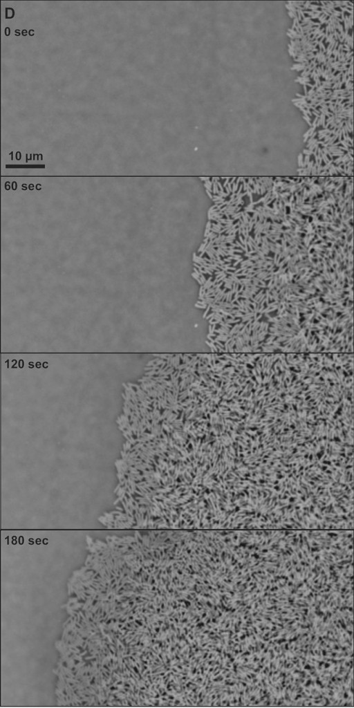
Swarming E. coli.

There are an estimated 1030 bacteria on Earth. The number of bacteria is greater than the number of stars in the Universe and is growing exponentially.

Bacteria are generally studied in the laboratory in Petri dishes under very well defined conditions. However, bacteria also thrive in more complex environments where the conditions are constantly varying. Some of these environmental changes are regular e.g. variations in light intensity from day to night, while others are random e.g. temperature, food availability and the presence of toxins or other bacteria.

Bacteria have developed a number of strategies to survive in these fluctuating environments. In the opening talk of the DPG spring meeting in Berlin last week, Stanislas Leibler from the Rockefeller University, New York and the Institute for Advance Studies, Princeton, discussed recent experimental and theoretical studies exploring the complex behaviour observed in bacterial colonies.

Consider a growing colony of bacteria. When an environmental change occurs one of two things may happen if the colony is to survive. (1) The bacteria ‘senses’ the change and changes to a state that is adapted for this new environment. This is known as responsive switching. (2) A small minority of the bacteria in the colony are poorly adapted to the initial environment. However, they become the most-adapted when the environment changes and survive while the rest are killed; the minority becomes the majority. This is known as stochastic switching.

So which is it? For colonies of bacteria with antibiotic persistence, experiments suggest that stochastic switching is the dominant behaviour. Leibler’s group added Ampicillin to growing colonies of Escherichia coli. The majority of the colony dies, but a few resistant bacteria survive. These resistant bacteria are able to grow, forming a new colony, once the antibiotic is removed. The persistent bacteria have a different phenotype to the rest of the colony. Under normal conditions, they grow much more slowly than the non-resistant bacteria, but are not killed when the antibiotics are added. Although the presence of these persistent cells leads to a lower population fitness, they act as an insurance policy and ensure that the colony can survive in the event of an antibiotic encounter. Leibler believes that this heterogeneity of bacterial populations is important for their ability to adapt to fluctuating environments and the persistence of bacterial infections.

While important when considering antibiotic resistant infections, these results may have much wider implications in areas ranging from cancer treatments, to models of financial investments, to information theory and statistical mechanics.

For more information see:

Balaban, N.Q. et al., Bacterial persistence as a phenotypic switch, Science, 2004.

Kussell, E. et al., Bacterial persistence: A model of survival in changing environments, Genetics, 2005.

Rivoire O, Leibler S, The value of information for populations in varying environments, J. Statist. Phys., 2011.

The image is taken from: Bacterial swarming: a model for studying dynamic self-assembly, Soft Matter, 2009, and shows a swarming colony of E. coli bacteria.

Digg This
Reddit This
Stumble Now!
Share on Facebook
Bookmark this on Delicious
Share on LinkedIn
Bookmark this on Technorati
Post on Twitter
Google Buzz (aka. Google Reader)

Embryonic polarisation

Growing up is a stressful process, particularly during embryonic development. The embryo starts out as a single symmetric cell. This cell can be considered as an active fluid bound by the cell membrane. Flow within this fluid leads to the build-up of stresses, the formation of patterns and asymmetries within the cell. Stephan Grill, at the Max-Planck Insitute for the Physics of complex systems and the Max-Planck Institute for Molecular Cell Biology and Genetics, Dresden, is interested in understanding how these flows lead to the polarisation observed in the Caenorhabditis elegans zygote.

Grill has shown that the changes in cell polarity are driven by myosin flow on the surface of the cell. The cortex can be considered as a dynamic self-contracting polymer gel surface, which lies underneath the cell membrane. This polymer gel layer behaves as a thin film of an active fluid. Passive advective transport of molecules – in this case myosin – embedded in the fluid can occur depending on the diffusivity and the flow velocities of the molecules.  In C. elegans the flows are fast enough that advection does play a role, influencing the distribution of the molecules as they diffuse on the cortex. Modelling shows that the passive advective transport by flow of such a mechanically active materials acts as a trigger for the segregation of the proteins, resulting in the polarisation of the zygote.

The movement of myosin across the surface of the cell also results in anisotropies in the cortical tension. These so called active stresses cause isolated sections of the cortex to self-contract. Grill has developed a novel method for locally determining the stresses, by cutting the cortex with a laser and measuring the recoil. The cortical tension is found to be greatest in the direction orthogonal to the flow.

Grill suggests that advective transport in active fluids is a general mechanism for the formation of patterns in developmental biology.

For more information see:

Goehring, N.W. et al., Polarization of PAR Proteins by Advective Triggering of a Pattern-Forming System, Science, 2011.

Bois, S, et al., Pattern Formation in Active Fluids, Phys. Rev. Lett., 2011.

Mayer. M, et al., Anisotropies in cortical tension reveal the physical basis of polarizing cortical flows, Nature, 2010.

The image above is taken from: Cyclodextrin/dextran based drug carriers for a controlled release of hydrophobic drugs in zebrafish embryos, Soft Matter, 2011, and shows C. elegans embryo 30hrs after fertilisation.

Digg This
Reddit This
Stumble Now!
Share on Facebook
Bookmark this on Delicious
Share on LinkedIn
Bookmark this on Technorati
Post on Twitter
Google Buzz (aka. Google Reader)

Saliva necessary for survival

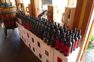
Tannins are well known to influence the taste of food and drinks, in particular red wine. This has to do with the astringent sensation that the tannins produce in the mouth; a dry and puckering effect. Tannins are water soluble polyphenols that interact with proteins and precipitate them out. They are produced by plants as part of their defence mechanism against parasitic invasion. The tannins bind to enzymes released by the parasite causing the enzyme to precipitate. The enzyme is rendered useless and the invasion halted.

The binding of tannins to proteins also occurs in our bodies when we eat or drink tannin-containing foods. In the mouth the interaction between tannins and saliva causes astringency. Tannins are also known to bind to our digestive enzymes resulting in a reduced ability to digest food. If consumed in large quantities tannins can lead to serious malnutrition. So why is it that we can drink, for example, red wine and not die? Bernard Cabane at the ESPCI Paris has been trying to answer exactly this.

Cabane and coworkers have been investigating the interactions between salivary proline-rich proteins and the tannins present in green tea. Their work concentrates on two salivary proteins, one glycosylated and one non-glycosylated, with the same polypeptidic backbone. For the non-glycosylated protein the tannins are observed to bind randomly along the protein chain. The chains have very extended conformations, which may make it more efficient at binding the tannins. Increasing the tannin concentration results in the formation of protein-tannin aggregates and precipitation of the proteins, once the concentration is high enough. The precipitation of the protein degrades the lubrication in the mouth resulting in an astringent sensation. Since precipitation only occurs once the threshold concentration of tannins to proteins is reached, Cabane suggests that it may act as a warning system telling us when the tannin levels in our body are too high.

For the glycosylated protein, on the other hand, no precipitation is observed in the presence of tannins. Instead globular aggregates, resembling micelles,  form with the hydrophilic sugars on the outside and the hydrophobic residues of the protein backbone, which bind the tannins, on the inside. These micelles act as tannin traps with roughly 1000 tannins locked within each micelle. The efficient binding of the tannins in the micelle, means that the tannins can enter the digestive system with no adverse effects on the body.

Cabane is also interested in the oxidisation of tannins and the effect it has on the flavour of foods.

H. Boze et al., Proline-rich salivary proteins have extended conformations, Biophysical Journal,  2010.

Pascal, C et al., Aggregation of a proline-rich protein induced by epigallocatechin gallate and condensed tannins: Effect of protein glycosylation, Journal of Agricultural and Food Chemistry, 2008.

Vernhet, A et al., Characterization of oxidized tannins: comparison of depolymerization methods, asymmetric flow field-flow fractionation and small-angle X-ray scattering, Analytic and Bioanalytical Chemistry, 2011.

Digg This
Reddit This
Stumble Now!
Share on Facebook
Bookmark this on Delicious
Share on LinkedIn
Bookmark this on Technorati
Post on Twitter
Google Buzz (aka. Google Reader)

Antidrip paint

Images of the drying process (a) taken from above and (b) from the side.

Watching paint dry can be fun and rather surprising! David Fairhurst from Nottingham Trent University has been looking at the structures that form when polymer droplets are left to dry.

Small droplets of aqueous poly(ethylene oxide) (PEO) are placed onto glass substrates and the water allowed to evaporate. Initially, pinned drying is observed; the radius of the droplet remains fixed while the height and contact angle decrease. When the polymer concentration, at the contact line, reaches the saturation concentration PEO starts to precipitate. A semi-crystalline solid forms and the contact line starts to recede. As the droplet dewets polymer is continually deposited at the contact line. The solid layer pushes the droplet back further in an autophobic like manner. This process continues until the height of the droplet starts to increase. Now the PEO is deposited on top of itself. The edge of the ever shrinking droplet is lifted and a conical structure forms. Any remaining water evaporates, leaving behind a semi-crystalline PEO tower.

The structure which forms is dependent on a number of parameters including: the initial polymer concentration in the drop, the relative humidity, temperature and pressure under which the droplet is dried and the contact angle of the drop when it is first deposited. The results, recently published in Soft Matter, are explained in terms of polymer flow within the drop due to evaporative flux and the diffusion of the polymer. Diffusion drives the homogenisation of the polymer within the droplet, while the evaporative flux induces an outward flow of the polymer. If the evaporative flux dominates, deposition at the contact line occurs and a pillar structure forms.

Various videos of the drying droplets can be found here and are well worth a look. Fairhurst suggested in his talk that these solutions could be used as potential antidrip paints. When left to dry on a slanted substrate the pillars form uphill of the initial drop. Missing corners when painting could be a thing of the past.
K. Baldwin, M.Granjard, D. I. Willmer, K. Sefiane and D. J. Fairhurst, Drying and deposition of poly(ethylene oxide) droplets determined by Péclet number, Soft Matter, 2011, 7, 7819-7826

Digg This
Reddit This
Stumble Now!
Share on Facebook
Bookmark this on Delicious
Share on LinkedIn
Bookmark this on Technorati
Post on Twitter
Google Buzz (aka. Google Reader)

Loopy veins

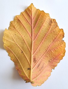
If someone asked you to draw the vein architecture of a leaf, your first thought might be to draw a tree-like structure; a central trunk with branches and twigs coming off it. While this picture may be correct for ancient and living fossils, it is not sufficient to describe the venation of most modern plants. For this loops are required. Eleni Katifori, Rockefeller University, is interested in why these loops have evolved and in understanding what purpose they serve.

On first sight, loops seem inefficient due to the redundancy inherent in a loopy structure. The veins in a leaf act as a transport system, delivering water and nutrients to the leaf. Assuming that the demand for nutrients is constant across the whole leaf, then yes a loopy structure is inefficient. However, this is not the case. Fluctuations occur across the leaf, not all stomata are open or closed at the same time. This leads to variation in water evaporation rates and photosynthetic activity. Loops allow the flow to be efficiently re-routed through the leaf in response to these fluctuations.

As well as improving efficiency, Katifori has found that ‘loopyness’ increases the resilience of the leaf to damage. Take a look at a nearby tree, almost every leaf on it will be damaged in some way or other. For a simple tree like network damage will halt flow. There is no way for veins on the other side of the injury to receive any nutrients or water. For a loopy network however, this is not the case. The nutrients can flow around the injury closing the loops and will eventually reach all parts of the leaf. Videos showing the two different cases can be found here.

Similar loopy architectures are seen in the veins of some insect wings, animal tissues such as the retina and the road networks of cities.

Katifori E., Szollosi G. J. and Magnasco M. O., Damage and fluctuations induce loops in optimal transport networks, Phys. Rev. Lett., 2010, 104, 048704.

Recent paper on leaves in Soft Matter:

Xiao, H., Chen, X., Modelling and simulation of curled dry leaves, Soft Matter, 2011, 7, 10794-10802.

Digg This
Reddit This
Stumble Now!
Share on Facebook
Bookmark this on Delicious
Share on LinkedIn
Bookmark this on Technorati
Post on Twitter
Google Buzz (aka. Google Reader)

Reptate

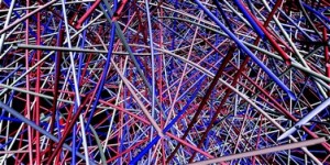
Image taken from: Mechanics of random fiber networks—a review, Soft Matter, 2011.

Rheology for Entangled Polymers: Toolkit for Analysis of Theory and Experiment.

Reptate is a software package developed as part of the Microscale Polymer Processing project with collaborators from the University of Leeds and the University of Reading. The main authors of the software were Jorge Ramierez and Alexi Likhtman. Reptate was highlighted by Tom McLeish in a recent talk at the Physical Aspects of Polymer Science conference,  as a wonderful tool for those studying the rheological behaviour of polymers.

Reptate provides a platform where experimental rheological data can be easily compared to theoretical predictions for the behaviour of entangled polymers. The software includes both classical and current theories of polymer dynamics. As well as a tool for understanding experimental data, Reptate could be used to design polymers for specific applications; the properties are chosen, the polymer architecture inferred and the polymer designed. Reptate is available online at reptate.com and is provided for free. More information can be found here.

Recent papers on understanding entangled polymer behaviour:

Linking models of polymerisation and dynamics to predict branched polymer structure and flow, Science (2011).

Counting polymer knots to find the entanglement length, Soft Matter (2011).

Microscopic origin of the terminal relaxation time in polymer nanocomposites: an experimental precedent, Soft Matter (2011).

Digg This
Reddit This
Stumble Now!
Share on Facebook
Bookmark this on Delicious
Share on LinkedIn
Bookmark this on Technorati
Post on Twitter
Google Buzz (aka. Google Reader)