The articles below are 10 of the most read Review articles and 10 of the most read Papers & Communications from Organic & Biomolecular Chemistry in July, August and September 2016.
Review Articles
Transition-metal catalyzed valorization of lignin: the key to a sustainable carbon-neutral future
Markus D. Kärkäs, Bryan S. Matsuura, Timothy M. Monos, Gabriel Magallanes and Corey R. J. Stephenson
DOI: 10.1039/C5OB02212F, Review Article
Selective chemical labeling of proteins
Xi Chen and Yao-Wen Wu
DOI: 10.1039/C6OB00126B, Review Article
Synthesis of substituted pyrenes by indirect methods
Juan M. Casas-Solvas, Joshua D. Howgego and Anthony P. Davis
DOI: 10.1039/C3OB41993B, Review Article
Modern advances in heterocyclic chemistry in drug discovery
Alexandria P. Taylor, Ralph P. Robinson, Yvette M. Fobian, David C. Blakemore, Lyn H. Jones and Olugbeminiyi Fadeyi
DOI: 10.1039/C6OB00936K, Review Article
Recent applications in natural product synthesis of dihydrofuran and -pyran formation by ring-closing alkene metathesis
Reece Jacques, Ritashree Pal, Nicholas A. Parker, Claire E. Sear, Peter W. Smith, Aubert Ribaucourt and David M. Hodgson
DOI: 10.1039/C6OB00593D, Review Article
Organic synthetic transformations using organic dyes as photoredox catalysts
Shunichi Fukuzumi and Kei Ohkubo
DOI: 10.1039/C4OB00843J, Review Article
Palladium catalysed meta-C–H functionalization reactions
Aniruddha Dey, Soumitra Agasti and Debabrata Maiti
DOI: 10.1039/C6OB00395H, Review Article
Design and synthesis of analogues of natural products
Martin E. Maier
DOI: 10.1039/C5OB00169B, Review Article
Biomineralization-inspired synthesis of functional organic/inorganic hybrid materials: organic molecular control of self-organization of hybrids
Atsushi Arakaki, Katsuhiko Shimizu, Mayumi Oda, Takeshi Sakamoto, Tatsuya Nishimura and Takashi Kato
DOI: 10.1039/C4OB01796J , Review Article
Synthesis of highly functionalized C60 fullerene derivatives and their applications in material and life sciences
Weibo Yan, Stefan M. Seifermann, Philippe Pierrat and Stefan Bräse
DOI: 10.1039/C4OB01663G, Review Article
Papers & Communications
A protocol for amide bond formation with electron deficient amines and sterically hindered substrates
Maria E. Due-Hansen, Sunil K. Pandey, Elisabeth Christiansen, Rikke Andersen, Steffen V. F. Hansen and Trond Ulven
DOI: 10.1039/C5OB02129D, Communication
5-Position-selective C–H trifluoromethylation of 8-aminoquinoline derivatives
Ben S. Pilgrim, Alice E. Gatland, Carlos H. A. Esteves, Charlie T. McTernan, Geraint R. Jones, Matthew R. Tatton, Panayiotis A. Procopiou and Timothy J. Donohoe
DOI: 10.1039/C6OB01325B, Paper
Enantioselective synthesis of 2,3-disubstituted trans-2,3-dihydrobenzofurans using a Brønsted base/thiourea bifunctional catalyst
Pan Gao, Li Liu, Zhuangzhi Shi and Yu Yuan
DOI: 10.1039/C6OB01326K, Paper
Iridium(III)-catalyzed regioselective direct arylation of sp2 C–H bonds with diaryliodonium salts
Estela Haldón, M. Carmen Nicasio and Pedro J. Pérez
DOI: 10.1039/C6OB01145D, Paper
Palladium-catalyzed enolate arylation as a key C–C bond-forming reaction for the synthesis of isoquinolines
Ben S. Pilgrim, Alice E. Gatland, Carlos H. A. Esteves, Charlie T. McTernan, Geraint R. Jones, Matthew R. Tatton, Panayiotis A. Procopiou and Timothy J. Donohoe
DOI: 10.1039/C5OB02320C, Paper
Facile one-pot synthesis of unsymmetrical ureas, carbamates, and thiocarbamates from Cbz-protected amines
Hee-Kwon Kim and Anna Lee
DOI: 10.1039/C6OB01290F, Paper
Insights into the catalytic mechanism of N-acetylglucosaminidase glycoside hydrolase from Bacillus subtilis: a QM/MM study
Hao Su, Xiang Sheng and Yongjun Liu
DOI: 10.1039/C6OB00320F, Paper
Scalable anti-Markovnikov hydrobromination of aliphatic and aromatic olefins
Marzia Galli, Catherine J. Fletcher, Marc del Pozo and Stephen M. Goldup
DOI: 10.1039/C6OB00692B, Communication
A rapid and efficient one-pot method for the reduction of N-protected α-amino acids to chiral α-amino aldehydes using CDI/DIBAL-H
Jakov Ivkovic, Christian Lembacher-Fadum and Rolf Breinbauer
DOI: 10.1039/C5OB01838B, Communication
Pd-catalyzed cascade allylic alkylation and dearomatization reactions of indoles with vinyloxirane
Run-Duo Gao, Qing-Long Xu, Li-Xin Dai and Shu-Li You
DOI: 10.1039/C6OB01523A, Communication
Keep up-to-date with the latest issues of Organic & Biomolecular Chemistry with our E-alerts
Comments Off on Most read articles in July – September 2016
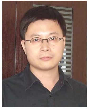
 Motomu Kanai was born in 1967 in Tokyo, Japan, and received his bachelor degree from The University of Tokyo (UTokyo) in 1989 under the direction of late Professor Kenji Koga. In the middle of his PhD course in UTokyo (in 1992), he obtained an assistant professor position in Professor Kiyoshi Tomioka’s group of Osaka University. He obtained his PhD from Osaka University in 1995 before moving to the University of Wisconsin, USA, for postdoctoral studies with Professor Laura L. Kiessling. In 1997 he returned to Japan and joined Professor Masakatsu Shibasaki’s group in UTokyo as an assistant professor, being a lecturer (2000~2003) and an associate professor (2003~2010). He is currently a professor at UTokyo and is a principle investigator of the ERATO Kanai Life Science Project (2011~2017). He has received The Pharmaceutical Society of Japan Award for Young Scientists (2001), Thieme Journals Award (2003), Merck-Banyu Lectureship Award (MBLA: 2005), Asian Core Program Lectureship Award (2008 and 2010), and Thomson-Reuters The 4th Research Front Award (2016). His research interests focus on the design and synthesis of functional (especially, biologically active) molecules.


