

Nanoscale Horizons is a leading journal for the publication of exceptionally high-quality, innovative nanoscience and nanotechnology. To celebrate the excellent articles that are published in the journal, we asked some of our authors to discuss their research in more detail.
In this post, we hear from Yu Qiu, Haijiao Xu and Hongda Wang as they discuss their recent article, ‘Actin-dependent regulation of RSV F-mediated cell–cell fusion revealed by visualizing its spatiotemporal dynamics‘.
Insights from the authors
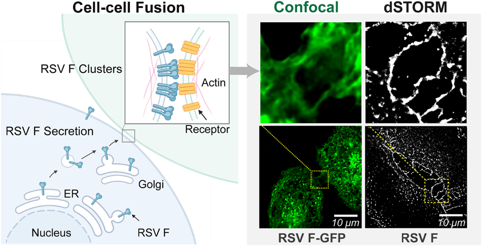
This study integrates live‑cell and super‑resolution microscopy to directly visualize the spatiotemporal dynamics of respiratory syncytial virus fusion (F) protein from its secretion to membrane assembly. It reveals that RSV F traffics efficiently to the plasma membrane and induces F‑actin‑enriched membrane protrusions that promote cell‑cell fusion, a process strictly dependent on branched actin remodeling. Beyond mechanical support, super‑resolution imaging uncovers that actin orchestrates the nanoscale reorganization of RSV F from dispersed clusters into near‑continuous ribbon‑like nanodomains specifically at cell–cell contact sites, establishing a stable fusion platform. This work thus demonstrates a dual role of actin—both in generating fusogenic protrusions and in spatially organizing the viral fusogen. Collectively, these findings define a key mechanism in RSV pathogenesis, reveal a nanoscale organizational target for antiviral intervention, and underscore the power of advanced nano-scale to unravel complex host‑pathogen interactions at the molecular level.
Highlights of the work
- We visualized, for the first time, the entire secretory trafficking of RSV F in living cells, showing its independent transport through the ER‑Golgi pathway to the plasma membrane without other viral proteins.
- Time‑lapse imaging captured the stepwise fusion pore formation, from filopodial bridges to pore nucleation and expansion, culminating in multinucleated syncytia.
- Pharmacological perturbations revealed that Arp2/3‑dependent branched actin, but not formin‑mediated parallel actin, is essential for RSV F‑induced syncytium formation, whereas global actin polymerization is indispensable.
- dSTORM super‑resolution imaging quantified the nanoscale clustering of RSV F on the plasma membrane and showed that actin disruption fragments these clusters, reducing localization density, cluster number and area.
- At cell–cell contacts, RSV F reorganizes into near‑continuous ribbon‑like nanodomains—a structure invisible to conventional microscopy—that are disrupted upon actin depolymerization, highlighting actin’s role in building a mature fusion platform.
Future perspectives
Future research will focus on the dynamic interplay between RSV F protein and the actin cytoskeleton, especially the physiological spatiotemporal evolution of nanoscale fusion platforms. While actin’s dual functions in force generation and spatial scaffolding have been established, the stability and dynamic remodeling of F protein nanoclusters during fusion remain poorly characterized. Transient rearrangement of viral fusogens at cell–cell contacts profoundly modulates fusion efficiency and is undetectable by static super-resolution imaging alone. Integrating live-cell dynamic monitoring with nanoscale static characterization is therefore a core research direction. Advanced live-cell super-resolution microscopy enables real-time tracking of actin remodeling and F protein assembly, facilitating precise kinetic analysis and the rational design of host-directed antiviral strategies. This nanoscale mechanistic insight offers a basis for developing broad-spectrum therapies targeting conserved host–virus interfaces to limit drug resistance, and provides a universal framework for studying cytoskeleton-dependent viral infections.
Meet the authors
 |
Yu Qiu is currently a postgraduate student at the University of Science and Technology of China, under the supervision of Researcher Hongda Wang. Her research focuses on the dynamic transport mechanisms of cell membrane systems.
|
 |
Haijiao Xu is an Associate Professor at Changchun Institute of Applied Chemistry, Chinese Academy of Sciences. Haijiao Xu received her Ph.D. degree in Analytical Chemistry from the same institute in 2021. Her research has long been dedicated to the field of single molecule biophysics, with a particular focus on the mechanistic relationships between membrane protein distribution patterns and dynamic vesicular trafficking.
|
 |
Hongda Wang is a Professor at Changchun Institute of Applied Chemistry, Chinese Academy of Sciences, and University of Science and Technology of China. His research revolves around single-molecule biophysics and biomembrane structural biology. He has pioneered two original membrane architecture models: the semi-mosaic model for erythrocyte membranes, as well as the protein layer-lipid-protein island model applicable to nucleated tissue cell membranes. Meanwhile, he has autonomously engineered topographic super-resolution fluorescence microscopes and high-precision motorized fluorescence microscopes optimized for biophysical imaging applications.
|
Actin-dependent regulation of RSV F-mediated cell–cell fusion revealed by visualizing its spatiotemporal dynamics
Yu Qiu; Xin Ji; Jinrui Zhang; Jing Gao; Yan Shi; Yangang Pan; Guanghua Che; Haijiao Xu; Hongda Wang
Nanoscale Horiz. (2026), https://doi.org/10.1039/d6nh00043f
 |
Nanoscale Horizons is a leading journal for the publication of exceptionally high-quality, innovative nanoscience and nanotechnology. The journal places an emphasis on original research that demonstrates a new concept or a new way of thinking (a conceptual advance), rather than primarily reporting technological improvements. However, outstanding articles featuring truly breakthrough developments such as record performance alone may also be published in the journal. |