


Classical immunology has centered on Pathogen‑Associated Molecular Patterns (PAMPs) — conserved chemical motifs recognized by pattern recognition receptors (PRRs) of the innate immune system (e.g., lipopolysaccharide, flagellin) that initiate immune responses.1-3 By contrast, recent theoretical concept suggests that higher‑order spatial geometry, conserved across viral and microbial structures, may represent another dimension of patterning, termed Pathogen‑Associated Geometric Patterns (PAGPs)4, that may influence how the immune system perceives and responds to pathogens beyond chemical identity. PAGPs has been proposed to describe evolutionarily conserved geometric signatures in pathogen architectures, for example, the polygonal arrangements seen in viral capsids. These geometric patterns are not random but represent highly conserved geometric configurations transcending single molecular motifs and reflect higher‑order spatial information that may be conserved across pathogen families, shaped by evolutionary pressures.
Viruses use icosahedral symmetry for capsid formation, which balances genome protection with mechanical stability. This symmetry often results in convergent evolution, where different virus families independently develop similar structural patterns due to similar selective pressures. This evolutionarily conserved architecture has led to the identification of PAGPs as key features recognized by the immune system.5 While PAGPs offer significant promise, the field is still in its early stages. One critical gap in current research is the mechanistic understanding of how immune receptors interact with geometric patterns at the molecular level. While it is clear that geometry plays a critical role, the spatial tolerance window—the exact spacing and orientation required for optimal immune recognition—remains underexplored.
The recent work of Li et al. provides a critical experimental entry point for understanding how the precise spatial organization of ligands governs receptor signaling, bringing us closer to explaining how geometry can transduce biological information at the immune synapse. Using DNA origami nanoplatforms, Li and colleagues achieved nanometer-level control over the presentation of physiologically relevant pMHC-I ligands to CD8⁺ T cells and demonstrated several core principles: T cell activation was highly sensitive to inter-ligand spacing, increasing by roughly eightfold when spacing was reduced to ~7.5 nm compared with ~22.5 nm; multivalency enhanced activation but exhibited a clear saturation point, with as few as six ligands sufficient to elicit robust responses and only marginal gains beyond that threshold; geometric patterning emerged as a decisive determinant of potency, with a hexagonal arrangement outperforming triangular, linear, and parallel configurations; and, importantly, ligand rigidity proved essential for efficient triggering, as flexible linkers weakened TCR signaling even when overall geometric patterns were maintained, underscoring that mechanical precision helps preserve and amplify the functional “readout” of spatial information. These results reveal that TCR triggering is not merely a function of ligand density but depends on spatial patterning and mechanical stability, defining a spatial tolerance window for productive receptor–ligand engagement at the immunological synapse.
The findings of Li et al. intersect conceptually with pathogen-associated geometric patterns (PAGPs) by demonstrating that nanoscale spatial patterning and symmetry can modulate receptor engagement and downstream signaling. In particular, the enhanced activation produced by hexagonally arranged pMHC-I clusters supports the broader idea that geometric symmetry can optimize multivalent interactions and stabilize signaling assemblies—an intuition that resonates with how many viruses deploy highly ordered, repetitive surface architectures. In the Caspar–Klug description of viral icosahedral capsids, the surface can be viewed as hexons wrapped onto a sphere that must incorporate exactly pentons at the curvature vertices, thus the most capsomers are typically hexameric.7-8 These parallels suggest that spatial geometry itself may be recognized as a biologically salient pattern, potentially akin to how the immune system processes molecular cues. The immune system can function as a spatial pattern–recognition system: nanoscale ligand spacing, symmetry, and multivalent organization can tune receptor engagement and signaling beyond molecular identity alone, highlighting geometry as a meaningful variable in immune responses.
Studies on viral capsid geometry demonstrate the importance of structural symmetry and repeated geometric motifs in pathogen life cycles and host interactions, showing that geometric defects or variations in capsids can affect accessibility to host factors and immunogenicity.9 These findings collectively support the idea that geometric patterning is not a passive feature but a biologically meaningful dimension influencing molecular engagement and immune outcomes.
However, the mechanisms by which geometric patterning of pathogens (e.g., capsid symmetry) influences innate and adaptive immune responses remain poorly understood. While the Li et al. study quantifies spatial parameters for pMHC‑TCR interactions, it does not directly address how pathogen geometry itself shapes immune detection in vivo. Future work should explore whether immune receptors, including PRRs and antigen receptors, are sensitive to global geometric cues on pathogen surfaces.
In summary, Li et al. provide a compelling demonstration that nanoscale spatial geometry critically governs TCR activation, revealing an underappreciated dimension of immune signaling.6 When viewed through the lens of Pathogen‑Associated Geometric Patterns, these findings suggest that spatial patterning is a biologically meaningful code — one that cells may read in both self–nonself discrimination and engineered immunomodulation. Integrating structural virology, pattern recognition, and immune biophysics, future research at this intersection holds promise for deepening our understanding of host–pathogen interactions and engineering next‑generation immunotherapies and vaccines that harness geometric wisdom from nature.
To find out more, please read:
Pattern and precision: DNA-based mapping of spatial rules for T cell activation
Shujie Li, Kaltrina Paloja and Maartje M. C. Bastings
Nanoscale Horiz. (2025) 10 (11): 3041–3050.
References
[1] C.A. Janeway, Jr., Approaching the asymptote? Evolution and revolution in immunology, Cold Spring Harb Symp Quant Biol, 54 Pt 1 (1989) 1-13.
[2] R. Medzhitov, Approaching the asymptote: 20 years later, Immunity, 30 (2009) 766-775.
[3] R. Medzhitov, Pattern recognition theory and the launch of modern innate immunity, J Immunol, 191 (2013) 4473-4474.
[4] M. Zheng, Pathogen-associated geometric patterns, Colloids Surf B Biointerfaces, 245 (2025) 114239.
[5] M. Zheng, Homologous biological structures: Design by natural selection and principle of Pareto optimality, The Innovation Life, 2 (2024) 100073.
[6] S. Li, K. Paloja, M.M.C. Bastings, Pattern and precision: DNA-based mapping of spatial rules for T cell activation, Nanoscale Horiz., 10 (2025) 3041-3050.
[7] D.L. Caspar, A. Klug, Physical principles in the construction of regular viruses, Cold Spring Harb Symp Quant Biol, 27 (1962) 1-24.
[8] R. Zandi, D. Reguera, R.F. Bruinsma, W.M. Gelbart, J. Rudnick, Origin of icosahedral symmetry in viruses, Proc Natl Acad Sci U S A, 101 (2004) 15556-15560.
[9] J.C. Wang, S. Mukhopadhyay, A. Zlotnick, Geometric Defects and Icosahedral Viruses, Viruses, 10 (2018).
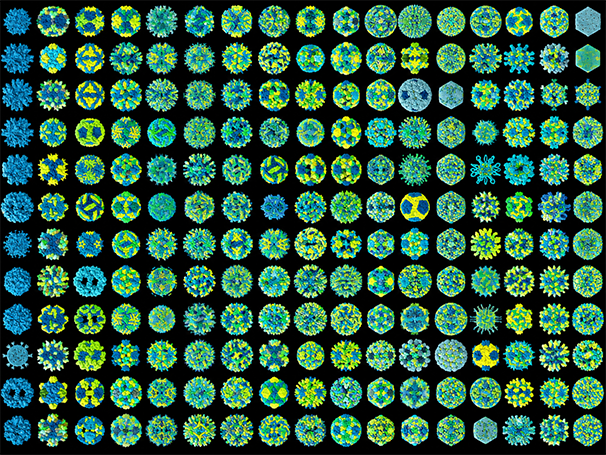
Image from the RSC PDB (RCSB.org) pf PDB ID pdb_000043mi (H.M. Berman, J. Westbrook, Z. Feng, G. Gilliland, T.N. Bhat, H. Weissig, I.N. Shindyalov, P.E. Bourne, The Protein Data Bank (2000) Nucleic Acids Research 28: 235-242 https://doi.org/10.1093/nar/28.1.235). The viral particle image is based on the experimentally determined viral structure obtained from the RCSB Protein Data Bank. AI-assisted image editing software (DeepSeek) was used exclusively to modify the colour palette, including the introduction of yellow and cyan tones, to align with the visual style of Nanoscale Horizons. The underlying viral structure was not AI-generated or otherwise altered.
About the blogger
 |
Dr. Ming Zheng, MD, PhD, is affiliated with the Human Disease Continuum Project Consortium (HDCPC) in Beijing, China. His research interests span immunology, structural virology, and nanobiotechnology, with a particular focus on pathogen-associated geometric patterns (PAGPs)—an emerging framework he introduced to explore how evolutionarily conserved geometric architectures of pathogens influence host–pathogen interactions and immune recognition. His recent work further develops this concept by investigating how particle symmetry, ligand valency, nanoscale spacing, orientation, and mechanical presentation shape molecular recognition and immune signaling. He is particularly interested in bridging structural virology and immune biophysics to understand how the immune system responds to spatial information, with potential applications in biomimetic vaccine design and next-generation immunomodulatory materials. |


