Free to access HOT articles!

These HOT articles have been recommended by our referees and are free to access for 4 weeks*

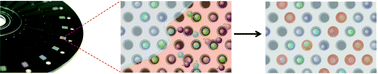
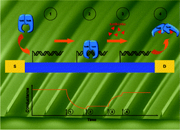
Multiplexed single molecule immunoassays
David M. Rissin, Cheuk W. Kan, Linan Song, Andrew J. Rivnak, Matthew W. Fishburn, Qichao Shao, Tomasz Piech, Evan P. Ferrell, Raymond E. Meyer, Todd G. Campbell, David R. Fournier and David C. Duffy
DOI: 10.1039/C3LC50416F

GA

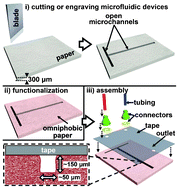
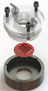
Rapid fabrication of pressure-driven open-channel microfluidic devices in omniphobic RF paper
Ana C. Glavan, Ramses V. Martinez, E. Jane Maxwell, Anand Bala Subramaniam, Rui M. D. Nunes, Siowling Soh and George M. Whitesides
DOI: 10.1039/C3LC50371B

GA

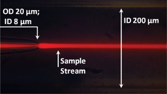
A simple three-dimensional-focusing, continuous-flow mixer for the study of fast protein dynamics
Kelly S. Burke, Dzmitry Parul, Michael J. Reddish and R. Brian Dyer
DOI: 10.1039/C3LC50497B

GA

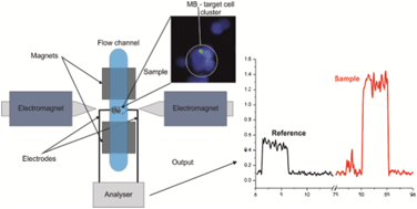
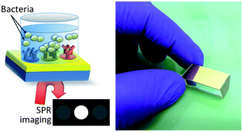
Assessment of pathogenic bacteria using periodic actuation
Sorin David, Cristina Polonschii, Mihaela Gheorghiu, Dumitru Bratu, Alin Dobre and Eugen Gheorghiu
DOI: 10.1039/C3LC50411E

GA

Microfluidic heart on a chip for higher throughput pharmacological studies
Ashutosh Agarwal, Josue Adrian Goss, Alexander Cho, Megan Laura McCain and Kevin Kit Parker
DOI: 10.1039/C3LC50350J

GA

Low-cost fabrication of centimetre-scale periodic arrays of single plasmid DNA molecules
Brett Kirkland, Zhibin Wang, Peipei Zhang, Shin-ichiro Takebayashi, Steven Lenhert, David M. Gilbert and Jingjiao Guan
DOI: 10.1039/C3LC50562F

GA

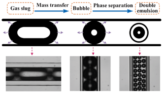
A novel microfluidic technology for the preparation of gas-in-oil-in-water emulsions
Lu Yang, Kai Wang, Sy Mak, Yankai Li and Guangsheng Luo
DOI: 10.1039/C3LC50652E

GA

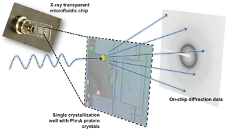
A microfluidic approach for protein structure determination at room temperature via on-chip anomalous diffraction
Sarah L. Perry, Sudipto Guha, Ashtamurthy S. Pawate, Amrit Bhaskarla, Vinayak Agarwal, Satish K. Nair and Paul J. A. Kenis
DOI: 10.1039/C3LC50276G

GA

Steam-on-a-chip for oil recovery: the role of alkaline additives in steam assisted gravity drainage
Thomas W. de Haas, Hossein Fadaei, Uriel Guerrero and David Sinton
DOI: 10.1039/C3LC50612F

GA

Out of the cleanroom, self-assembled magnetic artificial cilia
Ye Wang, Yang Gao, Hans Wyss, Patrick Anderson and Jaap den Toonder
DOI: 10.1039/C3LC50458A

GA

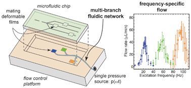
Flow switching in microfluidic networks using passive features and frequency tuning
Rachel R. Collino, Neil Reilly-Shapiro, Bryant Foresman, Kerui Xu, Marcel Utz, James P. Landers and Matthew R. Begley
DOI: 10.1039/C3LC50481F

GA

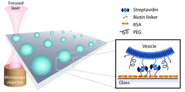
Single vesicle biochips for ultra-miniaturized nanoscale fluidics and single molecule bioscience
Andreas L. Christensen, Christina Lohr, Sune M. Christensen and Dimitrios Stamou
DOI: 10.1039/C3LC50492A

GA

Pinched-flow hydrodynamic stretching of single-cells
Jaideep S. Dudani, Daniel R. Gossett, Henry T. K. Tse and Dino Di Carlo
DOI: 10.1039/C3LC50649E

GA

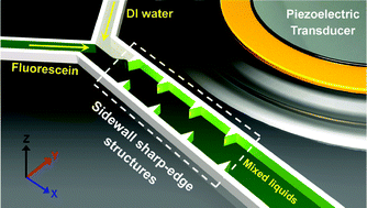
An acoustofluidic micromixer based on oscillating sidewall sharp-edges
Po-Hsun Huang, Yuliang Xie, Daniel Ahmed, Joseph Rufo, Nitesh Nama, Yuchao Chen, Chung Yu Chan and Tony Jun Huang
DOI: 10.1039/C3LC50568E

GA

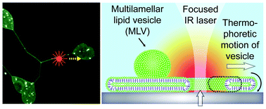
Thermal migration of molecular lipid films as a contactless fabrication strategy for lipid nanotube networks
Irep Gözen, Mehrnaz Shaali, Alar Ainla, Bahanur Örtmen, Inga Põldsalu, Kiryl Kustanovich, Gavin D. M. Jeffries, Zoran Konkoli, Paul Dommersnes and Aldo Jesorka
DOI: 10.1039/C3LC50391G

GA

On-chip microbial culture for the specific detection of very low levels of bacteria
Sihem Bouguelia, Yoann Roupioz, Sami Slimani, Laure Mondani, Maria G. Casabona, Claire Durmort, Thierry Vernet, Roberto Calemczuk and Thierry Livache
DOI: 10.1039/C3LC50473E

GA

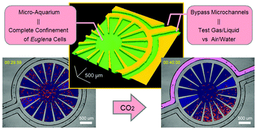
Gas/liquid sensing via chemotaxis of Euglena cells confined in an isolated micro-aquarium
Kazunari Ozasa, Jeesoo Lee, Simon Song, Masahiko Hara and Mizuo Maeda
DOI: 10.1039/C3LC50696G

GA

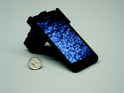
Smart-phone based computational microscopy using multi-frame contact imaging on a fiber-optic array
Isa Navruz, Ahmet F. Coskun, Justin Wong, Saqib Mohammad, Derek Tseng, Richie Nagi, Stephen Phillips and Aydogan Ozcan
DOI: 10.1039/C3LC50589H

GA

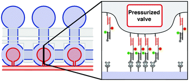
Protein–DNA force assay in a microfluidic format
Marcus Otten, Philip Wolf and Hermann E. Gaub
DOI: 10.1039/C3LC50830G

GA

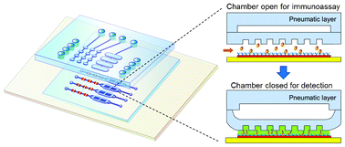
Ultrasensitive microfluidic solid-phase ELISA using an actuatable microwell-patterned PDMS chip
Tanyu Wang, Mohan Zhang, Dakota D. Dreher and Yong Zeng
DOI: 10.1039/C3LC50783A

GA

Detection of real-time dynamics of drug–target interactions by ultralong nanowalls
Andreas Menzel, Raphael J. Gübeli, Firat Güder, Wilfried Weber and Margit Zacharias
DOI: 10.1039/C3LC50694K

GA

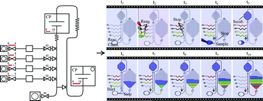
Capillarics: pre-programmed, self-powered microfluidic circuits built from capillary elements
Roozbeh Safavieh and David Juncker
DOI: 10.1039/C3LC50691F

GA

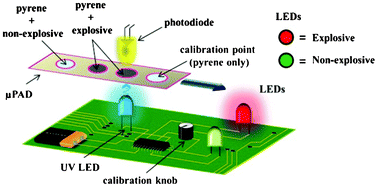
A portable explosive detector based on fluorescence quenching of pyrene deposited on coloured wax-printed μPADs
Regina Verena Taudte, Alison Beavis, Linzi Wilson-Wilde, Claude Roux, Philip Doble and Lucas Blanes
DOI: 10.1039/C3LC50609F

GA

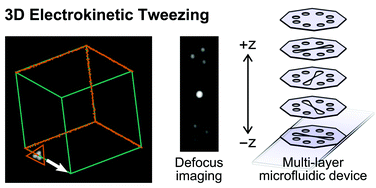
Electrokinetic tweezing: three-dimensional manipulation of microparticles by real-time imaging and flow control
Zachary Cummins, Roland Probst and Benjamin Shapiro
DOI: 10.1039/C3LC50674F

GA

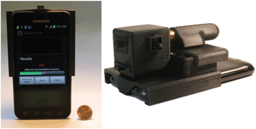
Albumin testing in urine using a smart-phone
Ahmet F. Coskun, Richie Nagi, Kayvon Sadeghi, Stephen Phillips and Aydogan Ozcan
DOI: 10.1039/C3LC50785H

GA

*Free access to individuals is provided through an RSC Publishing personal account. It’s quick, simple and more importantly – free – to register!

Digg This
Reddit This
Stumble Now!
Share on Facebook
Bookmark this on Delicious
Share on LinkedIn
Bookmark this on Technorati
Post on Twitter
Google Buzz (aka. Google Reader)

Personal kidney disease monitoring on your phone

Posted on behalf of Angharad Rosser-James, Publishing Editor | Lab on a Chip

Angharad Rosser-James, Publishing Editor in the Lab on a Chip editorial production team, recently wrote this fantastic article for Chemistry World. It focuses on a recent Lab on a Chip paper, and shows how the miniaturisation field can have a huge impact on our daily lives: 

A smart phone attachment and accompanying app that could be used by people in their own home to monitor the health of their kidneys have been developed by scientists in the US. The lightweight and cost-effective device contains a fluorescent assay which works with the phone’s existing camera to provide results within minutes. 

The lightweight and compact attachment is installed on the existing camera unit of a smart-phone

Millions of people die each year from chronic kidney disease with 11% of US adults thought to have some form of kidney-related problem. Early detection and treatment is the key to prevent or control kidney damage. Routine screening for kidney damage checks albumin levels in urine with high levels of the protein indicating a potential problem. These tests are currently carried out using bench-top urine analysers and require patients to make regular trips to a clinic or hospital. 

The Albumin Tester, a digital fluorescent tube reader accompanied by an android smart phone app devised by Aydogan Ozcan and colleagues at the University of California in Los Angeles could save patients from having to make so many of these trips. Weighing only 148 g, a similar weight to the smart phone itself, the whole device can be attached to the back of a smart phone. Urine is added to fluorescent assays confined within disposable test tubes and the smart phone’s camera collects images of the assays via an external plastic lens. The app converts the fluorescence signals into an albumin concentration value within 1 second. Its detection limit of 5–10 µg ml-1 is more than 3 times lower that the clinically accepted healthy threshold. 

The user-friendly app converts fluorescence signals into albumin concentrations within 1 s and can give daily or weekly reports

Ozcan envisions the device’s application in ‘the early diagnosis of kidney disease or for routine monitoring of high-risk patients, especially those suffering from chronic conditions such as diabetes, hypertension, and/or cardiovascular diseases.’ Govind Kaigala, who develops microsystems for biomolecule analysis at IBM Research in Switzerland agrees and says ‘the albumin tester is a gadget which holds the promise of a simple, rapid and low-cost test for regular use by the patient.’ 

‘This technology has the potential to make widespread impact on health care in developing as well as developed countries,’ says Olav Solgaard, an expert in optical microelectromechanical systems at Stanford University in the US. 

Ozcan anticipates that their next step is to make it possible to measure other kidney disease biomarkers, such as creatinine, using the same smart phone attachment. 

View this article on the Chemistry World website, or access the full paper: A F Coskun et alLab Chip, 2013, DOI: 10.1039/c3lc50785h

Digg This
Reddit This
Stumble Now!
Share on Facebook
Bookmark this on Delicious
Share on LinkedIn
Bookmark this on Technorati
Post on Twitter
Google Buzz (aka. Google Reader)

Making cilia without the bunny suit

Published on behalf of Aleksandra K. Denisin, Lab-on-a-Chip web writer.

Cilia are microscale ‘eyelash-like’ extensions of eukaryotic cells found in epithelial linings throughout the body. In the fallopian tubes, windpipe, and lungs, motile cilia beat rhythmically to move objects within the viscous liquid above. Non-motile cilia in the inner ear transduce mechanical vibrations to electrical signals to ultimately excite auditory nerves.

Previously, these appendages have been built using advanced microfabrication techniques. Now, for the first time, researchers at Eindhoven University of Technology in the Netherlands present a simple bench-top fabrication method for self-assembly of artificial cilia using magnetic beads and latex particles


Jaap den Toonder and his team created the artificial cilia sans cleanroom by coating magnetic beads with latex particles in a fluid cell using a magnetic field to control bead orientation. Latex particles were attracted to the beads by electrostatic forces and the whole structure was bonded together using a heating cycle. The completed artificial cilia were 3 μm in diameter and could be made into lengths of up to 33 μm by optimizing the magnetic field strength and protocol duration. The cilia were actuated by oscillating magnetic fields after fabrication to produce flow velocities of 3 μm/s.

Microfluidic devices operate under low Reynolds numbers where inertia is negligible, presenting a significant challenge to efficient mixing and moving of objects. Cilia and flagella evolved in organisms living in low Reynolds numbers to enable swimming by generating fluid flow using nonreciprocal (nonreversible) motions of beating and twisting.1, 2 The fabrication method presented in this work powerfully enables artificial cilia to be fabricated in situ in assembled platforms and “ship-in-a-bottle” constructed devices, thus facilitating practical applications for these structures in existing microfluidic platforms for bio-inspired fluid manipulation at the microscale.

Out of the cleanroom, self-assembled magnetic artificial cilia, Ye Wang, Yang Gao, Hans Wyss, Patrick Anderson, and Japp den Toonder, Lab Chip, 2013, 13, 3360-3366. DOI: 10.1039/C3LC50458A


References:
1. E. M. Purcell, AIP Conference Proceedings, 1976, 28, 49.
2. S. Khaderi, J. den Toonder and P. Onck, Biomicrofluidics, 2012, 6, 014106.

Digg This
Reddit This
Stumble Now!
Share on Facebook
Bookmark this on Delicious
Share on LinkedIn
Bookmark this on Technorati
Post on Twitter
Google Buzz (aka. Google Reader)

Liquid Chromatography – Mass Spectrometry: Start to Finish on One Microfluidic Chip

Published on behalf of Kathryn Mayer, Lab on a Chip web-writer.

Researchers at Virginia Tech have designed a microfluidic system that integrates optimized fluid handling, liquid chromatography, and a mass spectrometry sample platform – all in one small device.

Matrix-assisted laser desorption ionization (MALDI) mass spectrometry (MS) is a powerful analytical tool which enables the identification and quantification of thousands of proteins and peptides within a single complex sample. Liquid chromatography (LC) can be used to pre-sort sample contents by mass, increasing the sensitivity and selectivity of the MS measurements. A novel system designed by Iulia Lazar and Jarod Kabulski (view the full paper here) allows the entire process of LC-MS to be carried out on a microfluidic chip.

Lazar and Kabulski’s chip has several features that set it apart from previous microfluidic devices incorporating LC and MS.1 It comprises a unique microfluidic system2 driven by electro-osmotic pumps, an LC channel packed with microparticles, and a novel system of transverse microchannels to draw LC fractions from the main separation channel and into a series of reservoirs. The chip can then be directly loaded into the MALDI-MS instrument for analysis. Flow control in the LC channel was carefully optimized so that the solution components would be perfectly distributed along the channel length.

Figure: The LC-MS microfluidic chip. (Figure 2 from the original paper.)

Using the microfluidic LC-MS chip, the researchers were able to obtain results comparable to conventional LC-MS, for both peptide mixtures and cytoplasmic cell extract. Thus, the chips are very promising for practical high-throughput applications such as biomarker screening. The chip format will also enable samples to be collected and prepped by an MS non-expert at a remote location, then transported to a MS lab for analysis.

Learn more from this HOT article from Lab on a Chip!

Microfluidic LC device with orthogonal sample extraction for on-chip MALDI-MS detection
Iulia M. Lazar and Jarod L. Kabulski
DOI: 10.1039/c3lc50190f

  1. D. Gao et al., Lab on a Chip 13, 3309-3322, 2013.
  2. I. Lazar et al., Analytical Chemistry 78 (15), 5513-5524, 2006.
Digg This
Reddit This
Stumble Now!
Share on Facebook
Bookmark this on Delicious
Share on LinkedIn
Bookmark this on Technorati
Post on Twitter
Google Buzz (aka. Google Reader)

Finding a needle in a haystack

Published on behalf of Alphonsus Ng, Lab-on-a-Chip web writer

Many diseases, especially cancers (and recurrences), are not detected until it is too late for effective treatment. Often, this is because available tests lack the sensitivity to find the appropriate protein biomarker in the body.1 Consequently, ultrasensitive tools for measuring proteins are vital for early diagnosis of diseases, and monitoring the effectiveness of surgery or therapy.

For 40 years, scientists and clinicians have been using immunoassays for protein detection. This technique relies on the precise recognition of a target molecule (antigen) by a unique Y-shaped protein (antibody) among thousands of interfering species. However, to achieve ultrasensitive detection, the antigen must be effectively transported to the antibody on the detection surface.2

To tackle this problem, a research team led by Prof. Martin Gijs drew inspiration from the human immune system. In the body, white blood cells are transported to the site of injury with the help of adhesion molecules. In the presence of blood flow, weak adhesion molecules cause the cells to slow down and roll along the vessel wall. This “rolling adhesion” allows the cells to search the wall meticulously for a stop signal. If they come across this signal, the cells will adhere firmly to the wall and squeeze into the site of injury.

Taking cue from this phenomenon, the research team designed a microfluidic immunoassay using two sizes of magnetic beads coated with antibodies. First, ‘large’ (2.8 micrometer) beads are loaded with antigen (from the serum sample) in an on-chip mixing chamber. After unbound components are washed away, the large beads are flown over a surface decorated with a pattern of ‘small’ antibody-coated (1.0 micrometer) beads. In the presence of a magnet, the large beads will roll on the surface and interact closely with the small beads (via magnetic dipolar forces). Beads that are loaded with antigen will adhere firmly to the surface through antibody-antigen complexes, while beads with no antigen are washed away (via flow-induced drag forces).

The antigen concentration in the serum sample can be detected by simply counting the number of large beads in the detection area. This method can rapidly (<20 min) detect down to 200 proteins (Tumor necrosis factor-α) in a 5 microliter of sample (i.e. 60 attomolar), making it one of the fastest and most sensitive immunoassays ever reported. Such salient technique has the potential to improve treatment and outcome for cancer patients worldwide.

The above paper is part of our Lab on a Chip Top 10%, a collection of articles selected by the Editors at Lab on a Chip, from all our high quality papers, to be of exceptional significance for the miniaturisation community. Papers in this category will have received excellent reports during peer review, and demonstrate a breakthrough in device technology, methodology or demonstrate important new results for chemistry, physics, biology or bioengineering enabled by miniaturisation.

The full paper details are here: Attomolar protein detection using a magnetic bead surface coverage assay, H. Cumhur Tekin, Matteo Cornaglia and Martin A. M. Gijs*, Lab Chip, 2013, 13, 1053-1059. DOI: 10.1039/C3LC41285G

1. C. S. Thaxton, R. Elghanian, A. D. Thomas, S. I. Stoeva, J.-S. Lee, N. D. Smith, A. J. Schaeffer, H. Klocker, W. Horninger, G. Bartsch and C. A. Mirkin, Proceedings of the National Academy of Sciences, 2009, 106, 18437-18442.

2. T. M. Squires, R. J. Messinger and S. R. Manalis, Nature Biotechnology, 2008, 26, 417-426.

Digg This
Reddit This
Stumble Now!
Share on Facebook
Bookmark this on Delicious
Share on LinkedIn
Bookmark this on Technorati
Post on Twitter
Google Buzz (aka. Google Reader)

Lab-on-a-Chip: The Most Cited Articles of 2010 and 2011

Lab-on-a-Chip would like to present the most cited articles of 2010 and 2011! We would like to use this opportunity to highlight some of the excellent work that the miniaturisation community is producing right now, and to congratulate our authors on their fantastic achievements.    

As of now, all of the below articles will be free for 4 weeks (until Monday 16th Sept),* so make the most of this opportunity to download the full papers!    


Top 3 Cited Reviews:    

  1. CN Baroud et. al.: Dynamics of microfluidic droplets (DOI: 10.1039/c001191f).

    A critical review on the current understanding of the formation, transport and merging of drops in microfluidics. Baroud and colleagues discuss the physical ingredients that differentiate droplet microfluidics from single-phase microfluidics.


  2. YK Cho et. al.: Centrifugal microfluidics for biomedical applications (DOI: 10.1039/b924109d).

    A critical review on the biomedical applications of centrifugal microfluidics. Cho and colleagues review current sample-to-naswer systems and the challenges that must be faced before the centrifugal platform can be used as a new diagnostic platform.


  3.  GB Lee et. al.: Microfluidic cell culture systems for drug research (DOI: 10.1039/b921695b).

    A tutorial review on microfluidic cell cultures and their use in drug research. The review covers the issues of cell immobilisation, medium pumping and gradient generation, as well as providing examples of practical applications.


Top 10 Cited Research Papers:    

  1. GM Whitesides et. al.: Electrochemical sensing in paper-based microfluidic devices (DOI: 10.1039/b917150a).

    A paper on the fabrication and performance of microfluidic paper-based sensing devices. Whitesides and colleagues demonstrated that their paper-based electrochemical devices are capable of quantifying concentrations of various analytes, including heavy metal ions and glucose.


  2. D Di Carlo et. al.: Sheathless inertial cell ordering for extreme throughput flow cytometry (DOI: 10.1039/b919495a).

    A paper which demonstrates the use of a microfluidic device for flow-cytometry with extreme throughput. Di Carlo and colleagues demonstrated 86-97% cell counting sensitivity and specificity.


  3. A Ozcan et. al.: Compact, light-weight and cost-effective microscope based on lensless incoherent holography for telemedicine applications (DOI: 10.1039/c000453g).

    Ozcan and colleagues demonstrate a lensless on-chip microscope weighing only approx. 46 g and dimensions smaller than 5 cm3. The microscope achieves subcellular resolution and may offer a cost-effective tool in the development of portable medicine.


  4. D Di Carlo et. al.: Deformability-based cell classification and enrichment using inertial microfluidics (DOI: 10.1039/c0lc00595a)


  5. BJ Kirby et. al.: Capture of circulating tumor cells from whole blood of prostate cancer patients using geometrically enhanced differential immunocapture (GEDI) and a prostate-specific antibody (DOI: 10.1039/b924420d)


  6. A Ozcan et. al.: Lensfree microscopy on a cellphone (DOI: 10.1039/c003477k)


  7. CF Carlborg et. al.: A packaged optical slot-waveguide ring resonator sensor array for multiplex label-free assays in labs-on-chips (DOI: 10.1039/b914183a)


  8. T Franke et. al.: Surface acoustic wave actuated cell sorting (SAWACS) (DOI: 10.1039/b915522h)


  9. JL Osborn et. al.: Microfluidics without pumps: reinventing the T-sensor and H-filter in paper networks (DOI: 10.1039/c004821f)


  10. LG Griffith et. al.: Perfused multiwell plate for 3D liver tissue engineering (DOI: 10.1039/b913221j)


*free through an RSC publishing personal account  


Digg This
Reddit This
Stumble Now!
Share on Facebook
Bookmark this on Delicious
Share on LinkedIn
Bookmark this on Technorati
Post on Twitter
Google Buzz (aka. Google Reader)

Look Mum, no pumps!

Posted on behalf of Alexandra K. Denisin, Lab on a Chip web writer

GA

Richard Crooks and colleagues, researchers at the University of Texas, Austin developed a way to locally concentrate and move analytes using internal bipolar electrodes (bypassing the need for an outside driver of fluid flow). Electrodes printed on the bottom of the microfluidic channels create controllable gates which balance the convective and electrokinetic forces acting on charged sample molecules. A single DC power supply and controller box is needed to open/close these gates to deliver analytes to different regions of the chip.­­

Crooks and his group have extensively investigated bipolar electrochemistry theory and in this paper demonstrate the use of bipolar electrodes to separate, enrich, and transport ­­­­­bands of analytes in microfluidic channels. Electric potentials applied across a channel induce an electric field within the buffer whilst conductive substrates present on the floor of the microchannel also adopt a potential between their two poles. H+ ions within the buffer are partially neutralized by electrogenerated OH and thus regions of ion depletion appear. These depletion zones attract charged analytes from the solution to maintain the charge gradient induced by the electric field (the speed of electromigration of analytes to the depletion zone is proportional to the electric field). Bipolar electrodes on the channel bottom contain their own local field and so analytes concentrate in these areas, leading to enrichment near the electrodes.

In this work, Crooks and his team demonstrate separation and enrichment of two common fluorescent dyes:  BODIPY2- and MPTS3-. The two dye bands are then directed to two separate reservoirs. In previous papers, the group focused on optimizing enrichment and achieved enrichment rates of up to 0.57 BODIPY2 1, . The current extension and integration of online separation and enrichment achieves comparable rates of enrichment, 0.11 and 0.31 fold/second for BODIPY and MPTS, respectively, while also enabling control over separating analytes of different electromobility (μep) and transporting these bands to designated areas of the device.

To create the devices presented, the group used conventional photolithography techniques to pattern gold bipolar passive electrodes (BPEs) on glass and bonded PDMS channels on top of the regions. This method can be easily multiplexed as additional BPEs can be activated to guide separated and enriched analytes to different areas of the chip.

 References:

1 R. K. Anand, E. Sheridan, D. Hlushkou, U. Tallarek and R. M. Crooks, Lab on a Chip, 2011, 11, 518-527.

Electrochemically-gated delivery of analyte bands in microfluidic devices using bipolar electrodes
Karen Scida, Eoin Sheridan and Richard M. Crooks, Lab Chip, 2013, 13, 2292-2299.
DOI: 10.1039/c3lc50321f

Digg This
Reddit This
Stumble Now!
Share on Facebook
Bookmark this on Delicious
Share on LinkedIn
Bookmark this on Technorati
Post on Twitter
Google Buzz (aka. Google Reader)

Engineering the ‘microHuman’

Posted on behalf of Andries van der Meer, Lab on a Chip web writer

Research based on the use of organs-on-chips is rapidly expanding and developing. These microengineered devices are microfluidic physiological models of tissues and organs. All sorts of organs-on-chips have been reported, including lung-on-a-chip, heart-on-a-chip and even blood-brain barrier-on-a-chip. See http://dx.doi.org/10.1039/C2LC40089H for a Frontier article by Ingber et. al., which focuses on advances in these microengineered organs.

The models have great potential in the field of pharmacology and toxicology, where they can be used to chart the effects of candidate drugs. But what if we could connect all these organs-on-chips together, and create an actual ‘human-on-a-chip’? Could such a model be used to replace the popular animal models? Could we soon be testing our drug candidates not on mouse or rat, but on the microHuman?

What would a microHuman look like? The answer is tiny. It would be a million times lighter than a regular human, giving it a mass of merely 70 mg. At such an incredible size, would the microHuman be functional, like you or I?

In a Critical Review by John Wikswo and colleagues (http://dx.doi.org/10.1039/C3LC50243K), the scaling requirements of a microHuman are discussed. It is concluded that a simple scaling system is not enough; a microhuman would be completely dysfunctional if we applied basic scaling laws to it. For example, its breathing rate would be approximately 10 breaths per second. In addition, its capillaries would be so small that any naturally-occurring blood cell would be too large to traverse them.

In response to these barriers, Wikswo and colleagues provide detailed discussion of scaling in a number of organ systems in the microHuman. Additionally, they give an overview of structural and functional parameters to guide the scaling of organs-on-chips in a microHuman, which are based upon human and animal data.

Interestingly, whilst the potential of a microengineered human-on-a-chip is huge, Wikswo et. al. point out that, like any model system, the microHuman will never be perfect: “It is important to realize that [these] systems reside in a niche of abstraction that will improve constantly with technology but will never exactly recreate a full human, which represents approx. 109 years of evolutionary engineering”.

Organs-on-chips, and their potential for the development into humans-on-chips, are currently an incredibly hot topic area. If you want to find out more, in addition to Wikswo’s excellent review you can read more about this exciting field in a paper by Shuichi Takayama and colleagues, published recently in the RSC Journal Integrative Biology (http://dx.doi.org/10.1039/C3IB40040A).

Digg This
Reddit This
Stumble Now!
Share on Facebook
Bookmark this on Delicious
Share on LinkedIn
Bookmark this on Technorati
Post on Twitter
Google Buzz (aka. Google Reader)

New YouTube videos

View the new videos on the Lab on a Chip YouTube site using the links below:


Aqueous two-phase microdroplets with reversible phase transitions


Passive Droplet Sorting using Viscoelastic Flow Focusing 


Formation of polymersomes with double bilayers templated by quadruple emulsions


A doubly cross-linked nano-adhesive for the reliable sealing of flexible microfluidic devices


Ultrafast cell switching for recording cell surface transitions: new insights into epidermal growth factor receptor signalling

Flow-switching allows independently programmable, extremely stable, high-throughput diffusion-based gradients

Highly reproducible chronoamperometric analysis in microdroplets

Disaggregation of microparticle clusters by induced magnetic dipole–dipole repulsion near a surface

Exploring a direct injection method for microfluidic generation of polymer microgels

Droplet morphometry and velocimetry (DMV): a video processing software for time-resolved, label-free tracking of droplet parameters

Digg This
Reddit This
Stumble Now!
Share on Facebook
Bookmark this on Delicious
Share on LinkedIn
Bookmark this on Technorati
Post on Twitter
Google Buzz (aka. Google Reader)

Circulating cancer cells spiral towards separation

Repeated biopsies of tumours can be a painful and distressing procedure for cancer patients. A new biochip developed by researchers in Singapore can isolate tumour cells from blood samples, and may one day be an alternative to more invasive methods for tracking later stage cancers. 

Operating principle of circulating tumour cell enrichment by a spiral channel with trapezoid cross-section

Operating principle of circulating tumour cell enrichment by a spiral channel with trapezoid cross-section

Deaths from cancer generally occur after the cancer has spread. Cells detach from the primary tumour and travel through the blood, subsequently forming new tumours. Being able to isolate and characterise these circulating tumour cells (CTCs) can provide information about the original tumour. However, CTCs exist in very low numbers in the blood stream and hence require enrichment and separation before analysis. 

Read the full article in Chemistry World 

Slanted spiral microfluidics for the ultra-fast, label-free isolation of circulating tumor cells
E W Majid et al, Lab Chip, 2013, Accepted manuscript, Paper
DOI: 10.1039/C3LC50617G

Digg This
Reddit This
Stumble Now!
Share on Facebook
Bookmark this on Delicious
Share on LinkedIn
Bookmark this on Technorati
Post on Twitter
Google Buzz (aka. Google Reader)