Author Archive

Microfluidic valves and pumps for all

Over the years, the materials used to make microfluidic devices have dictated the progress of the field. The development of early silicon and glass devices progressed very slowly because the fabrication methods required to make these devices were prohibitively expensive and inaccessible.1 Since the arrival of polydimethylsiloxane (PDMS)-based devices made by elastomeric micromolding or “soft lithography” in 1998,2 the pace of microfluidic technology development has increased dramatically. For example, between 1998 and 2010, the number of microfluidic-related publications increased from hundreds to thousands per year.3 These developments were fueled by the simplicity of PDMS-soft lithography, and more importantly, the ability of PDMS to form pneumatic valves and pumps.4

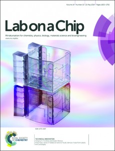
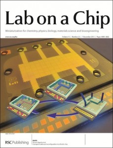
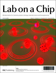
Although soft lithography has become one of the most popular methods for microfluidic fabrication, clean room processes are still needed to make a micromold, and PDMS is not compatible with existing high-throughput manufacturing methods.1 For these reasons and others, researchers are developing alternative methods for device fabrication. For example, Dr. Cooksey at the National Institute of Standards and Technology, Gaithersburg and Prof. Atencia at the University of Maryland developed techniques to create microfluidic devices from cut-off laminates and double-sided tapes.5 Their article, which featured as a cover in Lab on a Chip, showed how these film-based devices can be rapidly fabricated without a cleanroom using in-expensive materials and widely available equipment (e.g. razor cutter, laser cutter).

Like conventional PDMS-devices, these film-based devices can support pneumatic valves and pumps by sandwiching a thin layer of PDMS between two layers of film with cut-out channels. Accurate alignment between these layers is achieved using a self-alignment strategy, in which features of adjacent layers are mirrored across a folding line on a single piece of tape. To demonstrate valve functionality, Cooksey and Atencia created a device that uses 3 valves to control flow from 3 fluidic inputs, and one that uses 8 valves to control a 2-inlet rotary mixer.

One interesting feature of this technology is that very thin devices can be formed (less than 0.5 mm), which enables the fabrication of devices with many layers. For example, using the self-alignment strategy, the researchers fabricated a 6-layer device comprising a valve layer and five fluidic layers that form liquid chambers of varying heights.

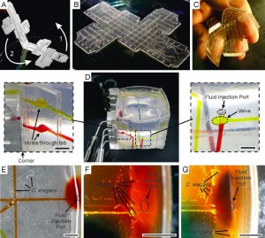
Perhaps the most fascinating trait of this technology is the ability to fold devices into 3D structures with fully functioning valves. As shown in the figure below, the researchers assembled a 3D microfluidic cube that can deliver reagents to specific locations on the cube using the fluid channels routed through the walls. The researchers filled the cube with agar and used it to study the chemotaxis of C. elegans. Within two hours, the worms migrated from the center of the cube toward the face introduced with food, and promptly moved away when the food was switched to a repellent.

In summary, Dr. Cooksey and Prof. Atencia developed a rapid prototyping technique that can create film-based devices with the similar valve functionalities as conventional PDMS-based devices. But because these devices are very thin, more complicated and unique devices structures can be created. This technology has the potential uncover new applications for microfluidics, and make microfluidic technologies more accessible to non-engineers (e.g. biologists and clinicians).


1.            E. K. Sackmann, A. L. Fulton and D. J. Beebe, Nature, 2014, 507, 181-189.

2.            D. C. Duffy, J. C. McDonald, O. J. A. Schueller and G. M. Whitesides, Analytical Chemistry, 1998, 70, 4974-4984.

3.            E. Berthier, E. W. K. Young and D. Beebe, Lab on a Chip, 2012, 12, 1224-1237.

4.            M. A. Unger, H.-P. Chou, T. Thorsen, A. Scherer and S. R. Quake, Science, 2000, 288, 113-116.

5.            Pneumatic valves in folded 2D and 3D fluidic devices made from plastic films and tapes, Gregory A. Cooksey and Javier Atencia, Lab on a Chip, 2-14, 14, 1665-1668

Digg This
Reddit This
Stumble Now!
Share on Facebook
Bookmark this on Delicious
Share on LinkedIn
Bookmark this on Technorati
Post on Twitter
Google Buzz (aka. Google Reader)

Dicty World Race 2014 – which cells will make it to the finish line first?

What?

The Dicty World Race is a cell engineering challenge – competitors must apply their knowledge of chemotaxis to engineer the ultimate chemotaxing cell line. The test is between Dictyostelium, HL60 cells and human neutrophils.

Cells will navigate a complex microfluidic maze to reach a pool of chemoattractant at the finish line. As the race goes on, chemoattractant will diffuse through the microfluidic device, creating a spatial gradient to guide cells along the shortest path.

When?

The date for the 2014 Dicty Race is set for Friday May 16

Why?

To show off your molecular skills!

If you need more encouragement to take part, the winning team will win $5,000 and 15 minutes of fame at the Annual Dicty Conference.

More info?

Visit the website for more details: https://sites.google.com/site/dictyworldrace2014/

Digg This
Reddit This
Stumble Now!
Share on Facebook
Bookmark this on Delicious
Share on LinkedIn
Bookmark this on Technorati
Post on Twitter
Google Buzz (aka. Google Reader)

A microfluidic chip for safekeeping

To accurately diagnose a disease or monitor the effectiveness of a treatment, patient samples (e.g. blood, saliva, or urine) must be analyzed in a central laboratory. After collection, the samples will begin to degrade due to chemical, bacterial, and enzymatic interactions.1 These changes can compromise the integrity of the analyte and introduce errors during analysis. Thus, stabilization techniques are necessary to preserve the diagnostic value of patient samples.

Typically, analyte stabilization is achieved by transporting and storing samples at low temperature using dry ice, refrigerators, freezers, or liquid nitrogen. Unfortunately, low-temperature preservation is costly to implement, requiring substantial infrastructure, specialized equipment, and trained personnel. It becomes particularly expensive for applications involving bio surveillance, long-term sample archiving, and clinical diagnostics in low-resource settings.

To address this challenge, a research team, led by Prof. Ismagilov at the California Institute of Technology, developed a microfluidic device that can preserve biological samples in the dry state. The device relies on pre-loaded desiccants to rapidly (~30 min) dry samples in a stabilization matrix, protecting the analyte from enzymatic degradation and light or heat activated reactions.2 Importantly, the device is simple to use, allowing minimally trained users to collect and preserve samples without worrying about the precision of the input volume.

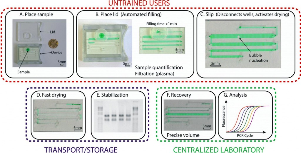
The device, based on SlipChip technology,3 was formed by stacking three layers of subassemblies: the top layer has through-holes for sample loading and recovery, the middle layer has channels for sample storage, and the bottom layer houses the desiccants for drying. With the help of a lubricant between each layer, the middle layer can be horizontally moved (slipped), relative to the other layers, allowing the device to be reconfigured into different states.

The device has three states, each with a specific function: loading, drying, and recovery (see figure 1). In the loading state, the loading inlet is connected to the sample storage channel.  An untrained user will place the sample in the inlet and close the lid to create a tight seal. This generates an air pressure that pushes the sample through the channels. To initiate the preservation process, the user will slip the device into the drying state. Here, the sample is disconnected from the inlet and is placed into vapor contact with the desiccant through a porous membrane. At this stage, the device can be stored or transported without the use of low-temperature preservation equipment. Just before analysis in the laboratory, a trained user will use a special tool to slip the device into the recovery state. Here, the sample is disconnected from the desiccant chamber and is connected to the recovery inlets. Using a pipette and water, the samples can be rehydrated and recovered for quantitative analysis.

As a validation, the device was subjected to an accelerated aging test (50oC for 5-weeks) while preserving samples containing control RNA or HIV-1 RNA. Remarkably, the RNA samples stored in the device were indistinguishable from the ones stored in the freezer at -80oC, as tested by electrophoresis and RT-qPCR. In contrast, significant degradation was observed in samples stored in the liquid state at 50oC. These results suggest that the RNA may be stable at room temperature in the device for at least up to 8 months.

In summary, the research team developed and validated a microfluidic device that can preserve biological samples in the dry state, eliminating the need for low-temperature equipment. The device is compact, easy to use, making it compatible for challenging applications such as clinical diagnostics in remote, resource-limited settings. The reduction of cost in transport, sample collection, and storage can open up many new possibilities in health care, diagnostics, and beyond.

To access the full article, click the following link: A microfluidic device for dry sample preservation in remote setting, Stefano Begolo, Feng Shen and Rustem F. Ismagilov.

1.            G. V. Iyengar, K. S. Subramanian and J. R. W. Woittiez, Element Analysis of Biological Samples: Principles and Practice, CRC Press, Boca Raton, New York, 1998.

2.            E. Wan, M. Akana, J. Pons, J. Chen, S. Musone, P. Y. Kwok and W. Liao, Current Issues in Molecular Biology, 2010, 12, 135-142.

3.            W. Du, L. Li, K. P. Nichols and R. F. Ismagilov, Lab on a Chip, 2009, 9, 2286-2292.

Digg This
Reddit This
Stumble Now!
Share on Facebook
Bookmark this on Delicious
Share on LinkedIn
Bookmark this on Technorati
Post on Twitter
Google Buzz (aka. Google Reader)

Emerging Technologies Competition 2014

The Royal Society of Chemistry is holding a competition to identify the latest technologies in chemical sciences which have significant potential impact on the UK economy.

The winner will receive one to one mentoring from renowned multinational companies and up to a £10,000 cash prize.

If you have an emerging technology that could be the next big chemical science revolution, submit your application by 1 March 2014!

Follow the link to find out more: http://rsc.li/LGCAwM


Digg This
Reddit This
Stumble Now!
Share on Facebook
Bookmark this on Delicious
Share on LinkedIn
Bookmark this on Technorati
Post on Twitter
Google Buzz (aka. Google Reader)

Art In Science Award – the contenders

Every year, Lab on a Chip sponsors the Art in Science award, titled: “Under the Looking Glass: Art from the World of Small Science”. This award, presented at the annual microfluidics conference MicroTas, aims to draw attention to the aesthetic value in scientific illustrations while still conveying scientific merit.

In 2013, the submissions were as fantastic as ever, so we must say a big well done to all of our contributors!

Have a look below at 2013’s winner, and other highly commended pieces…


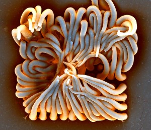
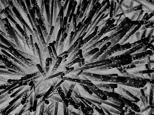
The Winner: “Artificial Life” by Ye Wang, Eindhoven University of Technology.


An SEM image of artificial cilia (microhairs) made with Polydimethylsiloxane and magnetic nanoparticles using a glass mold made by femtolaser modification and hydrofluoric acid etching.


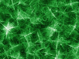
Highly Commended: “Trapping Trapping” by Satoru Ito, Nagoya University.

Fabricated ZnO nanowire (100 nm in diameter and 2-3 micrometer in length) trapping 100 nm beads by electrostatic interaction.


Highly Commended: “Nanoforest” by Sakon Rahong, Osaka University.

A colorised SEM micrograph showing Christmas-tree nano wires prepared by Vapour Liquid Solid (VLS) growth embedded in microchannel for fast DNA separation.


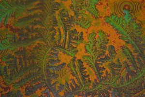
Highly Commended: “Van Gogh’s Wall Paper” by You-Ren Hsu, Institute of NanoEngineering and MicroSystems, NTHU.

Salt crystallization on a gold coated photonic crystal substrate. The salt crystallization changed the index of refraction on the surface, making the color tone.

Digg This
Reddit This
Stumble Now!
Share on Facebook
Bookmark this on Delicious
Share on LinkedIn
Bookmark this on Technorati
Post on Twitter
Google Buzz (aka. Google Reader)

MicroTas abstracts are now online!

Lab on a Chip presents uTAS Abstracts 2003 to Present:

The page link below gives the lab on a chip/microfluidics/uTAS communities FREE ACCESS to both current and archived content submitted to the uTAS conferences in the form of extended abstracts. It is hoped that this service will support workers in finding essential references and hence increase knowledge of past work in the field and assist with current and future research.

This archive includes abstracts presented at uTAS meetings from 2003 to present and essentially provides easy web access to the abstract discs supplied at the uTAS meetings.


CLICK HERE for abstracts! http://rsc.li/1eYWXQs

Digg This
Reddit This
Stumble Now!
Share on Facebook
Bookmark this on Delicious
Share on LinkedIn
Bookmark this on Technorati
Post on Twitter
Google Buzz (aka. Google Reader)

Re-Writable Microfluidics? New Device Points the Way

Researchers from the University of Tokyo and the CEA-Leti research institute in France have designed a platform that allows channels to be written and rewritten at will in a fluid layer atop a grid of electrodes.

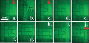
In their recent cover article in Lab on a Chip, Raphael Renaudot and colleagues describe this novel device, which is based on the Electrowetting on Dielectric (EWOD) and Liquid Dielectrophoresis (LDEP) phenomena1. Their goal was to create a flexible, reusable platform that would enable the creation of microfluidic devices without the use of expensive microfabrication techniques. The fluid layer of the device is filled with liquid paraffin, which can easily be solidified and re-melted via thermoelectric cooling and heating (its melting temperature is 35° C.) Water is injected into the paraffin layer, and its flow path is controlled via the underlying electrode layer, which is made up of a grid of 83 electrodes (the small squares seen in the figure). By tuning the interfacial tension between the water and the surface of each electrode via the independently controlled electrode voltages, it is possible to guide a channel (or “finger”) of water along the desired path on the grid (see figure).

After the water is guided into the desired path, the device is cooled, solidifying the paraffin and setting the channels in place. Later, the device can be reheated to erase the existing channels, and a new design can be drawn. The chip can be reused multiple times, reducing waste and making it very useful for prototype testing and low-cost applications. The authors demonstrated two chip designs using the reconfigurable platform: a droplet generator and a device for E. coli confinement within a fluidic cavity.

A water channel is drawn through a paraffin matrix via control of an underlying grid of electrodes (from Figure 2b)

Read this HOT article in Lab on a Chip today!

A Programmable and Reconfigurable Microfluidic Chip, Raphael Renaudot, Vincent Agache, Yves Fouillet, Guillaume Laffite, Emilie Bisceglia, Laurent Jalabert, Momoko Kumemura, Dominique Collard and Hiroyuki Fujita. DOI: 10.1039/c3lc50850a

References:

  1. T. B. Jones and K. L. Wang, Langmuir 2004, 20 (7), 2813–2818.
Digg This
Reddit This
Stumble Now!
Share on Facebook
Bookmark this on Delicious
Share on LinkedIn
Bookmark this on Technorati
Post on Twitter
Google Buzz (aka. Google Reader)

Seasons Greetings from Lab on a Chip!

The holidays are nearly here!!

We know everyone’s been working hard to finish off semesters and write up those papers. Here in Cambridge we’ve been working hard too, planning for the New Year and wrapping up 2013.

To spread the holiday cheer, we’ve chosen three highly accessed papers and made them *FREE TO ACCESS* for the next four weeks. Enjoy!

Merry Christmas from the LOC team!


Paper: Albumin testing in uring using a smartphone, by Aydogan Ozcan, UCLA

Critical Review: Paper-based microfluidic point-of-care diagnostic devices, by Ali Kemal Yetisen, Cambridge

Paper: Cholesterol testing on a smartphone, by David Erickson, Cornell


Access is free through a registered RSC account – click here to register

Digg This
Reddit This
Stumble Now!
Share on Facebook
Bookmark this on Delicious
Share on LinkedIn
Bookmark this on Technorati
Post on Twitter
Google Buzz (aka. Google Reader)

1024 samples analysed on a single chip

Researchers in Switzerland have developed a microfluidic platform able to measure four protein biomarkers in over 1000 blood samples on a single microfluidic chip. With a dramatic reduction in reagent consumption, time and cost, this new high-throughput technology could make early disease diagnosis more affordable.

Many clinical diagnostic tests measure the amount of a specific protein in a patient’s blood. If the levels of this protein are abnormal it is often an indicator for a disease. Currently these tests are very expensive and time-consuming and are only used once a certain condition is suspected, so the patient is already showing symptoms.

Jose Garcia-Cordero and Sebastian Maerkl at the Swiss Federal Institute of Technology in Lausanne hope to change this with their new microfluidic platform capable of measuring protein biomarkers from just 5nL of human blood with comparable results to a conventional enzyme-linked immunosorbent assay (ELISA), which typically uses a 50μL sample volume. The platform uses a microspotting technique to deliver a large number of blood samples to the chip combined with a microfluidic circuit that runs four parallel immunoassays.

The microfluidic device can perform over 4000 disease biomarker assays in a single run

‘The throughput of our device is roughly 10–100 times that of current microfluidic platforms with an estimated reagent cost of US$ 0.1 per chip,’ says Maerkl. ‘By drastically reducing the cost of diagnostic tests, we hope that everyone will be able to measure a number of biomarkers on a continuous basis, allowing people to take either preventive measures or to seek early treatment for diseases such as cancer.’

Michele Zagnoni, an expert in microfluidic techniques for cancer research at the University of Strathclyde in the UK, commends the ‘outstanding increase in analytical throughput’ and adds that ‘the choice of producing a microfluidic device which can be interfaced with robotic dispensers is an appealing one for pharmaceutical companies, offering a technology that can be easily integrated with existing industrial instrumentation and procedures.’

While their current platform is based on fluorescence to quantify biomarker levels, Maerkl hopes to move to an electronic readout to make smaller and cheaper point-of-care instruments.

Read this full Chemistry World story, by Michael Parkin, here.

Digg This
Reddit This
Stumble Now!
Share on Facebook
Bookmark this on Delicious
Share on LinkedIn
Bookmark this on Technorati
Post on Twitter
Google Buzz (aka. Google Reader)

DIY cholesterol monitoring

A team of scientists from the US have developed a simple system that will allow people to test their blood cholesterol levels at home, using a smartphone.

Cholesterol is an important organic molecule that performs a crucial role in modulating the permeability and fluidity of animal cell membranes, but having too much cholesterol in the blood can be a real problem. In particular, high blood cholesterol levels are known to be a risk factor for coronary heart disease. For many people, blood cholesterol levels can be controlled through diet, by eating less saturated fat. A simple system that enables people to routinely monitor their blood cholesterol levels, using a device that many people already own, would undoubtedly save lives.

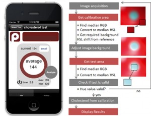
David Erickson and co-workers from Cornell University in New York may have developed just that. Their system consists of a small accessory device that attaches onto a smartphone, an app, and dry reagent test strips for measuring blood cholesterol levels that are already commercially available. A drop of blood is placed onto the test strip and an enzymatic, colorimetric reaction occurs. This strip is then placed into the accessory device and an image of the strip is generated using the camera on the phone. The app then quantifies the colour change and converts this into a blood cholesterol concentration using a calibration curve.

A screenshot of the cholesterol monitoring app (left) and the algorithm to process images of the test strips (right)

The achievement of Erickson and his colleagues should not be understated. Although what they have done may sound simple, developing a smartphone-based system that enables precise and reproducible diagnostic measurements to be taken is actually very difficult. The largest challenge comes from having to account for different lighting conditions and reaction times, differences between the cameras and camera settings in different types of phone, and the potential for misalignment of the test strip. The team overcame the lighting problem by using the accessory device to block out external light so that the test strip would be uniformly illuminated by the flash on the camera. Meanwhile, algorithms in the app account for the other potential variables.

‘This work is another excellent demonstration of cellphone-based sensing,’ says Aydogan Ozcan, a point-of-care diagnostics expert at the University of California in Los Angeles, US. Ozcan himself has developed numerous smartphone-based systems and he sees the value in this work: ‘it will provide numerous opportunities, especially for home monitoring and testing of chronic and elderly patients.’

Erickson and co-workers are now working to commercialise their system, so it may be available for the general public to purchase in the near future.

Click here to watch Erickson demonstrate the test in this great video from the Cornell Chronicle.

Or read this news article by Megan Tyler, and many more, on the Chemistry World website.

Digg This
Reddit This
Stumble Now!
Share on Facebook
Bookmark this on Delicious
Share on LinkedIn
Bookmark this on Technorati
Post on Twitter
Google Buzz (aka. Google Reader)