Top ten most accessed LOC articles in Q2 2013

This month sees the following articles in Lab on a Chip that are in the top ten most accessed April – July:-

Microfluidic approaches for cancer cell detection, characterization, and separation
Jian Chen, Jason Li and Yu Sun
Lab Chip, 2012,12, 1753-1767
DOI: 10.1039/C2LC21273K

Label-free biodetection using a smartphone
Dustin Gallegos, Kenneth D. Long, Hojeong Yu, Peter P. Clark, Yixiao Lin, Sherine George, Pabitra Nath and Brian T. Cunningham
Lab Chip, 2013,13, 2124-2132
DOI: 10.1039/C3LC40991K

Paper-based microfluidic point-of-care diagnostic devices
Ali Kemal Yetisen, Muhammad Safwan Akram and Christopher R. Lowe
Lab Chip, 2013,13, 2210-2251
DOI: 10.1039/C3LC50169H

Enrichment, detection and clinical significance of circulating tumor cells
Sunil K. Arya, Bing Lim and Abdur Rub Abdur Rahman
Lab Chip, 2013,13, 1995-2027
DOI: 10.1039/C3LC00009E

Nanotechnology for membranes, filters and sieves A series of mini-reviews covering new trends in fundamental and applied research, and potential applications of miniaturised technologies
Jan C. T. Eijkel and Albert van den Berg
Lab Chip, 2006,6, 19-23
DOI: 10.1039/B516903H

Microfluidic probes for use in life sciences and medicine
Mohammad A. Qasaimeh, Sébastien G. Ricoult and David Juncker
Lab Chip, 2013,13, 40-50
DOI: 10.1039/C2LC40898H

Droplet microfluidics
Shia-Yen Teh, Robert Lin, Lung-Hsin Hung and Abraham P. Lee
Lab Chip, 2008,8, 198-220
DOI: 10.1039/B715524G

Lab-on-a-chip technologies for single-molecule studies
Yanhui Zhao, Danqi Chen, Hongjun Yue, Jarrod B. French, Joseph Rufo, Stephen J. Benkovic and Tony Jun Huang
Lab Chip, 2013,13, 2183-2198
DOI: 10.1039/C3LC90042H

Rapid electrochemical detection on a mobile phone
Peter B. Lillehoj, Ming-Chun Huang, Newton Truong and Chih-Ming Ho
Lab Chip, 2013,13, 2950-2955
DOI: 10.1039/C3LC50306B

External cavity laser biosensor
Chun Ge, Meng Lu, Sherine George, Timothy A. Flood, Clark Wagner, Jie Zheng, Anusha Pokhriyal, J. Gary Eden, Paul J. Hergenrother and Brian T. Cunningham
Lab Chip, 2013,13, 1247-1256
DOI: 10.1039/C3LC41330F

Why not take a look at the articles today and blog your thoughts and comments below.

Fancy submitting an article to Lab on a Chip? Then why not submit to us today or alternatively email us your suggestions.

Digg This
Reddit This
Stumble Now!
Share on Facebook
Bookmark this on Delicious
Share on LinkedIn
Bookmark this on Technorati
Post on Twitter
Google Buzz (aka. Google Reader)

Cells in a pinch

Published on behalf of Aleksandra K. Denisin, web writer for Lab on a Chip

Figure

Researchers at the University of California, Los Angeles have developed a method to rapidly quantify the mechanical properties of single cells at high resolution using hydrodynamic and inertial fluid focusing. 

Cells undergo structural changes during disease states, particularly in certain cancer cell lines. For example, tumor cells with increased invasive potential exhibit increased deformability, which aids them during their migration into surrounding tissues1. Measuring the mechanical properties of single cells as a biomarker of cell health using conventional Atomic Force Microscopy (AFM) or micropipette aspiration is time-consuming and requires extensive sample prep, limiting the analysis to a few cells. Dino Di Carlo’s group and collaborators developed a way to apply fluids to stretch single cells along two axes using hydrodynamic flow and perpendicular cross-flows without waiting for cells to stop within the channel. This method, termed ‘hydropipetting’, is compatible with inline microfluidic rinsing, capable of processing 65000 cells/sec, and utilizes automatic image processing to rapidly derive the mechanical phenotype of cell populations. 

Hydropipetting starts with inertial fluid focusing of cells to position them precisely in a flow channel by balancing lift forces and secondary flows by fine control over the Reynolds number of the fluid. Cell-free liquid is then excluded and cells are deformed both parallel and perpendicular to the main channel flow. Automated image data analysis then extracts metrics of cell strain, viscosity, diameter and deformability from high speed observation during cell deformation. 

Di Carlo and his group demonstrated the hydropipetting technique on two cancer cell lines (HeLa and Jurkat cells) and observed increases in cell deformability upon drug treatment to increase invasiveness, metastatic potential, and when disrupting structural cellular filaments. 

References:
[1] S. E. Cross, Y. Jin, J. Rao and J. K. Gimzewski, Nature Nanotechnology, 2007, 2, 780-783. 

Pinched-flow hydrodynamic stretching of single-cells
Jaideep S. Dudani, Daniel R. Gossett, Henry T. K. Tse and Dino Di Carlo. Lab Chip, 2013, 13, 3728-3734.
DOI: 10.1039/C3LC50649E

Digg This
Reddit This
Stumble Now!
Share on Facebook
Bookmark this on Delicious
Share on LinkedIn
Bookmark this on Technorati
Post on Twitter
Google Buzz (aka. Google Reader)

Free to access HOT articles!

These HOT articles were recommended by our referees and are free to access for 4 weeks*

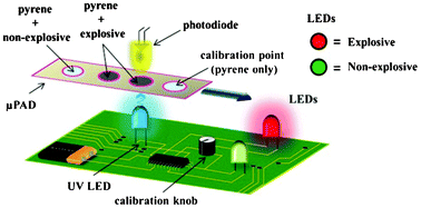
A portable explosive detector based on fluorescence quenching of pyrene deposited on coloured wax-printed μPADs
Regina Verena Taudte, Alison Beavis, Linzi Wilson-Wilde, Claude Roux, Philip Doble and Lucas Blanes  
DOI: 10.1039/C3LC50609F


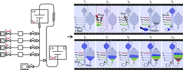
Capillarics: pre-programmed, self-powered microfluidic circuits built from capillary elements
Roozbeh Safavieha and David Juncker  
DOI: 10.1039/C3LC50691F

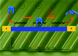
Detection of real-time dynamics of drug–target interactions by ultralong nanowalls
Andreas Menzel, Raphael J. Gübeli, Firat Güder, Wilfried Weber and Margit Zachariasa  
DOI: 10.1039/C3LC50694K

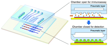
Ultrasensitive microfluidic solid-phase ELISA using an actuatable microwell-patterned PDMS chip
Tanyu Wang, Mohan Zhang, Dakota D. Dreher and Yong Zeng  
DOI: 10.1039/C3LC50783A

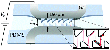
Protein–DNA force assay in a microfluidic format
Marcus Otten, Philip Wolf  and Hermann E. Gaub  
DOI: 10.1039/C3LC50830G

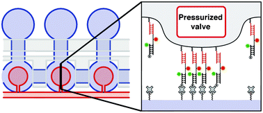
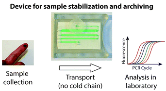
A microfluidic device for dry sample preservation in remote settings
Stefano Begolo, Feng Shen and Rustem F. Ismagilov  
DOI: 10.1039/C3LC50747E

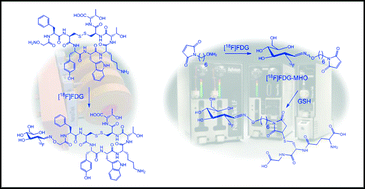
Application of [18F]FDG in radiolabeling reactions using microfluidic technology
Vincent R. Bouveta and Frank Wuest  
DOI: 10.1039/C3LC50797A

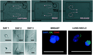
Size-selective collection of circulating tumor cells using Vortex technology
Elodie Sollier, Derek E. Go, James Che, Daniel R. Gossett, Sean O’Byrne, Westbrook M. Weaver, Nicolas Kummer,Matthew Rettig, Jonathan Goldman, Nicholas Nickols, Susan McCloskey, Rajan P. Kulkarni and Dino Di Carlo 
DOI: 10.1039/C3LC50689D

Ratcheted electrophoresis for rapid particle transport
Aaron M. Drews, Hee-Young Lee and Kyle J. M. Bishop  
DOI: 10.1039/C3LC50849H

Parallel measurements of reaction kinetics using ultralow-volumes
Etienne Fradet, Paul Abbyad, Marten H. Vos and Charles N. Baroud  
DOI: 10.1039/C3LC50768H

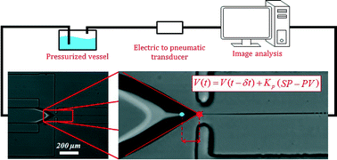
Controlling thread formation during tipstreaming through an active feedback control loop
Todd M. Moyle, Lynn M. Walker and Shelley L. Anna  
DOI: 10.1039/C3LC50946J

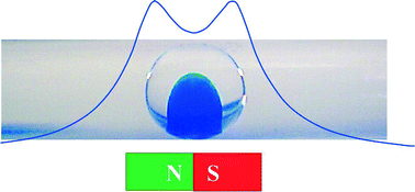
Micro magnetofluidics: droplet manipulation of double emulsions based on paramagnetic ionic liquids
Viktor Misuk, Andreas Mai, Konstantinos Giannopoulos, Falah Alobaid, Bernd Epple and Holger Loewe  
DOI: 10.1039/C3LC50897H

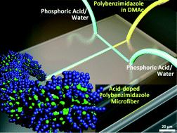
A microfluidic approach to synthesizing high-performance microfibers with tunable anhydrous proton conductivity Mohammad Mahdi Hasani-Sadrabadi,  Jules J. VanDersarl, Erfan Dashtimoghadam, Ghasem Bahlakeh, Fatemeh Sadat Majedi, Nassir Mokarram, Arnaud Bertsch, Karl I. Jacob and Philippe Renaud  
DOI: 10.1039/C3LC50862E

One-step microfluidic generation of pre-hatching embryo-like core–shell microcapsules for miniaturized 3D culture of pluripotent stem cells
Pranay Agarwal, Shuting Zhao, Peter Bielecki, Wei Rao, Jung Kyu Choi, Yi Zhao, Jianhua Yu, Wujie Zhang and Xiaoming He  
DOI: 10.1039/C3LC50678A

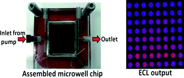
High-throughput metabolic genotoxicity screening with a fluidic microwell chip and electrochemiluminescence
Dhanuka P. Wasalathanthri, Spundana Malla, Itti Bist, Chi K. Tang, Ronaldo C. Fariaa and James F. Rusling  
DOI: 10.1039/C3LC50698C

*Free access to individuals is provided through an RSC Publishing personal account. It’s quick, simple and more importantly – free – to register!

Digg This
Reddit This
Stumble Now!
Share on Facebook
Bookmark this on Delicious
Share on LinkedIn
Bookmark this on Technorati
Post on Twitter
Google Buzz (aka. Google Reader)

Simplifying Microfluidic Flow Control

Published on behalf of Kathryn Mayer, web writer for Lab on a Chip

New research shows that it is possible to carry out flow switching in microfluidic devices via a single active hardware element: a tunable frequency periodic pressure source.

A common hindrance to the design of microfluidic systems is the large amount of unwieldy and expensive hardware (valves, actuators, etc.) required to control fluid flow rates in different parts of the chip. A team of engineers and chemists from UC Santa Barbara, the University of Virginia, and the University of Southampton has devised a method to address this problem. In the new method, a single variable-frequency pressure source communicates with the microfluidic chip via tubes connected to deformable films (“capacitors”) on the chip. The length of each tube is chosen such that the tubes will have well-separated resonance frequencies. Then, the frequency of the actuator is tuned to drive fluid flow in the desired channel only.

As demonstrated in their recent paper, the authors produced three separate devices with clearly differentiated excitation frequencies (see figure below). In addition, they demonstrated a single device with two separate flow channels that could be switched between by modulation of the driving frequency.

Fluid flow in the chip (left, from Figure 1E) is controlled by a periodic pressure source connected to a deformable film by a tube. By using tubes of different lengths, one can selectively drive fluid flow in a channel by tuning the pressure source to that channel’s resonant frequency (right, from Figure 2A).

Fluid flow in the chip (left, from Figure 1E) is controlled by a periodic pressure source connected to a deformable film by a tube. By using tubes of different lengths, one can selectively drive fluid flow in a channel by tuning the pressure source to that channel’s resonant frequency (right, from Figure 2A).

The authors state that by utilizing a large range of excitation frequencies it should be possible to independently control up to 10 flow channels on a single chip using their technique. They also project that it should be possible to control multiple channels simultaneously by employing an excitation signal incorporating multiple frequencies. Thus, this new flow control technique has the potential to be an elegant and low-cost solution for many types of diagnostic applications.

Read this article in Lab on a Chip today:

Flow switching in microfluidic networks using passive features and frequency tuning
Rachel R. Collino, Neil Reilly-Shapiro, Bryant Foresman, Kerui Xu, Marcel Utz, James P. Landers, and Matthew R. Begley
DOI: 10.1039/c3lc50481f

Digg This
Reddit This
Stumble Now!
Share on Facebook
Bookmark this on Delicious
Share on LinkedIn
Bookmark this on Technorati
Post on Twitter
Google Buzz (aka. Google Reader)

Insect-powered tweezers

This recent Lab on a Chip paper was covered in Chemistry World. Read the article, by Michael Parkin, below…

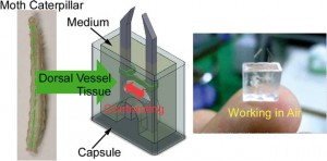
Scientists in Japan have developed the first biohybrid microdevice that can function in air. The microtweezers powered by insect muscle tissue could be used to handle cells and other fragile objects as part of a microelectromechanical system (MEMS).

cyborg jellyfish driven by rat heart cells is part of a recent flurry of research into devices that use mammalian muscle cells to provide motor power. Higher energy efficiency and the ability to self-repair are just some of the benefits of using cells in tiny devices. However, biohybrid devices don’t typically work outside cell culture medium. Now, a team lead by Keisuke Morishima at Osaka University has used insect muscle tissue to fabricate a microtweezer device that can operate in the open air.

Morishima‘s team assembled the microtweezers from polydimethylsiloxane and muscle cells taken from moth larvae and packaged them into a capsule containing a small amount of cell medium. The capsule’s clever design exploits a surface tension effect to stop medium from spilling even if the device is flipped upside down.

‘These results indicate that it will be possible to utilise insect muscle tissue and cells as microactuators both in a wet environment, such as in a microchannel, and on a dry substrate, such as on a silicon MEMS structure,’ says Morishima. When the team layered the medium surface with paraffin to slow evaporation from the opening of the device the microtweezers were functional for up to five days.


Shuichi Takayama a microfluidics and nanotechnology specialist at the University of Michigan in the US says that ‘while a variety of muscle-based actuators have been reported, this device is interesting as it is capable of operating in harsh environments. It is a great idea to combine robust muscle tissue from insects with clever packaging.’

The group now hopes to integrate muscle tissue that functions in both wet and dry environments into devices to expand their work into the nanorobotics field.

This article was taken from Chemistry World. To read this one, and many more, click here!

Digg This
Reddit This
Stumble Now!
Share on Facebook
Bookmark this on Delicious
Share on LinkedIn
Bookmark this on Technorati
Post on Twitter
Google Buzz (aka. Google Reader)

Reprogrammable microfluidic chips

A microfluidic chip with channels that can be programmed then reset and reconfigured has been developed by scientists from France and Japan.

Water is dispensed into chip reservoirs. By selectively switching on electrodes, water is manipulated to carve out the channels

Water is dispensed into chip reservoirs. By selectively switching on electrodes, water is manipulated to carve out the channels

In recent years, scientists from across of the globe have developed a plethora of microfluidic chips to perform a variety of tasks, from PCR to cell sorting. However, a serious drawback of microfluidic technologies is that each application requires a unique arrangement of inlets, outlets and microchannels, so microfluidic chips are usually specific to one particular purpose. This, combined with the time-consuming and costly manufacturing processes required to construct microfluidic devices, makes the idea of a reprogrammable chip very attractive.

Read the full article here at Chemistry World.

Programmable and reconfigurable microfluidic chip
Raphaël Renaudot, et al.
Lab Chip, 2013, Accepted Manuscript
DOI: 10.1039/C3LC50850A, Paper

Digg This
Reddit This
Stumble Now!
Share on Facebook
Bookmark this on Delicious
Share on LinkedIn
Bookmark this on Technorati
Post on Twitter
Google Buzz (aka. Google Reader)

Lab on a Chip Co-hosts EU-Korea Microfluidics Workshop

We are very pleased to announce that Lab on a Chip will once again Co-host the third EU-Korea Workshop on microfluidics, focusing on “Emerging Microfluidic Platform Technologies: From Biosciences to Applications”.

Please come along and see us at the meeting, which will be held in Postech International Centre, Pohang, Korea. The workshop takes place on October 3rd to 5th, 2013.

Meet the Editor and International speakers:

Jean-Louis Viovy, Institute Curie, France
Andreas Manz, KIST, Europe
Dongpyo Kim, Pohang, Koreas
Chris Abell, Cambridge, UK
Noo Li Jeon, Seoul, Korea
Sabeth Verpoorte, Groningen, Netherlands
Hywel Morgan, Southampton, UK
Petra Dittrich, ETH Zurich, Switzerland
Sanghyun Lee, FEMTOLAB, Korea
Samuel Sanchez, Max-Planck, Germany
Yoon Kyoung Cho, UNIST, Korea
Francois Leblanc, CEO Fluigent

Digg This
Reddit This
Stumble Now!
Share on Facebook
Bookmark this on Delicious
Share on LinkedIn
Bookmark this on Technorati
Post on Twitter
Google Buzz (aka. Google Reader)

Free to access HOT articles!

These HOT articles have been recommended by our referees and are free to access for 4 weeks*

Multiplexed single molecule immunoassays
David M. Rissin, Cheuk W. Kan, Linan Song, Andrew J. Rivnak, Matthew W. Fishburn, Qichao Shao, Tomasz Piech, Evan P. Ferrell, Raymond E. Meyer, Todd G. Campbell, David R. Fournier and David C. Duffy
DOI: 10.1039/C3LC50416F

GA

Rapid fabrication of pressure-driven open-channel microfluidic devices in omniphobic RF paper
Ana C. Glavan, Ramses V. Martinez, E. Jane Maxwell, Anand Bala Subramaniam, Rui M. D. Nunes, Siowling Soh and George M. Whitesides
DOI: 10.1039/C3LC50371B

GA

A simple three-dimensional-focusing, continuous-flow mixer for the study of fast protein dynamics
Kelly S. Burke, Dzmitry Parul, Michael J. Reddish and R. Brian Dyer
DOI: 10.1039/C3LC50497B

GA

Assessment of pathogenic bacteria using periodic actuation
Sorin David, Cristina Polonschii, Mihaela Gheorghiu, Dumitru Bratu, Alin Dobre and Eugen Gheorghiu
DOI: 10.1039/C3LC50411E

GA

Microfluidic heart on a chip for higher throughput pharmacological studies
Ashutosh Agarwal, Josue Adrian Goss, Alexander Cho, Megan Laura McCain and Kevin Kit Parker
DOI: 10.1039/C3LC50350J

GA

Low-cost fabrication of centimetre-scale periodic arrays of single plasmid DNA molecules
Brett Kirkland, Zhibin Wang, Peipei Zhang, Shin-ichiro Takebayashi, Steven Lenhert, David M. Gilbert and Jingjiao Guan
DOI: 10.1039/C3LC50562F

GA

A novel microfluidic technology for the preparation of gas-in-oil-in-water emulsions
Lu Yang, Kai Wang, Sy Mak, Yankai Li and Guangsheng Luo
DOI: 10.1039/C3LC50652E

GA

A microfluidic approach for protein structure determination at room temperature via on-chip anomalous diffraction
Sarah L. Perry, Sudipto Guha, Ashtamurthy S. Pawate, Amrit Bhaskarla, Vinayak Agarwal, Satish K. Nair and Paul J. A. Kenis
DOI: 10.1039/C3LC50276G

GA

Steam-on-a-chip for oil recovery: the role of alkaline additives in steam assisted gravity drainage
Thomas W. de Haas, Hossein Fadaei, Uriel Guerrero and David Sinton
DOI: 10.1039/C3LC50612F

GA

Out of the cleanroom, self-assembled magnetic artificial cilia
Ye Wang, Yang Gao, Hans Wyss, Patrick Anderson and Jaap den Toonder
DOI: 10.1039/C3LC50458A

GA

Flow switching in microfluidic networks using passive features and frequency tuning
Rachel R. Collino, Neil Reilly-Shapiro, Bryant Foresman, Kerui Xu, Marcel Utz, James P. Landers and Matthew R. Begley
DOI: 10.1039/C3LC50481F

GA

Single vesicle biochips for ultra-miniaturized nanoscale fluidics and single molecule bioscience
Andreas L. Christensen, Christina Lohr, Sune M. Christensen and Dimitrios Stamou
DOI: 10.1039/C3LC50492A

GA

Pinched-flow hydrodynamic stretching of single-cells
Jaideep S. Dudani, Daniel R. Gossett, Henry T. K. Tse and Dino Di Carlo
DOI: 10.1039/C3LC50649E

GA

An acoustofluidic micromixer based on oscillating sidewall sharp-edges
Po-Hsun Huang, Yuliang Xie, Daniel Ahmed, Joseph Rufo, Nitesh Nama, Yuchao Chen, Chung Yu Chan and Tony Jun Huang
DOI: 10.1039/C3LC50568E

GA

Thermal migration of molecular lipid films as a contactless fabrication strategy for lipid nanotube networks
Irep Gözen, Mehrnaz Shaali, Alar Ainla, Bahanur Örtmen, Inga Põldsalu, Kiryl Kustanovich, Gavin D. M. Jeffries, Zoran Konkoli, Paul Dommersnes and Aldo Jesorka
DOI: 10.1039/C3LC50391G

GA

On-chip microbial culture for the specific detection of very low levels of bacteria
Sihem Bouguelia, Yoann Roupioz, Sami Slimani, Laure Mondani, Maria G. Casabona, Claire Durmort, Thierry Vernet, Roberto Calemczuk and Thierry Livache
DOI: 10.1039/C3LC50473E

GA

Gas/liquid sensing via chemotaxis of Euglena cells confined in an isolated micro-aquarium
Kazunari Ozasa, Jeesoo Lee, Simon Song, Masahiko Hara and Mizuo Maeda
DOI: 10.1039/C3LC50696G

GA

Smart-phone based computational microscopy using multi-frame contact imaging on a fiber-optic array
Isa Navruz, Ahmet F. Coskun, Justin Wong, Saqib Mohammad, Derek Tseng, Richie Nagi, Stephen Phillips and Aydogan Ozcan
DOI: 10.1039/C3LC50589H

GA

Protein–DNA force assay in a microfluidic format
Marcus Otten, Philip Wolf and Hermann E. Gaub
DOI: 10.1039/C3LC50830G

GA

Ultrasensitive microfluidic solid-phase ELISA using an actuatable microwell-patterned PDMS chip
Tanyu Wang, Mohan Zhang, Dakota D. Dreher and Yong Zeng
DOI: 10.1039/C3LC50783A

GA

Detection of real-time dynamics of drug–target interactions by ultralong nanowalls
Andreas Menzel, Raphael J. Gübeli, Firat Güder, Wilfried Weber and Margit Zacharias
DOI: 10.1039/C3LC50694K

GA

Capillarics: pre-programmed, self-powered microfluidic circuits built from capillary elements
Roozbeh Safavieh and David Juncker
DOI: 10.1039/C3LC50691F

GA

A portable explosive detector based on fluorescence quenching of pyrene deposited on coloured wax-printed μPADs
Regina Verena Taudte, Alison Beavis, Linzi Wilson-Wilde, Claude Roux, Philip Doble and Lucas Blanes
DOI: 10.1039/C3LC50609F

GA

Electrokinetic tweezing: three-dimensional manipulation of microparticles by real-time imaging and flow control
Zachary Cummins, Roland Probst and Benjamin Shapiro
DOI: 10.1039/C3LC50674F

GA

Albumin testing in urine using a smart-phone
Ahmet F. Coskun, Richie Nagi, Kayvon Sadeghi, Stephen Phillips and Aydogan Ozcan
DOI: 10.1039/C3LC50785H

GA

*Free access to individuals is provided through an RSC Publishing personal account. It’s quick, simple and more importantly – free – to register!

Digg This
Reddit This
Stumble Now!
Share on Facebook
Bookmark this on Delicious
Share on LinkedIn
Bookmark this on Technorati
Post on Twitter
Google Buzz (aka. Google Reader)

Personal kidney disease monitoring on your phone

Posted on behalf of Angharad Rosser-James, Publishing Editor | Lab on a Chip

Angharad Rosser-James, Publishing Editor in the Lab on a Chip editorial production team, recently wrote this fantastic article for Chemistry World. It focuses on a recent Lab on a Chip paper, and shows how the miniaturisation field can have a huge impact on our daily lives: 

A smart phone attachment and accompanying app that could be used by people in their own home to monitor the health of their kidneys have been developed by scientists in the US. The lightweight and cost-effective device contains a fluorescent assay which works with the phone’s existing camera to provide results within minutes. 

The lightweight and compact attachment is installed on the existing camera unit of a smart-phone

Millions of people die each year from chronic kidney disease with 11% of US adults thought to have some form of kidney-related problem. Early detection and treatment is the key to prevent or control kidney damage. Routine screening for kidney damage checks albumin levels in urine with high levels of the protein indicating a potential problem. These tests are currently carried out using bench-top urine analysers and require patients to make regular trips to a clinic or hospital. 

The Albumin Tester, a digital fluorescent tube reader accompanied by an android smart phone app devised by Aydogan Ozcan and colleagues at the University of California in Los Angeles could save patients from having to make so many of these trips. Weighing only 148 g, a similar weight to the smart phone itself, the whole device can be attached to the back of a smart phone. Urine is added to fluorescent assays confined within disposable test tubes and the smart phone’s camera collects images of the assays via an external plastic lens. The app converts the fluorescence signals into an albumin concentration value within 1 second. Its detection limit of 5–10 µg ml-1 is more than 3 times lower that the clinically accepted healthy threshold. 

The user-friendly app converts fluorescence signals into albumin concentrations within 1 s and can give daily or weekly reports

Ozcan envisions the device’s application in ‘the early diagnosis of kidney disease or for routine monitoring of high-risk patients, especially those suffering from chronic conditions such as diabetes, hypertension, and/or cardiovascular diseases.’ Govind Kaigala, who develops microsystems for biomolecule analysis at IBM Research in Switzerland agrees and says ‘the albumin tester is a gadget which holds the promise of a simple, rapid and low-cost test for regular use by the patient.’ 

‘This technology has the potential to make widespread impact on health care in developing as well as developed countries,’ says Olav Solgaard, an expert in optical microelectromechanical systems at Stanford University in the US. 

Ozcan anticipates that their next step is to make it possible to measure other kidney disease biomarkers, such as creatinine, using the same smart phone attachment. 

View this article on the Chemistry World website, or access the full paper: A F Coskun et alLab Chip, 2013, DOI: 10.1039/c3lc50785h

Digg This
Reddit This
Stumble Now!
Share on Facebook
Bookmark this on Delicious
Share on LinkedIn
Bookmark this on Technorati
Post on Twitter
Google Buzz (aka. Google Reader)

Making cilia without the bunny suit

Published on behalf of Aleksandra K. Denisin, Lab-on-a-Chip web writer.

Cilia are microscale ‘eyelash-like’ extensions of eukaryotic cells found in epithelial linings throughout the body. In the fallopian tubes, windpipe, and lungs, motile cilia beat rhythmically to move objects within the viscous liquid above. Non-motile cilia in the inner ear transduce mechanical vibrations to electrical signals to ultimately excite auditory nerves.

Previously, these appendages have been built using advanced microfabrication techniques. Now, for the first time, researchers at Eindhoven University of Technology in the Netherlands present a simple bench-top fabrication method for self-assembly of artificial cilia using magnetic beads and latex particles


Jaap den Toonder and his team created the artificial cilia sans cleanroom by coating magnetic beads with latex particles in a fluid cell using a magnetic field to control bead orientation. Latex particles were attracted to the beads by electrostatic forces and the whole structure was bonded together using a heating cycle. The completed artificial cilia were 3 μm in diameter and could be made into lengths of up to 33 μm by optimizing the magnetic field strength and protocol duration. The cilia were actuated by oscillating magnetic fields after fabrication to produce flow velocities of 3 μm/s.

Microfluidic devices operate under low Reynolds numbers where inertia is negligible, presenting a significant challenge to efficient mixing and moving of objects. Cilia and flagella evolved in organisms living in low Reynolds numbers to enable swimming by generating fluid flow using nonreciprocal (nonreversible) motions of beating and twisting.1, 2 The fabrication method presented in this work powerfully enables artificial cilia to be fabricated in situ in assembled platforms and “ship-in-a-bottle” constructed devices, thus facilitating practical applications for these structures in existing microfluidic platforms for bio-inspired fluid manipulation at the microscale.

Out of the cleanroom, self-assembled magnetic artificial cilia, Ye Wang, Yang Gao, Hans Wyss, Patrick Anderson, and Japp den Toonder, Lab Chip, 2013, 13, 3360-3366. DOI: 10.1039/C3LC50458A


References:
1. E. M. Purcell, AIP Conference Proceedings, 1976, 28, 49.
2. S. Khaderi, J. den Toonder and P. Onck, Biomicrofluidics, 2012, 6, 014106.

Digg This
Reddit This
Stumble Now!
Share on Facebook
Bookmark this on Delicious
Share on LinkedIn
Bookmark this on Technorati
Post on Twitter
Google Buzz (aka. Google Reader)