DIY cholesterol monitoring

This news article was recently published in Chemistry World, and features a Lab on a Chip paper by David Erickson and colleagues from Cornell University, USA

A team of scientists from the US have developed a simple system that will allow people to test their blood cholesterol levels at home, using a smartphone.

Cholesterol is an important organic molecule that performs a crucial role in modulating the permeability and fluidity of animal cell membranes, but having too much cholesterol in the blood can be a real problem. In particular, high blood cholesterol levels are known to be a risk factor for coronary heart disease. For many people, blood cholesterol levels can be controlled through diet, by eating less saturated fat. A simple system that enables people to routinely monitor their blood cholesterol levels, using a device that many people already own, would undoubtedly save lives.

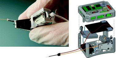
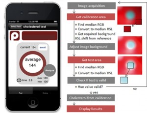
David Erickson and co-workers from Cornell University in New York may have developed just that. Their system consists of a small accessory device that attaches onto a smartphone, an app, and dry reagent test strips for measuring blood cholesterol levels that are already commercially available. A drop of blood is placed onto the test strip and an enzymatic, colorimetric reaction occurs. This strip is then placed into the accessory device and an image of the strip is generated using the camera on the phone. The app then quantifies the colour change and converts this into a blood cholesterol concentration using a calibration curve.

A screenshot of the cholesterol monitoring app (left) and the algorithm to process images of the test strips (right)

The achievement of Erickson and his colleagues should not be understated. Although what they have done may sound simple, developing a smartphone-based system that enables precise and reproducible diagnostic measurements to be taken is actually very difficult. The largest challenge comes from having to account for different lighting conditions and reaction times, differences between the cameras and camera settings in different types of phone, and the potential for misalignment of the test strip. The team overcame the lighting problem by using the accessory device to block out external light so that the test strip would be uniformly illuminated by the flash on the camera. Meanwhile, algorithms in the app account for the other potential variables.

‘This work is another excellent demonstration of cellphone-based sensing,’ says Aydogan Ozcan, a point-of-care diagnostics expert at the University of California in Los Angeles, US. Ozcan himself has developed numerous smartphone-based systems and he sees the value in this work: ‘it will provide numerous opportunities, especially for home monitoring and testing of chronic and elderly patients.’

Erickson and co-workers are now working to commercialise their system, so it may be available for the general public to purchase in the near future.

Click here to watch Erickson demonstrate the test in this great video from the Cornell Chronicle.

Or read this news article by Megan Tyler, and many more, on the Chemistry World website.

Digg This
Reddit This
Stumble Now!
Share on Facebook
Bookmark this on Delicious
Share on LinkedIn
Bookmark this on Technorati
Post on Twitter
Google Buzz (aka. Google Reader)

Altmetrics now featured on Lab on a Chip

 We are pleased to announce the inclusion of Altmetrics on Lab on a Chip.

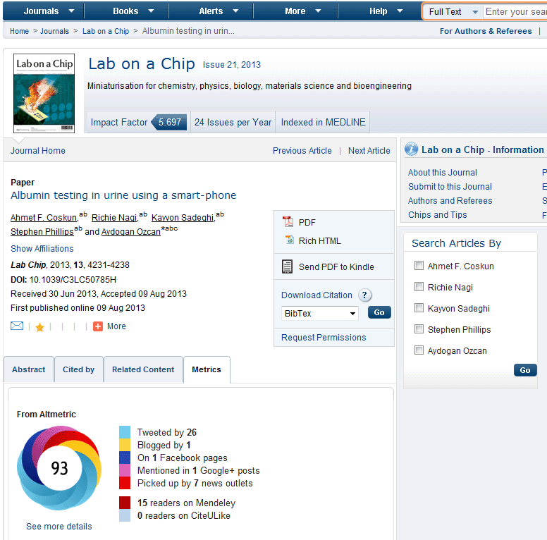
With a constantly changing publishing landscape and changes to the way people use scientific literature, altmetrics is a measure that can monitor the level of conversation and interest in a particular piece of research at the article level. Thus altmetrics provides an additional modern metric for our authors to measure the impact of their work, rather than rely solely on citations and impact factor.

To view altmetrics on Lab on a Chip articles, use the Metrics tab as pictured below on the article landing page.

 Altmetrics for LOC 

A press release from Altmetrics is available on our website.

What do you think? We are interested to hear your feedback on this new development and how you are utilising these new types of metrics. Please leave your comments below.

Digg This
Reddit This
Stumble Now!
Share on Facebook
Bookmark this on Delicious
Share on LinkedIn
Bookmark this on Technorati
Post on Twitter
Google Buzz (aka. Google Reader)

A New Method of Droplet Handling for Ultra-Low Volume Kinetics Studies

Published on behalf of Kathryn Mayer, web writer for Lab on a Chip

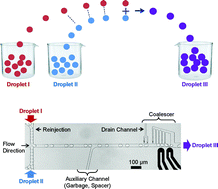
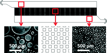
In a recent paper in Lab on a Chip, which featured as the Cover Article of Issue 22, Vol 13, researchers from the Ecole Polytechnique, Palaiseau, France describe a micro-droplet based method for measuring reaction kinetics using tiny (20 nL) sample volumes.


When working with precious reagents such as enzymes, minimizing sample volume is of the utmost importance.  Droplet-based microfluidic methods can greatly reduce sample volumes, but still waste considerable amounts of sample in fluid handling processes (e.g. pushing the sample through a channel).  A team led by Marten Vos and Charles Baroud has utilized a unique “rails and anchors” method1 of forming and controlling the movement of droplets to observe the kinetics of a chemical reaction using only 20 nL of each reagent solution.

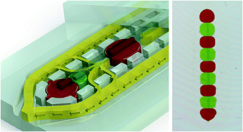
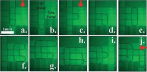
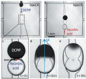
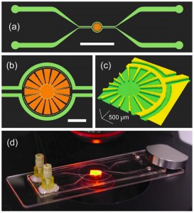
In this scheme, the flow chamber geometry is carefully designed so that when an aqueous solution is injected into the oil-filled chamber, droplets of a reproducible size are released and constrained between the top and bottom surfaces of the chamber.  Then, widening grooves (“rails”) in the bottom of the chamber guide the droplets via surface tension to “anchors” in the center.  (See figure a and b.)  In this way, no additional solution is wasted pushing droplets around.

Droplet-based scheme for observing reaction kinetics (Fig 2 from paper)

The authors used the “rails and anchors” to place two droplets, each containing a different reagent, in proximity, and then used an IR laser pulse to break the surface tension between them (figure c). The interface between the two solutions (in this case, DCPIP and ascorbic acid) can clearly be seen in figure d below.  By tracking the front of the color change that moves across the droplet as the reaction proceeds (figure e), it is possible to calculate the reaction rate (accounting also for the diffusion rates of the reactants).

The researchers also demonstrated the potential of multiplexing their technique by monitoring six different reactions in parallel droplets. They were able to obtain results in good agreement with a commercial stopped-flow spectrometer with greatly reduced cost and time.

Read this HOT article in Lab on a Chip today!

Parallel Measurements of Reaction Kinetics using Ultralow Volumes, Etienne Fradet, Paul Abbyad, Marten H. Vos, and Charles N. Baroud, DOI: 0.1039/c3lc50768h

References:

  1. P. Abbyad et al., Lab on a Chip 2011, 11, 813-821.
Digg This
Reddit This
Stumble Now!
Share on Facebook
Bookmark this on Delicious
Share on LinkedIn
Bookmark this on Technorati
Post on Twitter
Google Buzz (aka. Google Reader)

Free to access HOT articles!

These HOT articles were recommended by our referees and are free to access for 4 weeks*

One-step microfluidic generation of pre-hatching embryo-like core–shell microcapsules for miniaturized 3D culture of pluripotent stem cells
Pranay Agarwal, Shuting Zhao, Peter Bielecki, Wei Rao, Jung Kyu Choi, Yi Zhao, Jianhua Yu, Wujie Zhang and Xiaoming He  
Lab Chip, 2013,13, 4525-4533
DOI: 10.1039/C3LC50678A

A microfluidic approach to synthesizing high-performance microfibers with tunable anhydrous proton conductivity
Mohammad Mahdi Hasani-Sadrabadi, Jules J. VanDersarl, Erfan Dashtimoghadam, Ghasem Bahlakeh, Fatemeh Sadat Majedi, Nassir Mokarram, Arnaud Bertsch, Karl I. Jacob and Philippe Renaud  
Lab Chip, 2013,13, 4549-4553
DOI: 10.1039/C3LC50862E

The microfluidic Kelvin water dropper
Álvaro G. Marín, Wim van Hoeve, Pablo García-Sánchez, Lingling Shui, Yanbo Xie, Marco A. Fontelos,  Jan C. T. Eijkel, Albert van den Berg and Detlef Lohs  
Lab Chip, 2013,13, 4503-4506
DOI: 10.1039/C3LC50832C

A cell rolling cytometer reveals the correlation between mesenchymal stem cell dynamic adhesion and differentiation state
Sungyoung Choi, Oren Levy, Mónica B. Coelho, Joaquim M. S. Cabral, Jeffrey M. Karp and Rohit Karnik  
Lab Chip, 2013, Advance Article
DOI: 10.1039/C3LC50923K

Ultrahigh-throughput sorting of microfluidic drops with flow cytometry
Shaun W. Lim and Adam R. Abate  
Lab Chip, 2013,13, 4563-4572
DOI: 10.1039/C3LC50736J

A programmable and reconfigurable microfluidic chip
Raphael Renaudot, Vincent Agache, Yves Fouillet, Guillaume Laffite, Emilie Bisceglia, Laurent Jalabert, Momoko Kumemura,   Dominique Collard and Hiroyuki Fujita  
Lab Chip, 2013,13, 4517-4524
DOI: 10.1039/C3LC50850A

Digg This
Reddit This
Stumble Now!
Share on Facebook
Bookmark this on Delicious
Share on LinkedIn
Bookmark this on Technorati
Post on Twitter
Google Buzz (aka. Google Reader)

Capillarics – microfluidic circuits

Published on behalf of Andries Van Der Meer, Lab on a Chip web writer

The term ‘microfluidics’ immediately invokes the image of elegant, tiny devices with a small footprint in which fluids are manipulated with ease. It is an image that is most definitely correct. Still, every researcher in the field of microfluidics is aware of ‘the dirty little secret’ of these devices. Namely, that they often require bulky pumps and lots of tubing to be operated.

The requirement for macroscopic equipment to control microfluidic devices is a serious problem. It hinders the development of complex and integrated microfluidic circuits and it prevents the integration of the devices in small portable equipment.

Many researchers in the field are actively developing methods to get rid of all macroscopic equipment to control microfluidic devices. One such method is to build ‘autonomous’ devices, in which the flow is driven only by capillary forces and not by external pumps.

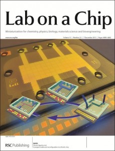
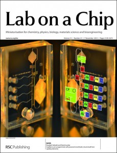
In a recent paper, which featured as the Cover article for Issue 21 of Lab on a Chip, Safavieh and Juncker give a solid overview of the various elements that can be used to autonomously control flow, such as pumps, vents and valves. Moreover, they propose a method called ‘capillarics’ in which autonomous fluidic circuits are designed in a way that is similar to electronic circuits. Finally, they demonstrate the power of their approach by rationally designing a ‘capillaric circuit’ and using it to perform a biochemical assay.

To learn more, read the full HOT article: Capillarics: pre-programmed, self-powered microfluidics circuits built from capillary elements, Roozbeh Safavieh and David Juncker, DOI: 10.1039/C3LC50691F

Digg This
Reddit This
Stumble Now!
Share on Facebook
Bookmark this on Delicious
Share on LinkedIn
Bookmark this on Technorati
Post on Twitter
Google Buzz (aka. Google Reader)

Follow Your Nose: A Micro-aquarium for Probing Bacterial Chemotaxis in Gas Gradients

Published on behalf of Lab on a Chip web writer Alphonsus Ng, post-graduate researcher at the University of Toronto

Many organisms can direct their movements according to their chemical environment. In bacteria, this chemical-directed migration (i.e. chemotaxis) is enabled by receptor-mediated signaling to the bacterial flagellar motors.1 Equipped with this remarkable sensory-motor system, bacteria can measure chemical gradients and swim towards nutritious substances (positive chemotaxis) or flee from cytotoxic chemicals (negative chemotaxis).

Bacterial chemotaxis is critical for various processes including biofilm formation, biomediation, and diseases pathogenesis.2 For example, at the onset of various digestive diseases, bacteria rely on chemotaxis to find a suitable colonization site in the human gastrointestinal tract. Thus, there is great interest in studying the rates and molecular mechanisms in these chemotactic processes.

In the past 10 years, microfluidic gradient-generators have improved the way scientists study bacterial chemotaxis;3 however, these studies have not addressed gas gradients (e.g. O2 and CO2), which are known to be important in bacterial motility.4 Microfluidics devices have enabled the generation of well-controlled chemical gradients and the measurement of bacterial response at high spatiotemporal resolution. Gradients are typically formed by flowing a sample liquid (e.g. aspartate in buffer) and a reference liquid (e.g. buffer) into the device.

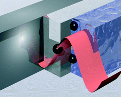
A team of researchers at the RIKEN Advanced Science Institute, Japan and Hanyang University, Korea carried out the world’s first microfluidic study of bacterial chemotaxis in gas gradients.5 The microfluidic device (see picture) comprises an isolated chamber (micro-aquarium) for bacterial culture and two bypass channels for sample and reference inputs. In this design, the sample molecules in the channel permeate through the PDMS walls (150 µm thick) and diffuse into the liquid of the micro-aquarium, facilitating the generation of a stable, shear-free gradient. The bacterial response can be monitored optically and quantified via image processing.

Using this device, the research team studied the chemotactic behaviour of Euglena gracilis microbial cells in the presence of CO2 gradients. These studies revealed that the chemotaxis of Euglena cells is rapid and is dependent on CO2 concentration. In particular, negative chemotaxis was observed at high concentrations (>15%) and positive chemotaxis was observed at low concentrations (<15%)— the maximum positive chemotaxis was observed at ~5% CO2, corresponding to the most favourable condition for photosynthesis. Interestingly, the team observed evidence of chemotactic “adaption” when they consecutively switched the sample and reference inputs.

The microfluidic device is also compatible with small molecule liquid samples such as ethanol, H2O2, and culture media. By continuously perfusing culture medium in the bypass channels, the viability of the Euglena cells in the micro-aquarium can be maintained for more than 2 months.

In summary, the research team created a microfluidic gradient-generator that is versatile and straightforward to use. The device is useful for bacterial chemotaxis studies in chemical gradients formed from gas or liquid samples. Furthermore, the device may potentially be useful for other applications including drug screening, toxicity assays, and environmental monitoring.

  1. H. C. Berg, Annual Review of Biochemistry, 2003, 72, 19-54.
  2. T. Ahmed, T. S. Shimizu and R. Stocker, Integrative Biology, 2010, 2, 604-629.
  3. J. Wu, X. Wu and F. Lin, Lab on a Chip, 2013, 13, 2484-2499.
  4. C. Douarche, A. Buguin, H. Salman and A. Libchaber, Physical Review Letters, 2009, 102, 198101.
  5. K. Ozasa, J. Lee, S. Song, M. Hara and M. Maeda, Gas/liquid sensing via chemotaxis of Euglena cells confined in an isolated micro-aquarium, Lab on a Chip, 2013, 13, 4033-4039
Digg This
Reddit This
Stumble Now!
Share on Facebook
Bookmark this on Delicious
Share on LinkedIn
Bookmark this on Technorati
Post on Twitter
Google Buzz (aka. Google Reader)

Top ten most accessed LOC articles in Q3 2013

This month sees the following articles in Lab on a Chip that are in the top ten most accessed July – September:-

Detection of waterborne parasites using field-portable and cost-effective lensfree microscopy
Onur Mudanyali, Cetin Oztoprak, Derek Tseng, Anthony Erlinger and Aydogan Ozcan
Lab Chip, 2010,10, 2419-2423
DOI: 10.1039/C004829A

Electrowetting-based actuation of droplets for integrated microfluidics
M. G. Pollack, A. D. Shenderov and R. B. Fair
Lab Chip, 2002,2, 96-101
DOI: 10.1039/B110474H

High-purity and label-free isolation of circulating tumor cells (CTCs) in a microfluidic platform by using optically-induced-dielectrophoretic (ODEP) force
Song-Bin Huang, Min-Hsien Wu, Yen-Heng Lin, Chia-Hsun Hsieh, Chih-Liang Yang, Hung-Chih Lin, Ching-Ping Tseng and Gwo-Bin Lee
Lab Chip, 2013,13, 1371-1383
DOI: 10.1039/C3LC41256C

Paper-based microfluidic point-of-care diagnostic devices
Ali Kemal Yetisen, Muhammad Safwan Akram and Christopher R. Lowe
Lab Chip, 2013,13, 2210-2251
DOI: 10.1039/C3LC50169H

Recent advances in microfluidics combined with mass spectrometry: technologies and applications
Dan Gao, Hongxia Liu, Yuyang Jiang and Jin-Ming Lin
Lab Chip, 2013,13, 3309-3322
DOI: 10.1039/c3lc50449b

Recent advances in particle and droplet manipulation for lab-on-a-chip devices based on surface acoustic waves
Zhuochen Wang and Jiang Zhe
Lab Chip, 2011,11, 1280-1285
DOI: 10.1039/C0lC00527D

Droplet microfluidics
Shia-Yen Teh, Robert Lin, Lung-Hsin Hung and Abraham P. Lee
Lab Chip, 2008,8, 198-220
DOI: 10.1039/B715524G

Probing cell–cell communication with microfluidic devices
Feng Guo, Jarrod B. French, Peng Li, Hong Zhao, Chung Yu Chan, James R. Fick, Stephen J. Benkovic and Tony Jun Huang
Lab Chip, 2013,13, 3152-3162
DOI: 10.1039/C3LC90067C

Albumin testing in urine using a smart-phone
Ahmet F. Coskun, Richie Nagi, Kayvon Sadeghi, Stephen Phillips and Aydogan Ozcan
Lab Chip, 2013,13, 4231-4238
DOI: 10.1039/C3LC50785H

Clear castable polyurethane elastomer for fabrication of microfluidic devices
Karel Domansky, Daniel C. Leslie, James McKinney, Jacob P. Fraser, Josiah D. Sliz, Tiama Hamkins-Indik, Geraldine A. Hamilton, Anthony Bahinski and Donald E. Ingber
Lab Chip, 2013,13, 3956-3964
DOI: 10.1039/C3LC50558H

Why not take a look at the articles today and blog your thoughts and comments below.

Fancy submitting an article to Lab on a Chip? Then why not submit to us today or alternatively email us your suggestions.

Digg This
Reddit This
Stumble Now!
Share on Facebook
Bookmark this on Delicious
Share on LinkedIn
Bookmark this on Technorati
Post on Twitter
Google Buzz (aka. Google Reader)

High-throughput chip for drug screening in 3D

Another Lab on a Chip paper makes it into Chemistry World! Read the article, by Megan Tyler, below…

A simple micro-array chip developed by scientists in China could sharpen the search for new drugs by enabling the high-throughput screening of drug candidates against cells cultured in three dimensions.

Developing new drugs for the treatment of complex diseases such as cancer is an expensive and time consuming process. Often, the first step in designing a new drug is to use high-throughput screening of large chemical libraries to identify compounds that have the desired medicinal effect.

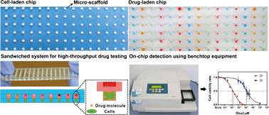
Most cell-based drug screening technologies currently rely on cell cultures grown in two dimensions (i.e. as a flat layer of cells) and require sophisticated robotic systems to seed the cells and administer the drugs. However, Yanan Du and colleagues from Tsinghua University may have revolutionised the process by developing a micro-array chip that can be used to culture three dimensional balls of cells, called spheroids, and administer candidate drugs on a high-throughput scale.

Du’s chip consists of two plates containing arrays of micro-wells. The micro-wells of the first plate contain porous gelatin sponges. Once cells and culture medium are added, these sponges act as a rudimentary extracellular matrix enabling the cells to form spheroids. Meanwhile, the micro-wells of the second plate are filled with potential drug candidates. Pushing the two plates together brings the drug into contact with the spheroids so the effect of the drug on the cells can be observed.

The technique should be accessible to any laboratory with basic cell culture facilities

There are two key advantages to this system. Firstly, cells that are cultured in 3D more closely mimic the tissues in the body than cells grown in 2D. Consequently, the effect that the drug has on these cells is likely to be more representative of the effect that the drug would actually have in the body, making the results of the screening process more reliable. Cells cultured in 3D are also more likely to develop drug resistance so these compounds can be weeded out at an earlier stage in the drug development process. The second advantage is that the technique doesn’t require any expensive or sophisticated equipment, and so should be accessible to any laboratory with basic cell culture facilities.

‘By using microwells they can scale-down the number of cells and the amount of drug they use whilst increasing throughput by having a device compatible with standard laboratory instrumentation,’ says Tony Cass from Imperial College London, UK, who also develops devices for high throughput analysis.

Mario Cabodi, an expert in microfabrication at Boston University in Massachusetts, US, says the chip potentially has a promising future: ‘3D cell culture systems, like the one described here, might be helpful in bridging the gap between in vitro tests and in vivo results.’

Du’s team are now in the process of standardising the chip and are collaborating with drug development companies to perform cell-based tests of potential anti-cancer drugs.

To see the original Chemistry World article, and many others, click here.

Alternatively, you can download the full paper here.

Digg This
Reddit This
Stumble Now!
Share on Facebook
Bookmark this on Delicious
Share on LinkedIn
Bookmark this on Technorati
Post on Twitter
Google Buzz (aka. Google Reader)

Molecular Force Assay on a Microfluidic Chip

Published on behalf of Kathryn Mayer, web writer for Lab on a Chip

Researchers in Munich have adapted their molecular force assay technique to a microfluidic system, allowing the strength of protein-DNA interactions to be measured in a chip format.

The measurement of intermolecular forces is a challenging and important task. Two well-known methods, atomic force microscopy and optical trapping, are capable of probing these forces1. However, these instrumentation-intensive techniques are not suitable for every application. A team from the Ludwig-Maximilians-Universität, Germany, headed by Hermann Gaub, a leader in the field of force spectroscopy, has now developed a technique, termed molecular force assay (MFA), to probe the strength of DNA hybridization and the effects of DNA-binding proteins2.

Schematic of the molecular force assay (adapted from Figure 1 of paper)

In MFA, a surface is functionalized with a carefully designed DNA construct, as seen in the left panel of the figure. This surface is brought into contact with another surface which is coated with neutravidin (center). The neutravidin binds the biotin at the end of the DNA construct, so when the two surfaces are separated, the DNA construct rips apart (dehybridization, right). The breakage can happen in one of two ways—if the hybridization of the “probe strand” is stronger than that of the “reference strand”, the reference strand will dehybridize, leaving behind a Cy3 tag. If the hybridization of the reference strand is stronger, the probe strand will dehybridize, leaving both a Cy3 tag and a Cy5 tag behind. Via the resulting fluorescence, it is possible to compare the hybridization strength of the probe strand to that of the known reference. DNA binding proteins which modify the hybridization strength of the probe strand can then be introduced.

In the original formulation of MFA, the surfaces were brought into contact using a piezoelectric device; however, in a recent paper in Lab on a Chip, the researchers have translated their assay into a microfluidic format. A microfluidic chip was used in which the lower layer contains sample wells and the upper layer contains control valves. By pressurizing button valves above each well, the top and bottom surfaces can be brought into contact and then separated. The authors demonstrated the effect of EcoRI binding on probe strand hybridization strength as a proof-of-concept, “paving the way for studies of currently unknown protein-DNA interactions, including those of transcription factors.”

Read this HOT article in Lab on a Chip today!

Protein–DNA force assay in a microfluidic format, Marcus Otten, Philip Wolf, and Hermann E. Gaub. DOI: 10.1039/C3LC50830G

References:

1. K. C. Neuman and A. Nagy, Nature Methods, 2008, 5, 491-505.

2. P. M. D. Severin et al., Lab on a Chip, 2011, 11, 856-862.

Digg This
Reddit This
Stumble Now!
Share on Facebook
Bookmark this on Delicious
Share on LinkedIn
Bookmark this on Technorati
Post on Twitter
Google Buzz (aka. Google Reader)

Paper device spots antibiotic-resistant bacteria

This recent Chemistry World article by Harriet Brewerton covers a Lab on a Chip paper. Read the article below…

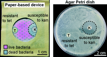
Scientists in Canada have developed a paper-based device that checks if bacteria are resistant to certain antibiotics. The simple system could help users in remote areas pick the most appropriate treatment for bacterial infections.

Testing bacteria to see which antibiotics will be effective against them is vital for properly medicating patients. Methods exist to assess bacteria but the equipment needed is often expensive and requires highly trained operators and laboratory conditions. Now, Ratmir Derda and colleagues at the University of Alberta have designed a portable device made from paper and other low-cost materials. The team sought the help of high school students to make and test the devices to show how easy they are to construct and use.

Derda’s device consists of a paper support with a clear plastic window over an area of nutritious media permeated into a sheet of thick blotting paper. This culture media has a uniform pattern of hydrophobic spots to ensure samples disperse evenly across it. Two zones of antibiotics are added onto the media before the device is sterilised in an autoclave. After autoclaving, a cell viability dye is dropped on the culture media and the bacterial sample is then added on top of the dye. Finally, the device is sealed and incubated overnight.

In areas with no bacterial growth the dye remains blue and in areas where bacteria do grown the dye turns pink. The colours are easy to see through the device’s plastic window – there is no need to open the device. A blue area around the antibiotic zones indicates that the bacteria are susceptible to the antibiotics. The hydrophobic spots on the culture media aid quantification of the blue area to give an idea of how sensitive the bacteria are to the antibiotics.

Preparing and using the device is simple. (M: culture media; A: antibiotic zone; C: culture zone)

‘This work shows that living microorganisms can be grown and screened for antibiotic resistance using paper devices that are small and light enough to store in a person’s pocket,’ says Marya Lieberman, an expert in paper-based sensors at the University of Notre Dame in Indiana, US.

The team also found that the device could easily be stored long-term. After assembling to the point that it contained the culture media and antibiotics, it could be left for up to 70 days in a sealed bag. This storage ability along with the device’s cheap components and ease of use make it very promising for use in remote areas.

Visit Chemistry World to read this article and others by clicking here.

Or read the full paper: Antimicrobial susceptibility assays in paper-based portable culture devices, DOI: 10.1039/C3LC50887K

Digg This
Reddit This
Stumble Now!
Share on Facebook
Bookmark this on Delicious
Share on LinkedIn
Bookmark this on Technorati
Post on Twitter
Google Buzz (aka. Google Reader)