Archive for the ‘News’ Category

Targeting organs with therapeutic carbon monoxide

Prolonged CO release for peptide-based gel compared to soluble peptide

Prolonged CO release for peptide-based gel compared to soluble peptide

Scientists in the US have created a gel that can be used to deliver therapeutic carbon monoxide gas to selected organs in the body.  

CO has a role in the body as a biological signalling molecule (as a neurotransmitter and a blood vessel relaxant, for example) and its delivery to tissues for therapeutic use for conditions such as cardiovascular disease and organ transplantation is done by inhalation therapy. This technique is risky, though, as overexposure to CO in this way can be poisonous, and the CO cannot be targeted to any organs other than the lungs.

More recently, small molecule CO-releasing molecules (CORMs) have been developed as an injectable targeted delivery method. However, the molecules – commonly used metal carbonyls, and the more recent polymeric micelles – have short half-lives and are not retained in tissues, which limits their use.

Samuel Stupp from Northwestern University, and colleagues, who previously developed self-assembling peptide-based materials to deliver another biological signalling molecule – NO – have now turned their attention to the problem of delivering CO. His team combined a peptide amphiphile (PA) designed to self assemble into a fibrous gel with a ruthenium carbonyl complex similar to a known CORM. They reacted the resulting compound with sodium methoxide to generate the CO-releasing PA. The PA self-assembled into nanofibres 8.2nm in diameter.

The CO release performance of the soluble PA gave a similar half-life to known CORMs.  The PA was, however, designed to form a gel, which was achieved by adding CaCl2 to the solution. ‘Gel formation slowed down CO release dramatically, from a half-life of 2.1 min for the soluble peptide to 17.8 min after gelation,’ says Stupp. This prolonged release could significantly improve the utility of CO therapy.

‘This work will add to the regenerative medicine toolbox by enabling researchers to modulate biological signalling through the delivery of a very simple diatomic gas,’ says Stupp.

Bing Xu, an expert in bionanomaterials for drug delivery from Brandeis University, US, says that the system’s future development could lead to a material that significantly outperforms current CORMs. ‘The demonstration of the delivery of therapeutic CO expands the horizon of PA applications,’ he adds.

A peptide-based material for therapeutic carbon monoxide delivery
John B. Matson, Matthew J. Webber, Vibha K. Tamboli, Benjamin Weber and Samuel I. Stupp
Soft Matter, 2012, Advance Article
DOI: 10.1039/C2SM25785H

Read the original Chemistry World article here.

Digg This
Reddit This
Stumble Now!
Share on Facebook
Bookmark this on Delicious
Share on LinkedIn
Bookmark this on Technorati
Post on Twitter
Google Buzz (aka. Google Reader)

Active soft matter

What does softness do for life? According to Zhigang Suo, Havard University, for life the important feature of soft materials is that they are easily deformable. In the human body the deformation of soft materials enables the heart to beat, the vocal folds to produce sound and the eye to focus. In all these examples, a stimulus is applied to the soft material and a large reversible deformation occurs. This deformation in turns provides a function.

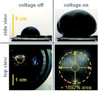
Soft active materials are not only important in life, but also have technological relevance for example in adaptive optics, self-regulated fluidics and soft robotics. Suo is interested in how mechanics, chemistry and electrostatics all work together to generate large deformations in soft materials. In soft dielectrics actuation can be readily observed by applying a voltage across the membrane, causing the thickness to reduce and the area to expand. In polymers, strains of up to 30% are easily achieved using this method.

To get higher deformations two limitations need to be overcome; electrical breakdown and electromechanical instability. An electromechanical instability is observed in compliant dielectrics such as elastomers, when the applied voltage causes the material to thin down excessively, amplifying the electric field. This is known as a snap-though instability and often occurs prior to electrical breakdown. Suo has shown, however, that if this electromechanical instability can be overcome, and the elastomer reaches a stable state without breakdown occurring, giant voltage-induced deformations of over 1000% are achievable.

Suo’s theory shows that the instability can be eliminated when pre-stretched or short-chain polymers, where the elastic strain is sufficiently low, are used. The elastomer is compliant at low deformations and stiffens rapidly as it is slowly stretched. This stiffening averts the rapid excessive deformation that would otherwise cause the material to fail. The elastomer survives the instability and reaches a steady state without breakdown occurring. Suo and his collaborators have used these ideas to carefully design and tailor the properties of soft materials realising area expansions of 1692%.

For more information see:

Keplinger et al., Harnessing snap-through instability in soft dielectrics to achieve giant voltage-triggered deformation, Soft Matter, 8,  285-288, 2012.

Lu et al., Dielectric elastomer actuators under equal-biaxial forces, uniaxial forces, and uniaxial constraint of stiff fibers, Soft Matter, 8, 6167-6173, 2012.

Zhao and Suo, Theory of Dielectric Elastomers Capable of Giant Deformation of Actuation, Phys. Rev. Lett., 104, 178302, 2010.

Digg This
Reddit This
Stumble Now!
Share on Facebook
Bookmark this on Delicious
Share on LinkedIn
Bookmark this on Technorati
Post on Twitter
Google Buzz (aka. Google Reader)

Using DNA to detect DNA

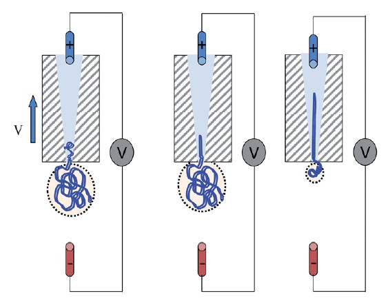
Schematic of DNA translocation in a glass capillary. Image taken from Soft Matter, 2012, doi:10.1039/C2SM25346A

Detecting single strands of DNA is a tricky business. One way in which it can be done is by creating a pore and sensing the DNA strand as it passes through the pore. This is what Ullrich Keyser from the University of Cambridge, UK and his group have been doing.

Keyser forms nanopores with diameters of less than 100nm from glass capillary tubes (the diameters can be as small as 20nm). These nanocapillaries act as single molecule sensors. Using electrophoresis, the negatively charged DNA is pulled towards a positively charged electrode inside the capillary, in a process known as DNA translocation. As the strand enters the capillary the resistance across the capillary pore changes, allowing the DNA to be detected. The change in ionic current is dependent not only on the presence of a DNA strand, but also on its folded state. This method offers a simple and cost-effective method for the detection of single molecules and for DNA sequencing.

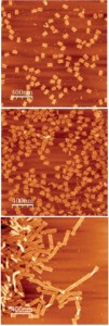
Examples of DNA origami. Image taken from Soft Matter, 2011, 7, 4636.

Full control over DNA translocation can be achieved using optical trapping, where the DNA is attached to a colloidal particle, held in place by an optical trap. The DNA can then be moved in and out of the capillary pore at will. Using this method, Keyser and his group have measured the capture force due to the electric field acting on the DNA. Their results show that the DNA capture force is linearly dependent on the number of strands captured in the capillary.

Whether using glass capillaries, or pores formed in silicon nitride membranes via focussed ion beam milling, control over the exact shape and functionality of the nanocavity can be problematic. Keyser has taken DNA detection yet another step further by using the DNA itself to create the nanopore. The shape into which a DNA strand folds can be controlled in a a process known as DNA origami; the DNA is synthesised such that it will self-assemble into a pre-designed three-dimensional shape. Using this origami, it is possible to design and fabricate virtually any nanosized shape that you want.

Keyser designed the DNA so that it folded into a funnel like shape with a long tail. This structure was then pulled through a pore in a  silicon nitride membrane to form a hybrid nanopore with a diameter of 7.5nm. The assembly of the hybrid pore is robust and easily reversible. These DNA/silicon nitride pores have been successfully used to detect single strands of DNA. The hybrid nanopores offer a novel way to change the size, shape and functionality of pores.

Relevant papers in SoftMatter:

Chen, Q. et al., How does a supercoiled DNA chain pass through a small conical glass pore? Soft Matter, 2012, Advanced Article.

Geerts, N., Eiser, E., DNA functionalized colloids: Physical properties and applications. Soft Matter, 2010, 6, 4647-4660.

Kim, K. N., et al., Comparison of methods for orienting and aligning DNA origami. Soft Matter, 2011, 7, 4636-4643.

Digg This
Reddit This
Stumble Now!
Share on Facebook
Bookmark this on Delicious
Share on LinkedIn
Bookmark this on Technorati
Post on Twitter
Google Buzz (aka. Google Reader)

Survival in the face of the unknown

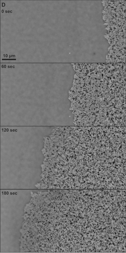
Swarming E. coli.

There are an estimated 1030 bacteria on Earth. The number of bacteria is greater than the number of stars in the Universe and is growing exponentially.

Bacteria are generally studied in the laboratory in Petri dishes under very well defined conditions. However, bacteria also thrive in more complex environments where the conditions are constantly varying. Some of these environmental changes are regular e.g. variations in light intensity from day to night, while others are random e.g. temperature, food availability and the presence of toxins or other bacteria.

Bacteria have developed a number of strategies to survive in these fluctuating environments. In the opening talk of the DPG spring meeting in Berlin last week, Stanislas Leibler from the Rockefeller University, New York and the Institute for Advance Studies, Princeton, discussed recent experimental and theoretical studies exploring the complex behaviour observed in bacterial colonies.

Consider a growing colony of bacteria. When an environmental change occurs one of two things may happen if the colony is to survive. (1) The bacteria ‘senses’ the change and changes to a state that is adapted for this new environment. This is known as responsive switching. (2) A small minority of the bacteria in the colony are poorly adapted to the initial environment. However, they become the most-adapted when the environment changes and survive while the rest are killed; the minority becomes the majority. This is known as stochastic switching.

So which is it? For colonies of bacteria with antibiotic persistence, experiments suggest that stochastic switching is the dominant behaviour. Leibler’s group added Ampicillin to growing colonies of Escherichia coli. The majority of the colony dies, but a few resistant bacteria survive. These resistant bacteria are able to grow, forming a new colony, once the antibiotic is removed. The persistent bacteria have a different phenotype to the rest of the colony. Under normal conditions, they grow much more slowly than the non-resistant bacteria, but are not killed when the antibiotics are added. Although the presence of these persistent cells leads to a lower population fitness, they act as an insurance policy and ensure that the colony can survive in the event of an antibiotic encounter. Leibler believes that this heterogeneity of bacterial populations is important for their ability to adapt to fluctuating environments and the persistence of bacterial infections.

While important when considering antibiotic resistant infections, these results may have much wider implications in areas ranging from cancer treatments, to models of financial investments, to information theory and statistical mechanics.

For more information see:

Balaban, N.Q. et al., Bacterial persistence as a phenotypic switch, Science, 2004.

Kussell, E. et al., Bacterial persistence: A model of survival in changing environments, Genetics, 2005.

Rivoire O, Leibler S, The value of information for populations in varying environments, J. Statist. Phys., 2011.

The image is taken from: Bacterial swarming: a model for studying dynamic self-assembly, Soft Matter, 2009, and shows a swarming colony of E. coli bacteria.

Digg This
Reddit This
Stumble Now!
Share on Facebook
Bookmark this on Delicious
Share on LinkedIn
Bookmark this on Technorati
Post on Twitter
Google Buzz (aka. Google Reader)

Martien Cohen Stuart talks to Soft Matter about his research on self assembly

Martien Cohen Stuart talks to Russell Johnson about his research on self assembly and what he thinks are the hot topics in soft matter research. Watch the video interview on YouTube here:

 Martien Cohen Stuart talks to Soft Matter about his research on self assembly

If you’re interested to know more about Professor Cohen Stuart’s research you can read a selection of his articles here:


To keep up-to-date with all the latest research, sign up for the Soft Matter e-Alert or RSS feeds or follow Soft Matter on Twitter or Facebook.

Digg This
Reddit This
Stumble Now!
Share on Facebook
Bookmark this on Delicious
Share on LinkedIn
Bookmark this on Technorati
Post on Twitter
Google Buzz (aka. Google Reader)

Soft Matter papered featured by the BBC

A recently published Soft Matter paper has been featured on the BBC website: ‘Avalanche research aids search for tastier ice cream’.

Avalanche experts were consulted for a study on how ice cream’s structure changes when it is stored in a household freezer. The structure within the ice cream is important for the taste of the food.

Read the original research here:
3D-characterization of three-phase systems using X-ray tomography: tracking the microstructural evolution in ice cream
B. R. Pinzer ,  A. Medebach ,  H. J. Limbach ,  C. Dubois ,  M. Stampanoni and M. Schneebeli
Soft Matter, 2012, Advance Article
DOI: 10.1039/C2SM00034B

Digg This
Reddit This
Stumble Now!
Share on Facebook
Bookmark this on Delicious
Share on LinkedIn
Bookmark this on Technorati
Post on Twitter
Google Buzz (aka. Google Reader)

Roll up, roll up!

Scientists in India – inspired by research on making water run uphill – have developed a technique that enables a polymer cylinder to not only autonomously climb an incline but carry a weight with it.

There are a number of different approaches towards limbless locomotion, but they focus on using mechanical or chemical pulses to drive a body forwards. Researchers from the Indian Institute of Technology in Kanpur have taken another approach: rolling.

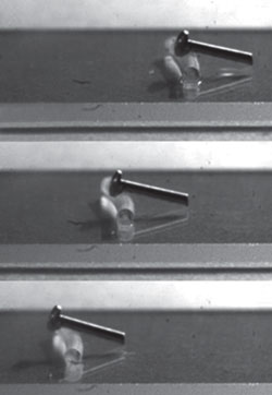
Polymer cylinder dragging a screw

A sequence of images captured at 0, 1.43 and 2.87 seconds (top to bottom) showing a 4mg polymer cylinder dragging a 18mg screw across a horizontal surface

The team, led by Animangsu Ghatak, used the swelling interactions of an elastomeric cylinder and a solvent to drive the cylinder forward. When the solvent is released, it spreads along the cylinder and swells at the contact points, particularly at the ends. This pushes the centre of mass forward and the rest of the cylinder moves to keep up via a rolling motion, which drags solvent with it. Because of this rolling motion, dry polymer is brought into contact with the solvent and swells in turn. The saturated polymer is exposed to the air and the solvent evaporates, forming a swelling-evaporation-de-swelling mechanism and enabling autonomous movement as long as the solvent lasts.

The most unusual aspect of the system, Ghatak explains, ‘is that gravity can be used to increase the asymmetric effect and thus the resultant increase in velocity’. By increasing the incline, ‘the solvent tends to accumulate more in the rear side of the cylinder than in the front and as a result, the asymmetric effect becomes more pronounced and the cylinder bends more’. This greater bend not only increases the velocity but creates enough torque for the cylinder to carry a dead weight with it.

Mark Geoghegan, from the University of Sheffield, UK, points out that although this technique will not scale up to the macro-scale, the field of self-powered locomotion is still very young. ‘This is an exciting area of research and one expects the benefits to be reaped only years in the future,’ he says.

Both Geoghegan and Ghatak can also see more immediate uses for the technology – despite the early days – and highlight potential roles in sensing or soft robotic components.

Read the original Chemistry World article, or watch the supplementary video

Digg This
Reddit This
Stumble Now!
Share on Facebook
Bookmark this on Delicious
Share on LinkedIn
Bookmark this on Technorati
Post on Twitter
Google Buzz (aka. Google Reader)

Most read Soft Matter Reviews in 2011

Read the most popular Review Articles of 2011 for free today

Self-assembly of amphiphilic peptides
I. W. Hamley
Soft Matter, 2011, 7, 4122-4138
DOI: 10.1039/C0SM01218A

Stimulus responsive core-shell nanoparticles: synthesis and applications of polymer based aqueous systems
Olivier J. Cayre , Nelly Chagneux and Simon Biggs
Soft Matter, 2011, 7, 2211-2234
DOI: 10.1039/C0SM01072C

Cellulose nanowhiskers: promising materials for advanced applications
Stephen J. Eichhorn
Soft Matter, 2011, 7, 303-315
DOI: 10.1039/C0SM00142B

Polylactide (PLA)-based amphiphilic block copolymers: synthesis, self-assembly, and biomedical applications
Jung Kwon Oh
Soft Matter, 2011, 7, 5096-5108
DOI: 10.1039/C0SM01539C

Morphology of polymer-based bulk heterojunction films for organic photovoltaics
Matthias A. Ruderer and Peter Müller-Buschbaum
Soft Matter, 2011, 7, 5482-5493
DOI: 10.1039/C0SM01502D

Stimulus responsive nanogels for drug delivery
Liusheng Zha , Brittany Banik and Frank Alexis
Soft Matter, 2011, 7, 5908-5916
DOI: 10.1039/C0SM01307B

Covalently cross-linked amphiphilic block copolymer micelles
Cornelus F. van Nostrum
Soft Matter, 2011, 7, 3246-3259
DOI: 10.1039/C0SM00999G

PNIPAM microgels for biomedical applications: from dispersed particles to 3D assemblies
Ying Guan and Yongjun Zhang
Soft Matter, 2011, 7, 6375-6384
DOI: 10.1039/C0SM01541E

Nanoparticles with targeting, triggered release, and imaging functionality for cancer applications
Kristin Loomis , Kathleen McNeeley and Ravi V. Bellamkonda
Soft Matter, 2011, 7, 839-856
DOI: 10.1039/C0SM00534G

Hydrophilic and superhydrophilic surfaces and materials
Jaroslaw Drelich , Emil Chibowski , Dennis Desheng Meng and Konrad Terpilowski
Soft Matter, 2011, 7, 9804-9828
DOI: 10.1039/C1SM05849E

To keep up-to-date with all the latest research, sign up for the Soft Matter e-Alert or RSS feeds or follow Soft Matter on Twitter or Facebook.

Digg This
Reddit This
Stumble Now!
Share on Facebook
Bookmark this on Delicious
Share on LinkedIn
Bookmark this on Technorati
Post on Twitter
Google Buzz (aka. Google Reader)

2012 Soft Matter Lectureship: Nominations Closing Soon!

Nominations for the 2012 Soft Matter Lectureship close 31 March

Now is your chance to nominate an early career researcher who has made signficant contributions to the field of soft matter.

This annual Lectureship was established by the journal in 2009, and last year’s winner was Michael J. Solomon, University of Michigan Ann Arbor.

Qualification

To be eligible for the Soft Matter Lectureship, the candidate should be in the earlier stages of their scientific career, typically within 15 years of attaining their doctorate or equivalent degree, and will have made a significant contribution to the field.

Description

The recipient of the Lectureship will be asked to present a lecture three times, one of which will be located in the home country of the recipient. The Soft Matter Editorial Office will provide the sum of £1000 to the recipient for travel and accommodation costs.

The recipient will be presented with the Lectureship at one of the three lectures. They will also be asked to contribute a lead article to the journal and will have their work showcased on the back cover of the issue in which their article is published.

Selection

The recipient of the Lectureship will be selected and endorsed by the Soft Matter Editorial Board.

Nominations

Those wishing to make a nomination should send details of the nominee, including a brief C.V. (no longer than 2 pages A4) together with a letter (no longer than 2 pages A4) supporting the nomination, to the Soft Matter Editorial Office (softmatter-rsc@rsc.org) by 31 March 2012.  Self nomination is not permitted.

Digg This
Reddit This
Stumble Now!
Share on Facebook
Bookmark this on Delicious
Share on LinkedIn
Bookmark this on Technorati
Post on Twitter
Google Buzz (aka. Google Reader)

Embryonic polarisation

Growing up is a stressful process, particularly during embryonic development. The embryo starts out as a single symmetric cell. This cell can be considered as an active fluid bound by the cell membrane. Flow within this fluid leads to the build-up of stresses, the formation of patterns and asymmetries within the cell. Stephan Grill, at the Max-Planck Insitute for the Physics of complex systems and the Max-Planck Institute for Molecular Cell Biology and Genetics, Dresden, is interested in understanding how these flows lead to the polarisation observed in the Caenorhabditis elegans zygote.

Grill has shown that the changes in cell polarity are driven by myosin flow on the surface of the cell. The cortex can be considered as a dynamic self-contracting polymer gel surface, which lies underneath the cell membrane. This polymer gel layer behaves as a thin film of an active fluid. Passive advective transport of molecules – in this case myosin – embedded in the fluid can occur depending on the diffusivity and the flow velocities of the molecules.  In C. elegans the flows are fast enough that advection does play a role, influencing the distribution of the molecules as they diffuse on the cortex. Modelling shows that the passive advective transport by flow of such a mechanically active materials acts as a trigger for the segregation of the proteins, resulting in the polarisation of the zygote.

The movement of myosin across the surface of the cell also results in anisotropies in the cortical tension. These so called active stresses cause isolated sections of the cortex to self-contract. Grill has developed a novel method for locally determining the stresses, by cutting the cortex with a laser and measuring the recoil. The cortical tension is found to be greatest in the direction orthogonal to the flow.

Grill suggests that advective transport in active fluids is a general mechanism for the formation of patterns in developmental biology.

For more information see:

Goehring, N.W. et al., Polarization of PAR Proteins by Advective Triggering of a Pattern-Forming System, Science, 2011.

Bois, S, et al., Pattern Formation in Active Fluids, Phys. Rev. Lett., 2011.

Mayer. M, et al., Anisotropies in cortical tension reveal the physical basis of polarizing cortical flows, Nature, 2010.

The image above is taken from: Cyclodextrin/dextran based drug carriers for a controlled release of hydrophobic drugs in zebrafish embryos, Soft Matter, 2011, and shows C. elegans embryo 30hrs after fertilisation.

Digg This
Reddit This
Stumble Now!
Share on Facebook
Bookmark this on Delicious
Share on LinkedIn
Bookmark this on Technorati
Post on Twitter
Google Buzz (aka. Google Reader)