Archive for the ‘Conference’ Category

APS March meeting 2011 – bacterial competition

The annual APS March meeting was held last week at the convention centre in Dallas. The conference consisted of over 660 session and 7350 contributed talks (according to my rough calculation) on subjects ranging from quantum computing to the physics of evolution to polymer dynamics. Due to the shear number of talks, I will split the conference into a series of blogs rather than one long one, which no-one would want to read!

Deadly competition between sibling bacteria coloniesAvraham Be’er, University of Texas, Austin.

In this invited talk, Avraham Be’er discussed the growth of competing bacterial sibling colonies. A single colony of bacteria grows with radial symmetry at a constant speed. However, for two colonies of P. dendritiformis, equidistant from the centre  and inoculated simultaneously, the dynamics differ. Initially the growth of each colony is radially outwards and independent of the other colony. However, while the distance is still large, growth in the centre, between the two colonies, decelerates and a gap forms between the two colonies. The colonies become asymmetric in shape and growth. (See Be’er’s website for pictures of the growing colonies.)

Be’er and co-workers have shown that the reason for this inhibition of growth is a toxic material secreted by the bacteria (doi:10.1073/pnas.0811816106). The toxin is lethal once it exceeds a well-defined threshold. Extracting the toxin and depositing it outside a growing single colony results in growth inhibition and cell death, which would otherwise not be seen. This toxin, termed ‘sibling lethal factor’ (Slf), lyses cells, rupturing them and is not limited to this bacteria (although the toxicity varies for different bacteria). The bacteria seem to have evolved to produce Slf and kill their own siblings, but only when there are two competing colonies. Slf is not secreted when there is only one colony.

Subtilisin was also found in the bacterial secretions. This protein is non-toxic. However, when Slf is exposed to subtilisin it is cleaved from a non-toxic protein of ~20 kDa to the toxic Slf ~12 kDa. The results suggest that subtilisin acts to regulate growth of the colony. Below a threshold value the subtilisin promotes growth and expansion of the colony. Above this threshold value Slf is secreted, reducing the density of cells (doi:10.1073/pnas.1001062107). The results also indicate that when the levels of Slf are small, rather than cell death occurring, the cells can instead enter a vegetative state. These vegetative cells ‘cocci’ are immobile, have a slow expansion and are spherical rather than rod-like in shape. These cocci cells are observed to switch back simultaneously and spontaneously to the healty rod-shaped cells, with growth continuing as before.

Related papers in Soft Matter

Variations in the nanomechanical properties of virulent and avirulent Listeria (doi:10.1039/B927260G)

Mechanical robustness of Pseudomonasaeruginosa biofilms (doi:10.1039/C0SM01467B)

Facile growth factor immobilization platform based on engineered phage matrices (doi:10.1039/C0SM01220C)

Digg This
Reddit This
Stumble Now!
Share on Facebook
Bookmark this on Delicious
Share on LinkedIn
Bookmark this on Technorati
Post on Twitter
Google Buzz (aka. Google Reader)

The potential of microfluidics

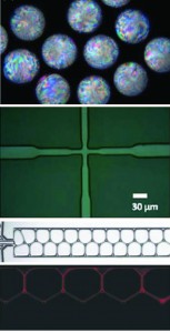
From top to bottom: Cholesteric particles formed via microfluidics. Flow-focusing microfluidic device. Gel emulsion in a channel. Fluorescence image of a dye-containing gel emulsion.

This week I attended a talk at the MPI for Dynamics and Self-Organisation in Goettingen, Germany by Nicolas Bremond from the EPSCI, Paris. The talk was titled “Microfluidic investigations of the destabilisation of emulsions via coalescence”. Nicolas discussed the use of microfluidic devices to study coalescence of emulsion droplets in the absence and presence of an ac electric field. The results have been published in Physical Review Letters and in the Journal of Fluid Mechanics.

In the absence of an electric field, a series of moving pairs of droplets in a microfluidic device were created. A widening of the channel, and therefore slowing of the fluid, was used to force the droplets together. Monitoring of the separation of the droplets showed that coalescence occurred just after closest proximity i.e. when they were separating. Nicolas believes that the separation of the droplets momentarily reduces the fluid pressure between the droplets, causing the high-pressure water in the droplets to burst through the barrier.

Amongst many of the movies shown in the talk was this one, which I have found on the web. It shows that this coalescence mechanism can also trigger coalescence in neighbouring droplets. This mechanism could perhaps be responsible for the clumping of industrial emulsion droplets.

My favourite video of the talk was one showing the behaviour of the emulsion droplets under an applied ac electric field. When the ratio of the radius of the droplet to the channel width is equal to 0.8 the droplets can be made to kiss each other; they come into contact with each other before separating again. On separation deformation of the droplets is observed. Unfortunately I haven’t found a video of this online, but a sequence of images can be seen in the Physical Review Letters paper.

Although not mentioned in the talk, Nicolas has also recently had his paper Formation of liquid-core capsules having a thin hydrogel membrane: liquid pearls published in Soft Matter.

Microfluidics offers an interesting method for studying the coalescence of emulsions under flow. However, this is not its only application as seen in the latest issue and advance articles of Soft Matter. Shashi Thutupalli et al. (doi:10.1039/c0sm00312c) demonstrate the use of microfluidics to self-assemble surfactant bilayer networks in water-oil emulsions. These bilayer membranes display a range of different electrical behaviours, which could be exploited to create wet circuitry. Formation of emulsions in this way, offers a feasible approach to construct complex devices out of molecular-sized components via controlled self-assembly.

Sara Abalde-Cela et al. demonstrate the use of a flow-focusing microfluidic device to form highly mono-disperse plasmonic agarose beads containing silver nanoparticles in their paper doi:10.1039/c0sm00601g, while Daniel Wenzlik et al. used microfluidics to prepare cholesteric liquid crystal particles from cellulose derivatives doi:10.1039/c0sm01368d.

Digg This
Reddit This
Stumble Now!
Share on Facebook
Bookmark this on Delicious
Share on LinkedIn
Bookmark this on Technorati
Post on Twitter
Google Buzz (aka. Google Reader)

Soft matter theme at CMMP10

The annual condensed matter and materials physics conference CMMP10 was held last week at the University of Warwick. This year the conference included a number of symposia relevant to the soft matter community including biological physics, polymer physics and soft matter in action. Soft matter plenary lectures were given by Prof. Marshall Stoneham “Where quantum physics meets biology”,  Prof. Dame Athene Donald “Self-assembly of proteins”, Prof. Christopher Ober “Will polymers be used to make the next generation nanoworld” and Prof. Mike Cates “Understanding liquid crystals using mesoscopic computer simulation”.

Below I have highlighted a couple of interesting talks. The conference proceedings will be published in the Journal of Physics: Conference Series (JPCS).

Magnetic alignment of anisotropic structures in solution.

Alex Holmes (University of Bristol) introduced the new Birmingham 17T cryomagnet designed for use in small angle neutron scattering (SANS) measurements. The magnet operates in the temperature range of 1.6K to 300K and magnetic field up to 17T with 0.1% uniformity over 10 nm.

Here Alex discussed initial experiments carried out at the Institut of Laue-Langevin, showing the viability of this technique for studying soft matter. The results show that the magnetic field can be used to align rod-like viruses, which behave as cholesteric liquid crystals at high concentrations. A transition from a multi-domain cholesteric structure at zero field, to a nematic phase at high field was observed.

The cryomagent offers an interesting technique to the soft matter community to study the behaviour of soft materials in the presence of high magnetic field and the group are open to collaborations. More information is available on their website.

AFM nanotools to investigate skin cells

In his talk, James Beard (University of Bath) discussed a novel approach to modifying AFM tips using electron beam induced deposition using a scanning electron microscopy (SEM). The secondary electrons interact with contaminants in the SEM leaving behind a deposit of amorphous carbon on surfaces exposed to the electron beam. A variety of highly durable ‘nanotools’ can be formed in this manner such as ‘nanoscapels’, ‘nanoneedles’ with lengths of 500nm and thicknesses of 10-50 nm and ‘nanotomes’.

James demonstrated the use of the nanoscapel probes for cutting biological samples with high precision and nanoneedles to investigate the mechanical properties of corneocyte cells using a nanoindentation technique. The results of the experiments were published in Nanotechnology doi: 10.1088/0957-4484/20/44/445302.

Micro-wrinkled bilayer structures with gradient wetting properties

Kevin Langley (University of Nottingham) demonstrated, in his talk, the use of Aluminium-elastomer bilayers to form micro-wrinkled structures. The Aluminium capping layer was thermally evaporated onto a thick pre-strained elastomer substrate. The strain was then released causing the bilayer to wrinkle with a well defined wavelength and amplitude, dependent on the capping layer thickness and the applied pre-strain.

Kevin was able to create gradient wavelength wrinkled surfaces. Such a surface provides anisotropic wetting properties and could be used as a gradient energy surface to, amongst other things, move water droplets. Similar surfaces have been fabricated through UV-ozone etching of elastomers (Soft Matter doi:10.1039/B705112C)  .

A short movie was played, demonstrating that the droplets did indeed move when placed onto these surfaces and vibrated close to their resonant frequency. These surfaces are an interesting candidate for low cost gradient energy surfaces. The results have been published in Langmuir doi: 10.1021/la1036212.

Digg This
Reddit This
Stumble Now!
Share on Facebook
Bookmark this on Delicious
Share on LinkedIn
Bookmark this on Technorati
Post on Twitter
Google Buzz (aka. Google Reader)