AC electric field induced droplet deformation in a microfluidic T-junction
Microfluidic Paper-based Biomolecule Preconcentrator Based on Ion Concentration Polarization


")
The 32nd International Symposium on Microscale Separations and Bioanalysis was held at Queen’s Landing in Niagara-on-the-Lake, Canada from April 3-7, 2016. This symposium has been running since 1989 and was originally founded by Professor Barry Karger (Northeastern University). Over the years the format of MSB 2016 has evolved into an interactive forum for the discussion of cutting-edge research on the frontiers of separation science.
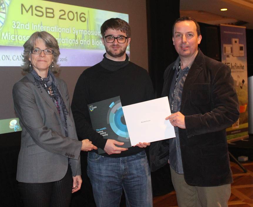
Several awards were on offer at MSB 2016, but we are happy to announce that the Lab on a Chip poster prize was won by Nicolas Drouin of the University of Genevea, Switzerland. He is pictured (above) receiving his prize from Philip Britz-McKibbin of McMaster University (Conference Chair) and Karen Waldron of Université de Montréal (Conference Co-organizer). Congratulations Nicolas!
More information on the symposium can be found here.
![]()
Lab on a Chip is proud to announce the third μTAS Video Competition, in partnership with Dolomite Microfluidics and supported by μTAS and Chemical and Biological Microsystems Society (CBMS).
We invite registered μTAS partipants to submit short videos (see terms and conditions below) that are either scientifically or educationally focused. Videos may be fun, artistic or just surprising and unusual in order to meet these criteria.
Dolomite Microfluidics, innovators in microfluidic solutions, are sponsoring this competition with the prize of €2500 worth of Dolomite equipment.
If you have an idea for a video that you would like to share with the μTAS community read the entry conditions below!
Terms and Conditions
Video Award Submission Process – Easy 3 Step Process
Step 1. Sign-In to the Electronic Form using your Registration Number
Please have your Registration Number accessible. If you are unable to locate your Registration Number, please contact info@microtas2016.org.
Step 2. Fill in information on Electronic Submission Form
Step 3. Upload Your Video
All entries are to be submitted online via this website as .mpg, .mp4, .mov, .avi or .wmv. Once your entry has been successfully uploaded and submitted, you will be given an entry number and you will be sent a confirmation email with the information your provided, minus the video. The ability to submit a video will close at the end of Monday, 10 October 2016 (Honolulu, Hawaii, USA time).
Good luck!
Previous winners:
MicroTAS 2015 Conference, Gyeongju, Korea
Spin Me Right Round
David Kinahan, Ducrée Labs, Dublin City University, Ireland
MicroTAS 2014 Conference, San Antonio, Texas, USA
Magnetotactic Bacteria
Tijmen Hageman, KIST Europe GmbH, Germany
Get your entries in before the deadline on 10th October 2016
![]()
The µTAS 2016 Conference will feature the ninth Art in Science competition entitled ‘Under the Looking Glass: Art from the World of Small Science‘, sponsored and supported by National Institute of Standards and Technology, Lab on a Chip, MicroTAS and the Chemical and Biological Microsystems Society.
Since the earliest publications of the scientific world, the aesthetic value of scientific illustrations and images has been critical to many researchers. The illustrations and diagrams of earlier scientists such as Galileo and Da Vinci have become iconic symbols of science and the scientific thought process.
In current scientific literature, many scientists consider the selection of a publication as a “cover article” in a prestigious journal to be very complimentary.
To draw attention to the aesthetic value in scientific illustration while still conveying scientific merit, NIST, LOC and CBMS are sponsoring this annual competition. Applications are encouraged from authors in attendance of the µTAS Conference and the winner will be selected by a panel of judges.
Applications must show a photograph, micrograph or other accurate representation of a system that would be of interest to the µTAS community and be represented in the final manuscript or presentation given at the Conference.
They must also contain a brief caption that describes the illustration’s content and its scientific merit. The winner will be selected on the basis of aesthetic eye appeal, artistic allure and scientific merit. In addition to having the image featured on the cover of Lab on a Chip, the winner will also receive a financial prize at the Conference.
Art in Science Competition Submission Process
Step 1. Sign-In to the Electronic Form Using Your Abstract/Manuscript Number
Step 2. Fill in Remaining Information on Electronic Submission Form
Step 3. Upload Your Image
Good Luck!
You can also take a look at the winners from last year on our blog.
an article by Claire Weston, PhD student at Imperial College London
Shefali Lathwal and Hadley Sikes at Massachusetts Institute of Technology have carried out an in-depth study published in Lab on a Chip comparing different colorimetric paper-based immunoassays (where a positive or negative result is shown by the appearance or absence of a certain colour). The assays used were all for the detection of an enzyme found in P. falciparum in order to diagnose malaria. The authors sought to identify the optimal readout times for the different methods in order to prevent false positives.

Time course for colour generation on negative and positive surfaces using different colorimetric methods
One of the main purposes of this study was to compare a new paper-based assay that had recently been developed by Sikes in collaboration with George Whitesides at Harvard (Lab on a Chip, 2015) with other state-of-the-art methods.
The new method in question is a colorimetric assay that utilises a photo-initiated polymerisation reaction to amplify the signal when the P. falciparum enzyme is present. The reaction only occurs when the sample is being irradiated, so by using an automated timing switch the reaction time can be accurately controlled and no further signal amplification will take place once the light has been turned off. In contrast, other methods require accurate manual time keeping as they are thermally rather than photochemically controlled. This means that if the sample is left too long, it may lead to false positives due to colour forming in a negative sample.
This can be seen in the figure on the right, where positive and negative controls detected using the different colorimetric methods were photographed at various time points. All the methods tested had the same binding events in order to allow a fair comparison (i.e., all assay steps were the same apart from the detection method). A diagram included in the manuscript (Scheme 2) shows the key steps to all the assays, and how the colour is formed.
Three of the methods were enzymatic amplifications; for these reactions t=0 was taken as when the substrate solution was added to the surface of the paper. Another method was silver deposition, and t=0 was taken as when the silver enhancement solution was added to the surface. For the polymerisation-based amplification (PBA) method, the aqueous monomer was added to the surface and after illumination a basic solution was added, which led to formation of colour in the positive samples; t=0 was taken as when the basic solution was added.
In all cases other than the photo-controlled reaction, colour developed in the negative controls over time, leading to very similar results as in the positive controls. These assays are usually used at the point of care in resource limited settings, therefore the readouts are carried out by eye and there is often not a negative control to compare to. Instead, the result is compared to a colour chart, making it even easier to obtain a false positive if the readout time is not correct.
For the enzymatic amplification and silver deposition the optimal readout times varied considerably and in some cases the time window was very narrow, in order to prevent false positives. In the PBA reaction however, no colour developed in the negative control over 40 minutes, and at all time intervals there was a clear difference between the positive and negative controls. In addition to this, the visual limit of detection for the PBA reaction was much higher than that of the enzymatic amplifications and silver deposition.
This study is the first to compare multiple colorimetric methods for paper-based immunoassays with carefully controlled variables. Previously, different binding reagents, imaging techniques and methods of quantification have meant that meaningful comparisons could not be obtained. The results clearly highlight the benefits of using a photo-controlled reaction, where the reaction time can be carefully controlled with an automated timer without the requirement of accurate manual time keeping.
To download the full article for free* click the link below:
Assessment of colorimetric amplification methods in a paper-based immunoassay for diagnosis of malaria
Shefali Lathwal and Hadley D. Sikes
Lab Chip, 2016, Advance Article
DOI: 10.1039/C6LC00058D
—————-

About the webwriter
Claire Weston is a PhD student in the Fuchter Group, at Imperial College London. Her work is focused on developing novel photoswitches and photoswitchable inhibitors.
—————-
*Access is free until 27/04/2016 through a registered RSC account – click here to register
an article by Burcu Gumuscu, PhD researcher at University of Twente
Water is a simple element found in abundance throughout nature, and for many, its commonplace nature may mask its importance. Even a single droplet of water is of high value in the science world: when manipulated with sound waves, a water droplet can be used in a tremendous number of applications. For example, this simple technique can contribute to reducing the high costs of diagnosis tools, ensure the correct dosage of many drugs for effective treatment, and detect contaminants or pathogenic threats in food industry. This actuation of droplets using sound waves on the micron-scale is known as acoustofluidics.
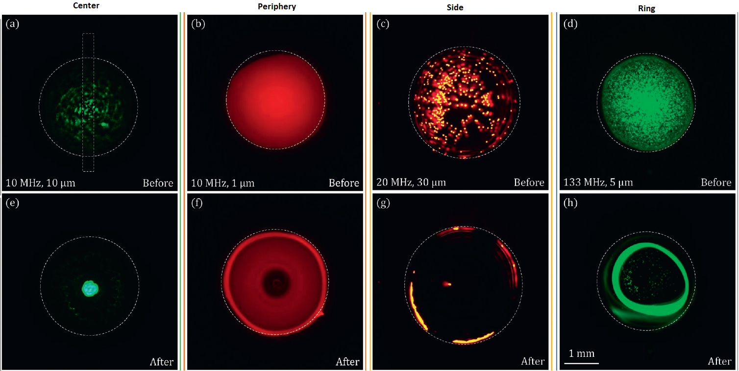
Fig. 1 Screenshots of the particle-laden sessile drops were captured before (a-d) and after (e-h) the SAW exposures
The science behind the above-mentioned advances lies in the handling of droplets with surface acoustic waves (SAW), produced by an interdigitated transducer interacting with a sessile water droplet. This interaction leads the droplets to dissipate the energy absorbed from SAW, giving rise to acoustic streaming flow (ASF) and acoustic radiation force (ARF). As a result, sound waves with different amplitudes travel across the droplet and create micro-streams. Mixing, merging, and even sorting of the suspended particles are therefore enabled due to these micro-streams. To achieve this, an in-depth understanding of the generation of these micro-streams and their effect on the suspended particles are crucial for better controlled manipulation of these on-demand applications.
Luckily, scientists in the Flow Control Laboratory at KAIST have recently published a comprehensive study in Lab on a Chip on the fate of different sized microparticles inside a droplet of water actuated by SAW. In this work, for the first time, polystyrene microparticles were reported to go under four different unexplored modes at high frequencies. The elastic character of the polystyrene particles exhibits significantly different behaviors under SAW applied with different frequencies. For example, large particles were found to concentrate at the center of the droplet while the smaller ones form a ring structure around the periphery.
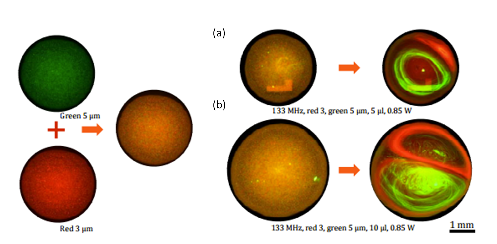
Fig. 2 Separation of red 3 and green 5 µm polystyrene particles inside a (a) 5 and (b) 10 µm droplets
Other intermediate modes include particle concentration at the side of the droplet and ring formation close to the droplet center: all dependent on the particle size, applied frequency, ASF, and ARF. Flow Control Laboratory further explored this interesting behavior using microparticles in the range of 1-30 µm at nominal frequencies of 10, 20, 80, and 133 MHz. Figure 1 shows before and after manipulation of the water droplets. Better yet, SAW applied at high frequencies allowed the separation of different-sized microparticles in a water droplet as seen in Figure 2.
For a real demonstration of what happens when a droplet of water meets with sound, the movie appended to the article and included below is well worth the watch. Thanks to the improved understanding of the physics behind the technique, this self-contained microcentrifuge technology seems promising to further widen our horizons in both clinical and biological applications.
To download the full article for free* click the link below:
Acoustofluidic particle manipulation inside a sessile droplet: four distinct regimes of particle concentration
Ghulam Destgeer, Hyunjun Cho, Byung Hang Ha, Jin Ho Jung, Jinsoo Park and Hyung Jin Sung
Lab Chip, 2016, 16, 660-667
DOI: 10.1039/C5LC01104C, Paper
—————-
About the webwriter
Burcu Gumuscu is a PhD researcher in BIOS Lab on a Chip Group at University of Twente in The Netherlands. Her research interests include development of microfluidic devices for second generation sequencing, organ-on-chip development, and desalination of water on the micron-scale.
—————-
*Access is free until 02/05/2016 through a registered RSC account.
Lab on a Chip and Corning Incorporated are proud to sponsor the eleventh Pioneers of Miniaturization Lectureship, to honour and support the up and coming, next generation of scientists who have significantly contributed to the understanding or development of miniaturised systems. This year’s Lectureship will be presented at the µTAS 2016 Conference in Dublin, Ireland, with the recipient receiving a prize of US$5,000.
Who should you nominate?
Early to mid-career scientists (maximum 15 years post completion of PhD).
Scientists who have demonstrated extraordinary contributions to the understanding or development of miniaturised systems.
How do you nominate?
Submit your nominations to Lab on a Chip Editor Sarah Ruthven at LOC-RSC@rsc.org
Nominations should include:
Nomination Deadline: 1 June 2016
Who has won the Pioneers of Miniaturisation Lectureship in the past?
Terms and Conditions
The Lectureship consists of the following elements:
The award is for early to mid-career scientists (maximum 15 years post completion of PhD).
The award is for extraordinary or outstanding contributions to the understanding or development of miniaturised systems. This will be judged mainly through their top 1-3 papers and/or an invention documented by patents/or a commercial product. Awards and honorary memberships may also be considered.
The winner will be expected to submit at least two significant publications to Lab on a Chip in the 12 months after the lectureship is awarded.
Nominations from students and self-nominations are not permissible.
The decision on the winner of the lectureship will be made by a panel of judges, and this decision will be final.
Sponsors
Corning Incorporated
Corning (www.corning.com) is one of the world’s leading innovators in materials science. For more than 160 years, Corning has applied its unparalleled expertise in specialty glass, ceramics, and optical physics to develop products that have created new industries and transformed people’s lives. Corning succeeds through sustained investment in R&D, a unique combination of material and process innovation, and close collaboration with customers to solve tough technology challenges. Corning’s businesses and markets are constantly evolving. Today, Corning’s products enable diverse industries such as consumer electronics, telecommunications, transportation, and life sciences. They include damage-resistant cover glass for smartphones and tablets; precision glass for advanced displays; optical fiber, wireless technologies, and connectivity solutions for high-speed communications networks; trusted products that accelerate drug discovery and manufacturing; and emissions-control products for cars, trucks, and off-road vehicles.
Lab on a Chip
The leading journal for miniaturization at the micro and nanoscale. Lab on a Chip supports research and development of miniaturization technologies and promotes interdisciplinary co-operation across all fields of science. The Journal also provides readers with a more fundamental understanding of miniaturization and related processes as well as the necessary tools for practical application of methods and devices.
From microgalaxies to micro-oceans to Warhol’s cellular images
The 19th International Conference of Miniaturized Systems for Chemistry and Life Sciences held in Gyeonju, South Korea on October 2015 saw the 8th Art in Science competition.
The judges thought the quality of submissions was really high and the Lab on a Chip team would like to thank all the contributors. Please join us at Lab on a Chip in congratulating all of our prize winners.
You can read more information about this competition and its winners on Darwin R. Reyes’s Editorial in Issue 8.
The Art in Science award is sponsored by NIST and supported by MicroTAS, the Chemical and Biological Microsystems Society (CBMS) and the Lab on a Chip journal. The award consists of a monetary prize ($2500), an award certificate, and the coveted front cover of the Lab on a Chip journal.
We encourage you to participate in the 2016 Art in Science competition
2015 Winner: Through Warhol’s eyepiece, by Matteo Cornaglia
Image winner of the MicroTAS 2015 Art in Science award titled Through Warhol’s eyepiece by Matteo Cornaglia (Laboratory of Microsystems, EPFL).
