Cerebellar neuron development on hybrid matrix constructs

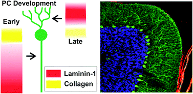
The cerebellum is a region in the brain responsible for regulating motor control and cognitive functions such as attention and language. During development of the cerebellum (and of other tissues), cells interact with the surrounding microenvironment known as the extracellular matrix (ECM). These neuron-ECM interactions regulate neuronal differentiation, growth, formation of synapses, and neurite outgrowth. Specifically, ECM components including collagen and laminin-1 (lam-1) are known to regulate the alignment, migration, and neurite outgrowth of Purkinje cells (PCs). However, the dual signaling roles of collagen and laminin-1 during cerebellar tissue development have not been fully explored.

Dr. Shantanu Sur, a postdoctoral fellow in Prof. Samuel Stupp’s laboratory at Northwestern University, has collaborated with Dr. Thomas Launey at the RIKEN Brain Science Institute in Japan to explore cerebellar tissue development using biomaterial hydrogels. Sur has developed an artificial matrix consisting of collagen and synthetic peptide amphiphile (PA) molecules presenting IKVAV, the peptide on laminin-1 responsible for cell adhesion and neurite outgrowth. Sur et al. evaluated the spatiotemporal expression of lam-1 and collagen in rat cerebellums during PC development (embryonic to post-natal) using histology and immunostaining. Given the changes in the ratio of lam-1 to collagen during PC formation and growth, these results suggest the dynamic involvement of these ECM proteins in forming the neural architecture of the cerebellum.

Using a biomimetic approach to mimic the critical lam-1 to collagen ratio, Sur proceeded to model the dynamic nature of the ECM during PC development with a synthetic collagen-PA hydrogel. Collagen (types I-V) and IKVAV-PA molecules were mixed together in solution at varying concentrations and gelled using ammonia vapor. This simple method allowed Sur to evaluate the density of PC growth, axon guidance, and dendrite morphology in gels using a wide array of collagen and IKVAV-PA concentrations. Strikingly, effects on PC phenotype were observed as a function of the collagen:IKVAV-PA ratio and not the absolute concentrations of each ECM component within the matrix.

Sur comments that the hybrid matrix provides an easily tunable environment to enable the in vitro testing of the role of ECM signals on neuronal maturation. “Our study shows that the optimal ECM-derived cues for neurons change at specific stages of development,” Sur says. “This observation will drive us to work on the design of a dynamic matrix where the extracellular signals delivered to the neuron can be tuned spatiotemporally.” Additionally, the collagen/IKVAV-PA gels may be used to identify cell-ECM interactions during the development of other tissue types, given the simplicity of the technique.

Collectively, this study demonstrates the exciting use of engineered matrices to evaluate spatiotemporal cell-ECM interactions, with the hopes to further elucidate mechanisms of tissue development.

Synergistic regulation of cerebellar Purkinje neuron development by laminin epitopes and collagen on an artificial hybrid matrix construct
Shantanu Sur, Mustafa O. Guler, Matthew J. Webber, Eugene T. Pashuck, Masao Ito, Samuel I. Stupp, and Thomas Launey
Biomater. Sci., 2014, Advance Article, DOI: 10.1039/C3BM60228A

Brian Aguado is currently a Ph.D. Candidate and NSF Fellow in the Biomedical Engineering department at Northwestern University. He holds a B.S. degree in Biomechanical Engineering from Stanford University and a M.S. degree in Biomedical Engineering from Northwestern University. Read more about Brian’s research publications here.

To keep up-to-date with all the latest research, sign-up to our RSS feed or Table of contents

Digg This
Reddit This
Stumble Now!
Share on Facebook
Bookmark this on Delicious
Share on LinkedIn
Bookmark this on Technorati
Post on Twitter
Google Buzz (aka. Google Reader)

Emerging Investigators themed issue now published

We are pleased to announce that the Biomaterials Science 2014 Emerging Investigators themed issue is now available to read online.

Edited by Phillip Messersmith and Norio Nakatsuji, co-Editors in Chief of Biomaterials Science, this issue highlights the exciting and important work being carried out by some of the most talented up-and-coming researchers in the field.  Read more about the issue in the Editorial.

Here is a sample of the reviews, communications and papers that feature in the Emerging Investigators themed issue:

On the cover

Fabrication of zeolite–polymer composite nanofibers for removal of uremic toxins from kidney failure patients Koki Namekawa, Makoto Tokoro Schreiber, Takao Aoyagi and Mitsuhiro Ebara 

 
Review
Smart hydrogels as functional biomimetic systems Han L. Lim, Yongsung Hwang, Mrityunjoy Kar and Shyni Varghese 
 
Minireviews
Peptoids for biomaterials science King Hang Aaron Lau 
 
Communications
 
Hyaluronic acid hydrogel stiffness and oxygen tension affect cancer cell fate and endothelial sprouting Yu-I Shen, Hasan E. Abaci, Yoni Krupski, Lien-Chun Weng, Jason A. Burdick and Sharon Gerecht
 
Papers
Translocation of flexible polymersomes across pores at the nanoscale Carla Pegoraro, Denis Cecchin, Jeppe Madsen, Nicholas Warren, Steven P. Armes, Sheila MacNeil, Andrew Lewis and Giuseppe Battaglia
 
Structural reinforcement of cell-laden hydrogels with microfabricated three dimensional scaffolds Chaenyung Cha, Pranav Soman, Wei Zhu, Mehdi Nikkhah, Gulden Camci-Unal, Shaochen Chen and Ali Khademhosseini
 
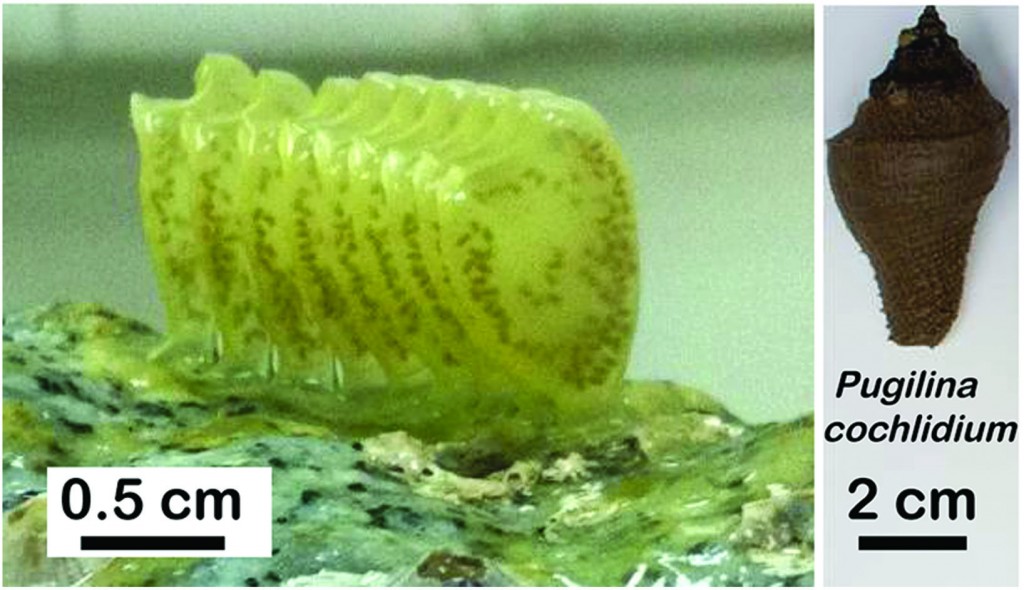
Integrative and comparative analysis of coiled-coil based marine snail egg cases – a model for biomimetic elastomers Paul A. Guerette, Gavin Z. Tay, Shawn Hoon, Jun Jie Loke, Arif F. Hermawan, Clemens N. Z. Schmitt, Matthew J. Harrington, Admir Masic, Angelo Karunaratne, Himadri S. Gupta, Koh Siang Tan, Andreas Schwaighofer, Christoph Nowak and Ali Miserez
 
 
Molecular farming of fluorescent virus-based nanoparticles for optical imaging in plants, human cells and mouse models S. Shukla, C. Dickmeis, A. S. Nagarajan, R. Fischer, U. Commandeur and N. F. Steinmetz 
 
More papers from the themed issue can be downloaded here.
 
Digg This
Reddit This
Stumble Now!
Share on Facebook
Bookmark this on Delicious
Share on LinkedIn
Bookmark this on Technorati
Post on Twitter
Google Buzz (aka. Google Reader)

Cells as Legos: Using cells for gelation of hydrophobically-modified polymers

Engineering tissue mimics in vitro using cells and materials is one of the core goals of biomaterials science. To develop a tailored cell culture environment, natural and/or synthetic hydrogels are used to mimic the extracellular matrix. Cells are embedded in a hydrogel matrix in a process known as “encapsulation.” During encapsulation, polymers in solution are chemically or physically cross-linked to immobilize cells in a mesh-like structure. The cell-loaded hydrogels are subsequently used for various regenerative medicine applications.

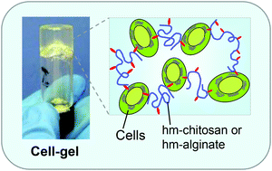
Researchers at the University of Maryland have developed a novel encapsulation technique for embedding cells in hydrogel materials. Instead of passively encapsulating cells in the polymer mesh, cells serve as “active structural elements” and are connected to the polymer chains. Using hydrophobically-modified (hm) alginate and chitosan, hydrophobic regions along the polymer chains are physically embedded in the hydrophobic cell membrane. The cells serve as struts to link together the polymer mesh. Rheological studies were performed to confirm the sol-gel transition when hm-polymers were combined to form gels (unmodified polymer controls did not form gels). To show the versatility of the system, various cell types were used to form gels with hm-alginate and hm-chitosan, including human umbilical vein endothelial cells (HUVECs), MCF7 breast cancer cells, and blood cells.

Analogous to building block toys such as Legos, the non-covalent hm-polymer and cell interactions are reversible with the addition of excess hydrophobic binding “pockets.” Using α-cyclodextrin, the gels are immediately transformed into free-flowing solutions (confirmed using rheology). The α-cyclodextrin serves to sequester the hydrophobic tails of the hm-polymer and gently release the cells from the mesh. Interestingly, α-cyclodextrin does not affect cell viability because the molecule is not large enough to bind two-tailed lipids of the cell membrane. Viability of the cells was confirmed before and after the gentle release from the gel, indicating that cell membranes remained unharmed during the gel reversal process.

Taken together, hm-polymers could serve as a unique technique to embed and release cells from a hydrogel matrix. Using cells as building blocks is highly desirable for several applications including 3D cell culture models and injectable cell therapies.

Reversible gelation of cells using self-assembling hydrophobically-modified biopolymers: toward self-assembly of tissue
Vishal Javvaji, Matthew B. Dowling, Hyuntaek Oh, Ian M. White and Srinivasa R. Raghavan
Biomater. Sci., 2014, Advance Article, DOI: 10.1039/C4BM00017J

Brian Aguado is currently a Ph.D. Candidate and NSF Fellow in the Biomedical Engineering department at Northwestern University. He holds a B.S. degree in Biomechanical Engineering from Stanford University and a M.S. degree in Biomedical Engineering from Northwestern University. Read more about Brian’s research publications here.

To keep up-to-date with all the latest research, sign-up to our RSS feed or Table of contents

Digg This
Reddit This
Stumble Now!
Share on Facebook
Bookmark this on Delicious
Share on LinkedIn
Bookmark this on Technorati
Post on Twitter
Google Buzz (aka. Google Reader)

Third Symposium on Innovative Polymers for Controlled Delivery Sponsorship

We are pleased to announce that The 3rd Symposium on Innovative Polymers for Controlled Delivery (SIPCD 2014)

In Honor of Prof. Dr. Jan Feijen’s 70th Birthday

will be held on September 16-19, 2014 in the Garden Hotel, Suzhou, China

The symposium is co-organized by Soochow University and Changchun Institute of Applied Chemistry, Chinese Academy of Sciences. SIPCD 2014 will welcome approximately 450 delegates, with 31 invited lectures and 220 poster presentations. The whole symposium is organized in one single session. It will also include a stimulating Debate on Drug Delivery.

Invited speakers include Biomaterials Science board members Jianjun Cheng and Jun Wang. A full list of International Plenary Speakers can be found here.

Please see the conference website to register your attendance or submit an abstract.

Digg This
Reddit This
Stumble Now!
Share on Facebook
Bookmark this on Delicious
Share on LinkedIn
Bookmark this on Technorati
Post on Twitter
Google Buzz (aka. Google Reader)

Controlling neuronal behavior with nano-topography

Morphological and cellular changes in neurons in response to different nanotopographies

The ability to control neuronal behavior and growth is highly sought among researchers interested in neuro-regenerative medicine.  Over the past two decades, it has become widely accepted that modifications in a substrate’s physical surface topography can influence the growth patterns of seeded neurites.  However, the advent of techniques that are capable of fabricating nano-textured surfaces has revealed that the magnitude of this influence may be much greater than originally thought.  In this paper, a group of scientists from KAIST review a number of recent advances in the development of nano-topographies which can influence neuronal behavior.

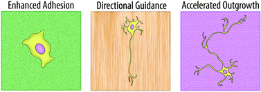
Many of these advances revolve around improving neural adhesion, providing directional guidance for axonal growth, and accelerating the speed of neurite outgrowth.  Numerous groups have become interested in using nanowire arrays to improve neuron adhesion, eliminating the need for any additional surface coating.  In a 2010 Nano Letters publication, Xie et al. were able to demonstrate the creation of a silicon nanopillar array that could selectively pin cortical neurons to desired locations.  Any cells that were seeded onto, or later came into contact with the nanopillars became immobilized, enabling a long-term observational study of their electrical activity.  Other groups have found that nanofiber bundles make excellent scaffolding materials for neurons and can be used to direct and align neurite growth.  In addition, when compared to planar surfaces, nanofiber-based substrates were shown to increase the speed at which initial neurite formation occurred.

Although the benefits of nano-textured substrates are clear, the mechanism by which neurons translate these physical cues into biological signals is still something of a mystery.  However, many of the reviewed papers suggest that actin filaments and focal adhesions (FA) play a major role.  One group found that f-actin often forms networks which resemble the shape of the underlying surface topography and that the impairment of f-actin polymerization completely erases any ability neurons have to respond to nanotopographical cues.  Other groups have demonstrated that the size of FA is proportional to the degree of neuronal alignment with underlying surface structures.  An increase in alignment leads to a larger FA which ultimately results in a more stable neurite attachment.

Regardless of the mechanism, it has become clear in recent years that nanotopography is an important parameter for the controlled manipulation of neuronal behavior.  In this mini-review, Kim et al. are able to provide an interesting overview on the progress that has already been made within this area.

Neurons on nanometric topographies: insights into neuronal behaviors in vitro
M. Kim, M. Park, K. Kang, and I.S. Choi.
Biomater. Sci., 2014, 2, 148-155 DOI: 10.1039/C3BM60255A

Ellen Tworkoski is a guest web-writer for Biomaterials Science.  She is currently a graduate student in the biomedical engineering department at Northwestern University (Evanston, IL, USA).

Follow the latest journal news on Twitter @BioMaterSci or go to our Facebook page.

To keep up-to-date with all the latest research, sign-up to our RSS feed or Table of contents alert.

Digg This
Reddit This
Stumble Now!
Share on Facebook
Bookmark this on Delicious
Share on LinkedIn
Bookmark this on Technorati
Post on Twitter
Google Buzz (aka. Google Reader)

NanoBio Australia 2014

Incorporating the 5th International NanoBio Conference & 3rd International Conference on BioNano Innovation (ICBNI), NanoBio Australia 2014 will take place on 6-10th July 2014 in Brisbane, Australia.

The intersection of Biology with Nanoscience and Nanotechnology currently represents one of the most exciting wellsprings of scientific innovation, and is also a major stimulus for a plethora of high value technologies and industries. By combining the two largest conferences in the world which focus on this scientific frontier, NanoBio Australia 2014 will feature a diverse array of multi-disciplinary science designed to connect world-leading scientists, engineers and entrepreneurs working in this space.

International Plenary Speakers:

Please see the conference website to register your attendance or submit an abstract.

Digg This
Reddit This
Stumble Now!
Share on Facebook
Bookmark this on Delicious
Share on LinkedIn
Bookmark this on Technorati
Post on Twitter
Google Buzz (aka. Google Reader)

Lectureship nominations close on Friday 7th March!

Time is running out to make your nominations for the Biomaterials Science Lectureship! Please submit your nominations by Friday 7th March 2014.

A reminder of the details…

Qualification

To be eligible for the Biomaterials Science Lectureship, the candidate should be in the earlier stages of their scientific career, typically within 15 years of attaining their doctorate or equivalent degree, and will have made a significant contribution to the field.

Description

The recipient of the award will be asked to present a lecture three times, one of which will be located in the home country of the recipient. The Biomaterials Science Editorial Office will provide the sum of £1000 to the recipient for travel and accommodation costs.

The award recipient will be presented with the award at one of the three award lectures. They will also be asked to contribute a lead article to the journal and will have their work showcased on the back cover of the issue in which their article is published.

Selection

The recipient of the award will be selected and endorsed by the Biomaterials Science Editorial Board.

Nominations

Those wishing to make a nomination should send details of the nominee, including a brief C.V. (no longer than 2 pages A4) together with a letter (no longer than 2 pages A4) supporting the nomination, to the Biomaterials Science Editorial Office (biomaterialsscience-rsc@rsc.org ) by 7th March 2014.  Self-nomination is not permitted.

We look forward to receiving your nominations!

Digg This
Reddit This
Stumble Now!
Share on Facebook
Bookmark this on Delicious
Share on LinkedIn
Bookmark this on Technorati
Post on Twitter
Google Buzz (aka. Google Reader)

Introducing new Editorial Board Member Adah Almutairi

We are very pleased to welcome Professor Adah Almutairi to the Biomaterials Science Editorial Board.

Adah Almutairi is co-director of the joint KACST-UC San Diego Center for Excellence in Nanomedicine and Engineering and an associate professor in the Skaggs School of Pharmacy and Pharmaceutical Sciences at UC San Diego, with secondary appointments in NanoEngineering and Materials Science. Her own research group, the Laboratory for Bioresponsive Materials, creates novel smart materials for on-demand drug delivery, regeneration of damaged tissue, and safe image-based diagnosis. She came to UC San Diego in 2008 from UC Berkeley, where she worked with Professor Jean Fréchet to develop several nanoprobes for in vivo imaging of pH and angiogenesis.  Prof. Almutairi is the recipient of an NIH New Innovator Award and has been recognized as a rising star in the field of polymeric materials by Chemical Communications and the ACS Division of Polymeric Materials Science and Engineering.

Adah’s recent papers include:

Increasing materials’ response to two-photon NIR light via self-immolative dendritic scaffolds
Nadezda Fomina, Cathryn L. McFearin and Adah Almutairi  
Chem. Commun., 2012, 48, 9138-9140 DOI: 10.1039/C2CC00072E

Metal chelating crosslinkers form nanogels with high chelation stability
Jacques Lux, Minnie Chan, Luce Vander Elst, Eric Schopf, Enas Mahmoud, Sophie Laurent and Adah Almutairi  
J. Mater. Chem. B, 2013, 1, 6359-6364 DOI: 10.1039/C3TB21104E, Paper

Antigen-loaded pH-sensitive hydrogel microparticles are taken up by dendritic cells with no requirement for targeting antibodies
Laura E. Ruff, Enas A. Mahmoud, Jagadis Sankaranarayanan, José M. Morachis, Carol D. Katayama, Maripat Corr, Stephen M. Hedrick and Adah Almutairi  
Integr. Biol., 2013, 5, 195-203 DOI: 10.1039/C2IB20109G

Digg This
Reddit This
Stumble Now!
Share on Facebook
Bookmark this on Delicious
Share on LinkedIn
Bookmark this on Technorati
Post on Twitter
Google Buzz (aka. Google Reader)

Zeolite–polymer composite adsorbs uremic toxins

Scientists in Japan have developed a nanofibre mesh that can adsorb creatinine from blood with the hope that it can eventually be developed into a wearable blood-cleaning device for patients with kidney failure.

It is hoped the mesh can eventually be developed into a wearable blood-cleaning device

It is hoped the mesh can eventually be developed into a wearable blood-cleaning device

Kidney failure causes dangerous concentrations of waste products, such as potassium, urea and creatinine, to build-up in the body. Apart from having a kidney transplant, the next best solution for patients is dialysis. Dialysis, however, is far from ideal. It is time-consuming and relies on access to specialist equipment, clean water, electricity, dialysate, and, usually, a hospital. Often these requirements aren’t accessible in rural parts of developing countries and disaster areas.

Read the full article at Chemistry World.

Fabrication of zeolite–polymer composite nanofibers for removal of uremic toxins from kidney failure patients
Mitsuhiro Ebara  
Biomater. Sci., 2014, Advance Article
DOI: 10.1039/C3BM60263J

Digg This
Reddit This
Stumble Now!
Share on Facebook
Bookmark this on Delicious
Share on LinkedIn
Bookmark this on Technorati
Post on Twitter
Google Buzz (aka. Google Reader)

Mending a broken heart: Myocardial matrix hydrogels for cardiac tissue engineering

According to the World Health Organization, cardiovascular disease causes 17.3 million deaths worldwide, with projections reaching 23.3 million deaths by the year 2030. Unfortunately, heart attack patients still have limited therapeutic options, commonly relying on left ventricular assist devices (LVADs) and heart transplantation. To provide more modern therapies, physicians have turned to tissue engineers to develop biomaterials that enable local regeneration of the heart to restore function and ideally improve the quality of life for the patient.

Professor Karen Christman’s lab at the University of California – San Diego (UCSD) is exploring methods to fabricate injectable hydrogels for cardiac repair. In the present study, the lab has developed human myocardial matrix (HMM), and compared the material properties of HMM to previously fabricated porcine myocardial matrix (PMM). The materials are made directly from the decellularized extracellular matrix (ECM) from human and porcine hearts. The decellularized matrices are composed of the natural structural proteins found in the heart, which make the ideal environment needed to promote cardiac cell growth and maturation.

To produce HMM, seven human hearts with a patient age range of 41-69 years were decellularized using sodium dodecyl sulfate (SDS) and a series of lyophilization, milling, and digestion steps.  Due to the dramatic patient-to-patient variability between hearts, over 50% of the HMM solutions were not able to self-assemble into hydrogels at physiological conditions, unlike self-assembling PMM hydrogels. The irreproducibility of HMM hydrogel fabrication is likely due to differences in protein composition between HMM and PMM. Using mass spectrometry to identify the proteins present in the decellularized matrices, the authors showed that porcine and human hearts have inherent differences in their matrix composition.

Although HMM did not produce hydrogels reproducibly, the matrix was still useful for in vitro cell culture protocols. Using PMM and HMM as coatings for cell culture plates, increased proliferation of rat aortic smooth muscle cells (RASMCs) and human coronary artery endothelial cells (HCAECs) was observed on HMM coated plates compared to PMM coated plates. Additionally, cell cultured on both HMM and PMM matrices showed increased expression of early cardiac transcription factor markers in human fetal cardiomyocyte progenitor cells (hCMPCs). This result indicates that biochemical cues from the HMM and PMM proteins may enhance early stages of cardiomyocyte differentiation.

Even though HMM was shown to not be a likely candidate for clinical translation due to large variability between samples, PMM injectable hydrogels are still a promising alternative for improving cardiac repair in vivo. Additionally, HMM materials may be used for future in vitro cell culture and cardiomyocyte differentiation protocols.

Human versus porcine tissue sourcing for an injectable myocardial matrix hydrogel
Todd D. Johnson, Jessica A. DeQuach, Roberto Gaetani, Jessica Ungerleider, Dean Elhag, Vishal Nigam, Atta Behfar, and Karen L. Christman
Biomater. Sci., 2014, Advance Article, DOI: 10.1039/C3BM60283D

Brian Aguado is currently a Ph.D. Candidate and NSF Fellow in the Biomedical Engineering department at Northwestern University. He holds a B.S. degree in Biomechanical Engineering from Stanford University and a M.S. degree in Biomedical Engineering from Northwestern University. Read more about Brian’s research publications here.

To keep up-to-date with all the latest research, sign-up to our RSS feed or Table of contents

Digg This
Reddit This
Stumble Now!
Share on Facebook
Bookmark this on Delicious
Share on LinkedIn
Bookmark this on Technorati
Post on Twitter
Google Buzz (aka. Google Reader)