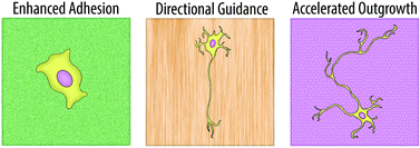
The ability to control neuronal behavior and growth is highly sought among researchers interested in neuro-regenerative medicine. Over the past two decades, it has become widely accepted that modifications in a substrate’s physical surface topography can influence the growth patterns of seeded neurites. However, the advent of techniques that are capable of fabricating nano-textured surfaces has revealed that the magnitude of this influence may be much greater than originally thought. In this paper, a group of scientists from KAIST review a number of recent advances in the development of nano-topographies which can influence neuronal behavior.
Many of these advances revolve around improving neural adhesion, providing directional guidance for axonal growth, and accelerating the speed of neurite outgrowth. Numerous groups have become interested in using nanowire arrays to improve neuron adhesion, eliminating the need for any additional surface coating. In a 2010 Nano Letters publication, Xie et al. were able to demonstrate the creation of a silicon nanopillar array that could selectively pin cortical neurons to desired locations. Any cells that were seeded onto, or later came into contact with the nanopillars became immobilized, enabling a long-term observational study of their electrical activity. Other groups have found that nanofiber bundles make excellent scaffolding materials for neurons and can be used to direct and align neurite growth. In addition, when compared to planar surfaces, nanofiber-based substrates were shown to increase the speed at which initial neurite formation occurred.
Although the benefits of nano-textured substrates are clear, the mechanism by which neurons translate these physical cues into biological signals is still something of a mystery. However, many of the reviewed papers suggest that actin filaments and focal adhesions (FA) play a major role. One group found that f-actin often forms networks which resemble the shape of the underlying surface topography and that the impairment of f-actin polymerization completely erases any ability neurons have to respond to nanotopographical cues. Other groups have demonstrated that the size of FA is proportional to the degree of neuronal alignment with underlying surface structures. An increase in alignment leads to a larger FA which ultimately results in a more stable neurite attachment.
Regardless of the mechanism, it has become clear in recent years that nanotopography is an important parameter for the controlled manipulation of neuronal behavior. In this mini-review, Kim et al. are able to provide an interesting overview on the progress that has already been made within this area.
Neurons on nanometric topographies: insights into neuronal behaviors in vitro
M. Kim, M. Park, K. Kang, and I.S. Choi.
Biomater. Sci., 2014, 2, 148-155 DOI: 10.1039/C3BM60255A
Ellen Tworkoski is a guest web-writer for Biomaterials Science. She is currently a graduate student in the biomedical engineering department at Northwestern University (Evanston, IL, USA).
Follow the latest journal news on Twitter @BioMaterSci or go to our Facebook page.
To keep up-to-date with all the latest research, sign-up to our RSS feed or Table of contents alert.


