Author Archive

A new PLS method for studying nonlinearity

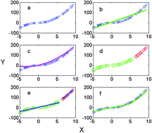
Statistical methods such as partial least squares (PLS) and principle component analysis (PCA) have been used to process vibrational spectra in order to understand certain trends between samples and their spectra. However, these methods and similar ones break down when the variables demonstrate a nonlinear relationship.

Guangzaho Huang and researchers at Wenzhou University explored updating a genetic algorithm to process nonlinear data. They found that this genetic algorithm PLS (GS-PLS) model performed more effectively than other PLS models did, particularly when nonlinearity was the primary restricting factor.  The GS-PLS worked in both simulated data sets and using near infrared spectra from different materials.

To read more about this new method, click the link below. It will be free to read until May 11th.

A segmented PLS method based on genetic algorithm
Guangzao Huang,  Xiukai Ruan,  Xiaojing Chen,  Dongxiu Lina and  Wenbin Liua
Anal. Methods, 2014, 6, 2900-2908
DOI: 10.1039/C3AY41765D

Digg This
Reddit This
Stumble Now!
Share on Facebook
Bookmark this on Delicious
Share on LinkedIn
Bookmark this on Technorati
Post on Twitter
Google Buzz (aka. Google Reader)

Probing tissue calcifications for cancer

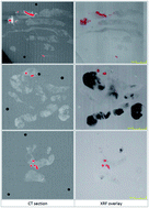
The presence of calcifications is an early indicator of breast cancer and is detected using a mammography to diagnose the disease. However, both malignant and benign tissues present these calcifications, and the small chemical differences in both cases have been difficult to distinguish. Vibrational spectroscopy has been used to study these differences, but the difficulty arises in trying to locate them deep in tissue.

Researchers in the UK at Gloucestershire Royal Hospital, Cranfield University and the University of Exeter compared micro CT and X-ray fluorescence (XRF) in locating calcifications embedded within wax biopsy tissue samples. Both techniques successfully located calcifications, and CT had some advantages in resolution, speed and depth. Furthermore, typical collection, such as the dyes used in histological staining can affect the collected spectra. An alternative method such as CT coupled with spectroscopy could potentially find calcifications and detect cancer.

To read more about this study, click the link below which is free until April 19th.

Locating microcalcifications in breast histopathology sections using micro CT and XRF mapping
Robert Scott, Catherine Kendall, Nicholas Stone and Keith Rogers
Anal. Methods, 2014, Advance Article
DOI: 10.1039/C3AY42158A

Digg This
Reddit This
Stumble Now!
Share on Facebook
Bookmark this on Delicious
Share on LinkedIn
Bookmark this on Technorati
Post on Twitter
Google Buzz (aka. Google Reader)

Binding Affinity of DNA Plasmids Using Surface Plasmon Resonance

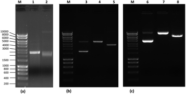
Different plasmind isoforms on agarose gel

Nucleic acids have emerged as a vital tool for targeted gene therapy treatments. In particular, plasmid DNA (pDNA), has become important in pharmaceutical research. Typically, pDNA exists in supercoiled, linear, and open-circular isoforms, and affinity chromatography can separate the supercoiled from these other impurities. Although the capture ligand used in affinity chromatography demonstrates high specificity, it cannot determine the binding strength of the plasmid to the ligand.

Researchers at University of Beira Interior in Portugal measured the binding kinetics of pDNA using surface plasmon resonance spectroscopy (SPR). They successfully calculated dissociation constants for all three types of plasmid isoforms, with supercoiled showing the tightest binding to an affinity chromatography mimic support.

To know more about the study, please read the full article below. It will be free for you to enjoy until April 2nd .

Affinity analysis between immobilized L-arginine and plasmid isoforms provided by surface plasmon resonance
C. Cruz ,  A. Sousa ,  F. Sousa and João A. Queiroz
Anal. Methods, 2013, Advance Article
DOI: 10.1039/C3AY40213D

Digg This
Reddit This
Stumble Now!
Share on Facebook
Bookmark this on Delicious
Share on LinkedIn
Bookmark this on Technorati
Post on Twitter
Google Buzz (aka. Google Reader)

Evaluating Procedures for Lead Detection

Trace lead analysis performed using ID-CP-MS

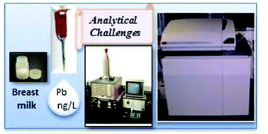
A toxin or heavy metal in a mother’s body can transfer to her baby, and even though lead is primarily stored in bones, trace amounts have the potential to enter breast milk and create a dangerous health risk.

Although multiple testing procedures are available, they have demonstrated a wide range of sensitivities arising from sample contamination and preparation issues. Adrienne Ettinger and researchers at Harvard School of Public Health, USA,  evaluated three digestion procedures for lead analysis using isotope dilution inductively coupled mass spectrometry (ID-ICP-MS).  The primary goal was to see if they could achieve 100% efficiency in any of the digestion processes in the complex media of breast milk, which contains high levels of contaminants such as fat and calcium, and trace levels of lead. Of these, the high pressure asher demonstrated complete sample digestion and had the most consistent lead concentration measurement. By removing analyte contaminants and maximizing the preparation efficiency, they detected trace levels of lead down to 0.2 ng mL-1.

To read more about this research, please access the link below. This paper will be free to read until March 8th.

Comparison of digestion procedures and methods for quantification of trace lead in breast milk by isotope dilution inductively coupled plasma mass spectrometry
Chitra J. Amarasiriwardena ,  Innocent Jayawardene ,  Nicola Lupoli ,  Ramon M. Barnes ,  Mauricio Hernandez-Avila ,  Howard Hu and Adrienne S. Ettinger
Anal. Methods, 2013, Advance Article
DOI: 10.1039/C3AY26321E

Digg This
Reddit This
Stumble Now!
Share on Facebook
Bookmark this on Delicious
Share on LinkedIn
Bookmark this on Technorati
Post on Twitter
Google Buzz (aka. Google Reader)

A Portable HPLC for On Site Water Testing

Antibiotics provide vital treatment in fighting bacterial infection, but over time some have become phased out or even toxic to humans.

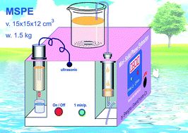
Miniaturized device for detection of chloramphenicol in water

One of these is chloramphenicol. Although banned in the European Union, chloramphenicol continues to be used as a cheap remedy in veterinary medicine and can lead to contaminated water and food for our consumption. One technique to test for this contaminant, solid phase extraction (SPE), purifies samples effectively, but requires a pump and a laboratory setting.

Alternatively, Sheng Liu, from the Chinese Academy of Sciences, and colleagues developed a simple yet portable miniaturized SPE (MSPE) device capable of detecting chloramphenicol in water, which has been difficult to test using other methods. A macroporous resin captures and concentrates the chloramphenicol before it passes through an HPLC, all within a 1.5 kg mass instrument. This new technique directly detects low concentrations of chloramphenicol in water, and demonstrates the capacity to function at contaminated locations.

To know more about the study, please click on the link below. This paper will be free to read for the next two weeks.

On-site solid phase extraction and HPLC determination of chloramphenicol in surface water and sewage
Sheng Liu ,  Xian-Zheng Wu ,  Zi-Hui Gao and Fang Jiao
Anal. Methods, 2013, Advance Article
DOI: 10.1039/C2AY26162F

Digg This
Reddit This
Stumble Now!
Share on Facebook
Bookmark this on Delicious
Share on LinkedIn
Bookmark this on Technorati
Post on Twitter
Google Buzz (aka. Google Reader)

Novel Separation Method for Cadmium Detection

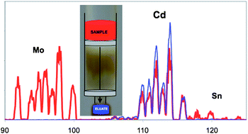
Trace concentrations of cadmium in water, soil and industrial settings can have devastating health effects if not detected. Furthermore, measuring accurate cadmium levels in these complex samples is hindered by signal interference from other metals such as tin, molybdenum, and zirconium.

Robert Thompson and Steven Christopher in the United States developed a four-step separation methodology to remove these metals, and then used inductively coupled plasma mass spectrometry to measure cadmium concentrations. For this procedure, commercially available solid phase extraction cartridges isolate most of cadmium and strong ion exchange chromatography removes tin and molybdenum. This unique sample preparation facilitates the detection of low cadmium concentrations in marine sediment and rice flour which contain high levels of tin and molybdenum respectively.

Determination of cadmium in samples containing molybdenum and tin

To know more about this study, please access the link below. This paper will be free to read until February 18th.

Novel separation for the determination of cadmium by isotope dilution ICP-MS in samples containing high concentrations of molybdenum and tin
Robert Q. Thompson and Steven J. Christopher
Anal. Methods, 2013, Advance Article
DOI: 10.1039/C2AY26212F

Digg This
Reddit This
Stumble Now!
Share on Facebook
Bookmark this on Delicious
Share on LinkedIn
Bookmark this on Technorati
Post on Twitter
Google Buzz (aka. Google Reader)

A Novel Assay for Studying DNA Repair

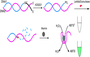
A novel DNAzyme-based colorimetric assay for the detection of hOGG1 activity with lambda exonuclease cleavage

Yu et al., Anal. Methods, 2013, Advance Article

Mutation, apoptosis and cancer can develop from damaged DNA, however the cell produces proteins capable of repairing DNA to prevent these dangerous effects. One of these proteins produced by cells, 8-oxoG DNA glycosylase/AP lyase (hOGG1), targets DNA lesions caused by reactive oxygen species. Typically studying this glycosylase requires large instrumentation, such as HPLC, or radioactive labeling. Ru-Qin Yu and coworkers at Hunan University developed a system, where the hOGG1 cleaves at the mismatch site on a DNA strand, and releases a DNAzyme mimetic, which produces a colored signal. This rapid and sensitive colorimetric assay measured the activity of hOGG1 for a range of concentrations, with an affordable DNA probe, and user-friendly instrumentation.

This paper will be free to read until Nov 30th.

A novel DNAzyme-based colorimetric assay for the detection of hOGG1 activity with lambda exonuclease cleavage
Shu-Cheng Liu, Hui-Wang Wu, Jian-hui Jiang, Guo-Li Shen and Ru-Qin Yu
Anal. Methods, 2013, Advance Article
DOI: 10.1039/C2AY26018B

Digg This
Reddit This
Stumble Now!
Share on Facebook
Bookmark this on Delicious
Share on LinkedIn
Bookmark this on Technorati
Post on Twitter
Google Buzz (aka. Google Reader)