Emerging Investigator Series – Joo H. Kang

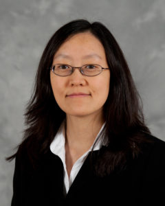
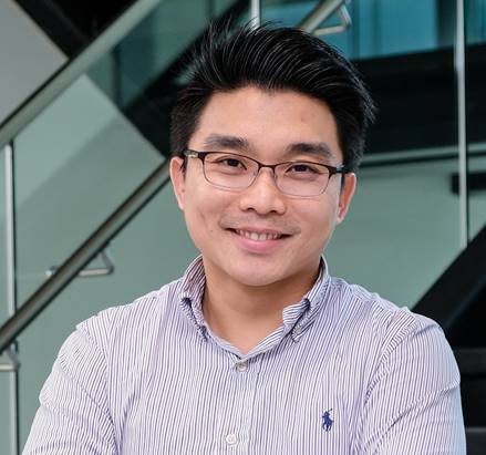
Dr. Joo H. Kang is currently an Assistant Professor of the Department of Biomedical Engineering, School of Life Sciences at Ulsan National Institute of Science and Technology (UNIST), Korea. He received double Bachelor’s degrees in Chemical Engineering and Life Science from Sogang University in 2002 and his M.S. and Ph.D. in Bioengineering from Korea Advanced Institute of Science and Technology (KAIST) in 2004 and 2008, respectively. He joined Children’s Hospital Boston, Harvard Medical School as a research fellow in 2009, and he continued his work at the Wyss Institute, Harvard University as a Wyss Technology Development Fellow from 2012-2016. He received several awards in his early career, including Postdoctoral Award for Professional Development from Harvard University, Wyss Technology Development Fellowship from Harvard University, Baxter Young Investigator Award from Baxter Inc., and Young Frontiers in Bio and Braining Engineering from KAIST. His research interests include multiscale biofluidic approaches for tackling infectious diseases and cancer, and miniaturized organ-mimicking microsystems.

Read Joo H. Kang’s Emerging Investigator article “Measurement of the magnetic susceptibility of subtle paramagnetic solutions using the diamagnetic repulsion of polymer microparticles” and find out more about him in the interview below:


Your recent Emerging Investigator Series paper focuses on measuring magnetic susceptibility of subtle paramagnetic solution using diamagnetic repulsion of polymer microparticles. How has your research evolved from your first article to this most recent article?

One of the research topics that interested me was to discriminate the subtle differences in the magnetic susceptibility of materials in a microfluidic regime. The first paper I published in regards to this (Kang,JH, et al., JACS, 2009) demonstrated the capability of discriminating the magnetic susceptibility of “solid microparticles” where they are diamagnetically forced to be located at a quasi-isomagnetic position in a microfluidic channel (a position where the differences of the magnetic susceptibility between the solid particles and surrounding media become nearly zero). When I was invited to make contribution to the Emerging Investigator Series of Lab on a Chip last year, I wanted to revisit this, and this time I aimed to assess the subtle magnetic susceptibility of “surrounding paramagnetic solutions”.

What aspect of your work are you most excited about at the moment?

As for the paper, I was surprised of the sensitivity of the device that can discriminate the magnetic susceptibility. We compared our results with those assessed by a conventional superconducting quantum interference device (SQUID), and found that our approach is even more sensitive than the conventional one. Likewise, we can unveil various scientific approaches when exploring fluidic regimes at the micro and nanoscale, and this is the most exciting aspect as being a part of the research community in this field.

In your opinion, what applications can your current approach be used for?

Because this is a platform technology, various applications are possible where we need to measure the magnetic susceptibility of paramagnetic solutions. Assessment of residual magnetic nanoparticles in biological samples, for examples, would be one of the potential uses. We could also use this platform to evaluate metal contamination of drinking water, such as chromium or iron oxide, which alters the magnetic susceptibility of water.

What do you find most challenging about your research?

Taking research from the “bench to products”. Since I started my independent research career, I realized that I have to make considerable efforts to get this happen while playing multiple roles at the same time. But I am enjoying it.

In which upcoming conferences or events may our readers meet you?

I am planning to attend microTAS 2019 that will be held in Basel, Switzerland this year.

How do you spend your spare time?

I am spending my time with my family, hiking, swimming, and playing soccer or games with my little son and daughter.

Which profession would you choose if you were not a scientist?

Probably an architect. This was one of the paths I was thinking of when I was a high school student.

Can you share one piece of career-related advice or wisdom with other early career scientists?

A clear vision on your own research and collaborators who you can share your vision with.

Digg This
Reddit This
Stumble Now!
Share on Facebook
Bookmark this on Delicious
Share on LinkedIn
Bookmark this on Technorati
Post on Twitter
Google Buzz (aka. Google Reader)

Emerging Investigator Series – Mei He

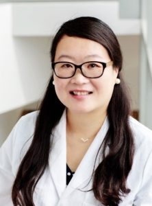
Dr. He is a tenure-track assistant professor at the University of Kansas. She received her PhD degree from the University of Alberta with professor Jed Harrison, and postdoctoral training from the University of California, Berkeley with professor Amy Herr. She is the vice chair of the ASABE Biosensor program and the Councilor of the American Electrophoresis Society. Dr. He is also the founder of Clara Biotech Inc. and the founder committee for the MidWest 3D technology society. Dr. He Received NIH Maximizing Investgator’s Research Award for Early Stage Investigators in 2019. She also received the Lab on Chip Outstanding Reviewer for the year of 2018. One of her publications also received the 2018 SLAS Technology Readers Choice Award. Her research interests include biomedical microfluidic devices and sensing approaches, 3D biomaterials, and nanodelivery, employed in programming and monitoring biomimetic immunity associated with extracellular vesicles.

Read Mei He’s Emerging Investigator article “3D-printing enabled micro-assembly of a microfluidic electroporation system for 3D tissue engineering” and find out more about her in the interview below:


Your recent Emerging Investigator Series paper focuses on “3D-printing Enabled Micro-assembly of Microfluidic Electroporation System for 3D Tissue Engineering”. How has your research evolved from your first article to this most recent article?

My first article observed the microscale evolution of porous polymer materials in the microfluidic channel when I was a PhD student. I found very interesting phenomena in the microfluidic device which actually inspired me to explore more surrounding dimensions, surface chemistry, and scales. Till to my recent article focusing on 3D geometric influence on cellular behavior and their extracellular vesicles secretion dynamics, the 3D dimension in microscale is intriguing in the biological system.

What aspect of your work are you most excited about at the moment?

I am very excited to take the microfluidic technology and phenomena into the biology world, as it will bring new investigation and discovery. Biology is still in its infancy stage, I am very excited to see how microfluidic technology could advance this growth.

In your opinion, what is the biggest impact your microfluidic electroporation system will have in tissue engineering?

Intracellular delivery of regulatory or therapeutic targets into the cell is very crucial in the field of tissue engineering and regenerative medicine. Current existing electro-transfection systems, including microfluidic platforms and commercial benchtop systems, are only able to study monolayer cell suspensions in vitro, which is incapable of clinical translation within in vivo tissue microenvironment. So developing a 3D, in vivo like tissue microenvironment with effective electro-transfection is very important to move to the clinical study in the future. We actually are more interested in downstream, precise control and manipulation of cellular machinery for secreting exosomes and extracellular vesicles under the transfection-induced stimulus, such technology is not existing yet but very important for understanding the interconnection of cargo internalization with cellular level responses elicited by exosomes delivery pathway.

What do you find most challenging about your research?

Building up an in vivo like tissue system with precise control is not straightforward. The environment in a controlled lab setting is totally different than in an in vivo biological system. So the analyzed information actually is not representative of the real situation in the human in vivo system. There are huge heterogeneities present in the cell population as well as human individuals, which poses the challenges for correctly understanding cellular system regulation, such as immunity, in our human body. Mimicking in vivo living system is very challenging, but crucial for understanding quite a few of mechanism and disease pathogenesis. Our research introduces new microfluidic technology and material solutions to solve such challenges.

In which upcoming conferences or events may our readers meet you?

I will attend next year Gorden Research Conference in Bioanalytical Sensors as well as the MicroTAS annual meeting.

How do you spend your spare time?

I have a 7-year-old boy and expecting a new baby girl this year. My spare time definitely is occupied by kids and watching them growing.

Which profession would you choose if you were not a scientist?

I always like to discover new things since I was a child. If I am not a scientist, I would like to be a greeting card designer or paleontologist.

Can you share one piece of career-related advice or wisdom with other early career scientists?

Being a life-long learner and always keeping strong scientific curiosity will definitely help with your research development. Get good mentors around you and you will appreciate their advice.

Digg This
Reddit This
Stumble Now!
Share on Facebook
Bookmark this on Delicious
Share on LinkedIn
Bookmark this on Technorati
Post on Twitter
Google Buzz (aka. Google Reader)

Through a cheaper ‘book of life’

Mapping the human genome project has been one of the world’s largest scientific collaborations. Completing the full genome sequencing for “the book of life” took more than 10 years with the efforts of 1000’s scientists and a budget of $3 billion. About 20 years after the finalization of this enormous project, it is now possible to complete a full human genome sequencing within 8 days for about $1,000 thanks to more advanced sequencing tools. Further improvements in genome sequencing tools are still warranted today because the genome sequencing field has been embraced by many more applications, including forensics, disease modeling & identification, and personalized medicine (e.g., identifying the genes that cause a medicine to work in one patient but not in another).

Initial sequencing technologies relied on standard DNA electrophoresis techniques such as slab gels and capillaries, allowing for the preparation of only small numbers of samples at a time. The sample preparation limitation was the primary reason for the increased costs and processing duration during the human genome project. Many efforts have been directed towards improving sample preparation techniques in the last decades. As the first step, electrophoresis techniques have been optimized to boost the sample throughput with user-friendly, smaller, and functional platforms. Traditional DNA separation gels, which have been used as the golden standard for many decades, have been replaced by microfabricated post arrays and nanometer-scale deterministic lateral displacement arrays.

A nice example of nanometer-scale deterministic lateral displacement arrays has been demonstrated recently by researchers from IBM T.J. Watson Research Center and Icahn School of Medicine at Mount Sinai in New York, USA. In this work, the researchers fractioned DNA in the range of 100-10.000 base pairs with a size-selective resolution of 200 base pairs. To achieve that, four different microchip configurations were fabricated on silicon wafers, where the array and nanopillar sizes were tuned for each configuration to obtain the optimum separation performance for the selected range of DNA fragments. Each configuration contained several separate arrays to conduct independent runs in a single chip. Instead of applying an electric field, the researchers applied pressure-driven force to separate the fragments. This strategy is particularly useful for separating non-charged species without being affected by buffer conditions (e.g., ionic strength).

The separation mechanism in the nanometer-scale deterministic lateral displacement array is simple: If the size of a DNA fragment is larger than the diameter of the pillars, the fragment is deflected towards the collection wall at a large angle, also called the bump mode. If the size of a DNA fragment is smaller than the diameter of the pillars, the fragment migrates at an angle nominally zero, termed as the zig zag mode. In such a system, diffusion of DNA fragments lead to intermediate migration angles, termed as the partial-bump mode. Different sizes of DNA fragments could be separated in the array since the fragments will follow distinct trajectories thanks to the existence of different modes. Figure 1 summarizes the separation mechanisms and gives an outline for the nanometer-scale deterministic lateral displacement array.

In the nanometer-scale deterministic lateral displacement array, the gap sizes were tuned from microscale to nanoscale only, without application of any other molecules that could change the DNA diffusion behavior, ionic strength (changing the effective gap distances). In such a setting, the researchers identified, for the first time, the flow velocity-dependence of different fragment lengths. Mainly, changing flow velocity caused a transition between bump and zig zag modes for the given size range of different DNA fragments: Slow speeds lead to partial-bump mode, and high speeds lead to the collapse of all DNA fragments to zigzag mode. The nanometer-scale deterministic lateral displacement array could also be used as a purification tool with 75% recovery and 3-fold concentration enhancement of DNA fragments. This tool could be used effectively for preparing next-generation sequencing libraries, on-chip DNA characterization, and circulating DNA characterization applications.


Figure 1. The nanometer-scale deterministic lateral displacement array and DNA separation mechanism at different flow velocities.

To download the full article for free* click the link below:

Gel-on-a-chip: continuous, velocity-dependent DNA separation using nanoscale lateral displacement

Benjamin H. Wunsch, Sung-Cheol Kim, Stacey M. Gifford, Yann Astier, Chao Wang, Robert L. Bruce, Jyotica V. Patel, Elizabeth A. Duch, Simon Dawes, Gustavo Stolovitzky and  Joshua T. Smith, Lab Chip, 2018, Lab on a Chip Articles

DOI: 10.1039/c8lc01053f


About the Webwriter

Burcu Gumuscu is a researcher in Mesoscale Chemical Systems Group at the University of Twente in the Netherlands. Her research interests include the development of microfluidic devices for quantitative analysis of proteins from single-cells, next-generation sequencing, compartmentalized organ-on-chip studies, and desalination of water on the microscale.


Digg This
Reddit This
Stumble Now!
Share on Facebook
Bookmark this on Delicious
Share on LinkedIn
Bookmark this on Technorati
Post on Twitter
Google Buzz (aka. Google Reader)

Lab on a Chip thematic collection on droplet-based single cell sequencing

Lab on a Chip is delighted to share with you our Thematic Collection on droplet-based single-cell sequencing.

The droplet-based single-cell sequencing field is advancing very rapidly. Large numbers of studies are underway to collect and explore the new information that is now accessible with single-cell RNA-seq. Improvements to the microfluidics are also advancing rapidly. This collection of papers and reviews focusses on the between the technological advancements and high impact applications of droplet-based single-cell sequencing.

This topical and exciting collection is collated by Thought leader Dave Weitz and the Lab on a Chip Editorial Board. The collection is introduced in a perspective on single cell sequencing by the Thought leader Dave Weitz, and in two editorials, one on “InDrops and Drop-seq” by Allon Klein and Evan Macosko and one on “an engineer and business person’s perspective” by businessman and engineer Mark Gilligan.

Read the full collection at: http://rsc.li/drop-sc-seq

Below is a selection of content highlights featured in the collection. In addition, all papers are free to read until 31st May*

Perspective

Droplet-based single cell RNAseq tools: a practical guide

Robert Salomon, David Gallego-Ortega, et al.

Critical Review

Finding a helix in a haystack: nucleic acid cytometry with droplet microfluidics

Iain C. Clark and Adam R. Abate

Paper

High throughput gene expression profiling of yeast colonies with microgel-culture Drop-seq

Leqian Liu, Adam R. Abate, et al.

Paper

Simplified Drop-seq workflow with minimized bead loss using a bead capture and processing microfluidic chip

Marjan Biočanin, Bart Deplancke, et al.

Lab on a Chip is the leading journal publishing significant and original work related to miniaturisation, at the micro- and nano-scale, of interest to a multidisciplinary readership with an Journal Impact Factor of 5.995**. The journal is guided by Editor-in-Chief Abraham (Abe) Lee (University of California, Irvine) who is supported by our team of Associate Editors (Yoon-Kyoung Cho, Petra Dittrich, Hang Lu, Jianhua Qin, Manabu Tokeshi, Joel Voldman and Aaron Wheeler).

We hope you enjoy reading the papers within this Thematic Collection and we welcome future submissions on droplet-based single-cell sequencing.

Dolomite/Lab on a Chip Pioneers of Miniaturization Lectureshipdeadline approaching-nominate a colleague now!

Organ-on a-chip systems- translating concept into practice thematic collectionSubmit now

Organ-,body- and disease-on-a-chip thematic collectionRead now

Personalised medicine:liquid biopsyRead now

Lab on a Chip Emerging Investigator Series Apply now

Digg This
Reddit This
Stumble Now!
Share on Facebook
Bookmark this on Delicious
Share on LinkedIn
Bookmark this on Technorati
Post on Twitter
Google Buzz (aka. Google Reader)

Lab on a Chip thematic collection on personalised medicine-liquid biopsy

Lab on a Chip is delighted to share with you our Thematic Collection on personalised medicine-liquid biopsy.

This collection of papers and reviews focusses on the interface between the technological advancements and high impact applications of liquid biopsy technologies. This collection is collated by Thought Leaders Mehmet Toner and Stefanie Jeffrey and the Lab on a Chip Editorial Board and is introduced in an Editorial by the Thought Leaders on Liquid biopsy: a perspective for probing blood for cancer

Read the full collection at: http://rsc.li/liquid-biopsy

Below is a selection of content highlights featured in the collection. In addition, all papers are free to read until 31st May*

Tutorial Review

Cancer diagnosis: from tumor to liquid biopsy and beyond

Ramanathan Vaidyanathan, Chwee Teck Lim, et al.

Critical Review

Circulating tumor DNA and liquid biopsy: opportunities, challenges, and recent advances in detection technologies

Lena Gorgannezhad, Nam-Trung Nguyen, Muhammad J. A. Shiddiky et al.

Paper

Dynamic CTC phenotypes in metastatic prostate cancer models visualized using magnetic ranking cytometry

Leyla Kermanshah, Shana O. Kelley et al.

Paper

An ultrasensitive test for profiling circulating tumor DNA using integrated comprehensive droplet digital detection

Chen-Yin Ou, Timothy J. Abram, Weian Zhao et al.

Paper

Cancer marker-free enrichment and direct mutation detection in rare cancer cells by combining multi-property isolation and microfluidic concentration

Soo Hyeon Kim, Teruo Fujii et al.

Paper

Urine-based liquid biopsy: non-invasive and sensitive AR-V7 detection in urinary EVs from patients with prostate cancer

Hyun-Kyung Woo, Hong Koo Ha, Yoon-Kyoung Cho et al.


Lab on a Chip is the leading journal publishing significant and original work related to miniaturisation, at the micro- and nano-scale, of interest to a multidisciplinary readership with an Journal Impact Factor of 5.995**. The journal is guided by Editor-in-Chief Abraham (Abe) Lee (University of California, Irvine) who is supported by our team of Associate Editors (Yoon-Kyoung Cho, Petra Dittrich, Hang Lu, Jianhua Qin, Manabu Tokeshi, Joel Voldman and Aaron Wheeler).

We hope you enjoy reading the papers within this Thematic Collection!

Keep up to date with Lab on a Chip throughout the year by signing up for free table of contents alerts and monthly e-newsletters.

Dolomite/Lab on a Chip Pioneers of Miniaturization Lectureshipdeadline approaching-nominate a colleague now!

Organ-on a-chip systems- translating concept into practice thematic collectionSubmit now

Organ-,body- and disease-on-a-chip thematic collectionRead now

Droplet-based single-cell sequencingRead now

Lab on a Chip Emerging Investigator Series Apply now

Digg This
Reddit This
Stumble Now!
Share on Facebook
Bookmark this on Delicious
Share on LinkedIn
Bookmark this on Technorati
Post on Twitter
Google Buzz (aka. Google Reader)

Emerging Investigator Series – Mathieu Odijk

Mathieu Odijk is an associate Professor at the University of Twente running his own research theme on Micro- and Nanodevices for Chemical Analysis. He received his PhD in Electrical Engineering from the University of Twente under the guidance of Albert van den Berg for his work on electrochemical microreactors for drug screening and proteomics applications in 2011. He has broaden his scope by various research visits at EPFL Lausanne (2012), the Wyss Institute at Harvard (2013), and MIT (2014).

The common aim of his research is to design novel devices to measure chemical quantities, pushing boundaries in applications to explore unknown territory. Often, this relates to faster, or better spatially resolved measurements at lower concentrations in small volumes. Micro- and nanofabrication techniques are used to enhance electrochemical, optical or mass spectrometric readout. The ultimate goal is to create new, yet robust tools for routine use in the lab or point-of-care applications.

Read Mathieu Odijk ’s Emerging Investigator article “A miniaturized push–pull-perfusion probe for few-second sampling of neurotransmitters in the mouse brain” and find out more about him in the interview below:

How has your research evolved from your first article to your most recent Emerging Investigator article?

I have nice memories about my first paper, as it was also submitted to Lab on Chip and immediately accepted without revisions (only one small question from 1 of the referee’s). The topic of that first paper was about the design of an electrochemical microreactor to study oxidative conversions in drug metabolism studies. We have been quite successful with that topic, now extending it also in the direction of studying electrochemical oxidative protein cleavage, and disulphide bond reduction using e.g. spectroelectrochemical means.

Many “ingredients” included in that first paper also are present in current projects such as microfluidics, advanced cleanroom fabrication, and analytical chemistry. These ingredients also form a key component of the focus area of my own research group (Micro- and nanodevices for Chemical Analysis).

 What aspect of your work are you most excited about at the moment?

It is my aim to push the boundaries of existing analytical tools with respect to limit detection, spatial or temporal resolution, or enhancing the number of repeats using high-throughput technology. I’m really excited about this latest paper demonstrating a miniaturized push-pull perfusion probe, as it is indeed improving both the spatial and temporal resolution by at least 1 order of magnitude compared to commercially available probes. As such, it is a nice showcase of what can be achieved by microfluidics.

In your opinion, what are the most important questions to be asked/answered in this field of research? 

I think it is really important to focus on the final application, and find good collaboration partners. If I take this push-pull perfusion probe as example, this research originated from discussions with neuroscientist who complained about a lack of temporal information from their existing microdialysis probes. However, quite a number of papers that describe probes with microfluidic channels only demonstrate in-vitro results. As we also found out in our project, bridging the gap towards in-vivo is certainly not trivial. It requires compromises in the technological area which you would not address if you stick to in-vitro experiments.

More generally I believe that the field has matured; lab on chip technology has become a means to achieve a higher goal. In this case this higher goal is to study neurochemical processes in the brain in more detail. However, I think this project also clearly demonstrates that there can be a lot of science in engineering. In this case we had to overcome challenges in microfabrication, fluid dynamics, mass-transport, protein chemistry, and adsorption kinetics.

What do you find most challenging about your research?

What I find really interesting is that my research is very multi-disciplinary in nature, crossing traditional boundaries such as “chemistry”, “physics”, or “biology”. However, that also poses a challenge as it is easy to develop a blind spot if you are exploring a new field of research. Again I would like to stress the importance of a good collaboration with experts in these fields to prevent failures at an early stage.

In which upcoming conferences or events may our readers meet you?

That is easy: I always try to attend MicroTAS.

How do you spend your spare time?

I’m a father of two small children, aged 1 and 4. If they leave me some spare time (and energy), I like to do woodworking, cycling, and indoor climbing. I also really like outdoors ice skating, but global warming is unfortunately interfering with the number of days ice skating is possible in the Netherlands.

Which profession would you choose if you were not a scientist?

I always wanted to be an inventor, with teacher as a close runner-up. I guess becoming a scientist is actually pretty close to that childhood dream. Any alternative profession should allow me to be able to either create new things, or educate other people (or both).

Can you share one piece of career-related advice or wisdom with other early career scientists?

At various points in my tenure track, I felt pressure to perform. This can be stressful and is most definitely counter-productive. Try to keep seeing/finding the fun in science, e.g. by asking your PhD students to share their Eureka moments in the lab with you. All the rest is of lesser importance.

Digg This
Reddit This
Stumble Now!
Share on Facebook
Bookmark this on Delicious
Share on LinkedIn
Bookmark this on Technorati
Post on Twitter
Google Buzz (aka. Google Reader)

Emerging Investigator Series – Han Wei Hou

Dr. Han Wei Hou is currently an Assistant Professor at the School of Mechanical and Aerospace Engineering and the Lee Kong Chian School of Medicine (LKCMedicine), Nanyang Technological University (NTU), Singapore. He received his BEng (First Class Hons) and PhD degree in Biomedical Engineering from the National University of Singapore in 2008 and 2012, respectively. Upon graduation, he did his postdoctoral training at Massachusetts Institute of Technology (MIT), and subsequently joined LKCMedicine at NTU as the inaugural LKCMedicine Postdoctoral Fellow in 2014.

Dr. Hou has over 30 peer-reviewed scientific publications, and his work has been featured in online science (ScienceDaily, TheScientist, Cancerforall and Genomeweb), healthcare (News Medical), as well as technology magazines (Gizmag, Nanowerk). He has received several scientific awards including the Singapore-MIT Alliance for Research and Technology (SMART) Graduate Fellowship (2009), Young Investigator Award at the 6th World Congress of Biomechanics (2010), and LKCMedicine Postdoctoral Fellowship (2014).


His current research focus on developing novel microfluidics point-of-care testing, and biomimetic organ-on-chip technologies for translational diabetes and cardiovascular diseases research. (Research group website: www.hwhoulab.com)

Read Han Wei Hou’s Emerging Investigator article “Integrated inertial-impedance cytometry for rapid label-free leukocyte isolation and profiling of neutrophil extracellular traps (NETs)” and find out more about him in the interview below:

Your recent Emerging Investigator Series paper focuses on Integrated inertial-impedance cytometry for rapid label-free leukocyte isolation and profiling of neutrophil extracellular traps (NETs). How has your research evolved from your first article to your most recent Emerging Investigator article?

My first article when I was an undergraduate student was on the study of cancer biomechanics using microfluidics. Since then, I worked on other blood-related diseases such as malaria, sepsis and diabetes, and gradually became more interested towards microfluidics-enabled studies of host inflammation and immune responses in metabolic diseases. Regardless of disease type, our key idea is to develop integrated label-free cell sorting and biosensing approaches so that it can be cheap, fast and readily translated to clinical use. In my opinion, this work is a nice combination of all aspects.

What aspect of your work are you most excited about at the moment?

With this paper, we can now use a drop of blood to assess immune heath within minutes in a single-step user operation. We believe this work has great translational potential, and we are actively seeking new collaborators to test other diseases with immune dysfunctions.

In your opinion, what is the next step from creating your device to it being used for point-of-care testing in diabetes? and what are the most important questions to be asked/answered in this field of research?

Through this work and other recent work by our group, we have showed that diabetic leukocytes have distinct dielectric differences which can be used for immune health risk stratification. The next few important questions to ask is why are they different, and how we can further develop our technologies/assays to improve prognostic capabilities.

What do you find most challenging about your research?

As our work is highly interdisciplinary, the most challenging aspects are about finding the right people (collaborators, students etc.) and asking the right scientific questions (not too basic science, not too clinical and not too engineering)!

In which upcoming conferences or events may our readers meet you?

MicroTAS 2019 (Basel) and Microfluidics & Organ-on-a-Chip Asia Conference 2019 (Tokyo)

How do you spend your spare time?

Family time! Nowadays I enjoy spending time with my 18-month-old daughter Hannah, who never fails to amuse me or tire me out. If time permits, I will try to catch some US late-night talk shows too!

Which profession would you choose if you were not a scientist?

Tough choice! I’m torn between being a Lego/toy designer and a pilot.

Can you share one piece of career-related advice or wisdom with other early career scientists?

Talk to people outside your research disciplines. Learn to unlearn things if necessary because science and technology is advancing so fast.


Digg This
Reddit This
Stumble Now!
Share on Facebook
Bookmark this on Delicious
Share on LinkedIn
Bookmark this on Technorati
Post on Twitter
Google Buzz (aka. Google Reader)

Microfluidics for improving the natural gas extraction process


shale rock

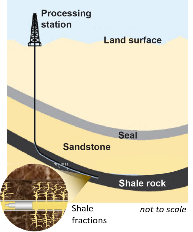
Figure 1. Natural gas extraction from shale rock.

Shale is a type of fine-grained rock that contains silt, clay, mineral particles, and pores ranging from meter to nanometer scales. The high organic material content in shale rock is used in natural gas extraction, for which shale reservoirs are mechanically stimulated to create permeability in the pores. A preferred stimulation method is called hydraulic fracturing, where a pressurized fluid fractures shale stone and keeps the fractures open for gas extraction (Figure 1). Natural gas extraction from shale rock is a relatively new process compared to existing energy sources. It has attracted growing interest in America and Asia especially after the 2000s because of being an environmentally friendly alternative to other consumable energy sources. On the other hand, the gas industry currently struggles with optimizing the use of pore space and fractions for efficient extraction of the gas. In a newly opened shale rock reservoir, volatile components vaporize from meter to micrometer-scale pores first, leaving heavier components in hard-to-access nanometer-scale pores. Extraction of the remaining components is necessary for full utilization of the reservoirs but poses a hard-to-solve problem for the industry.


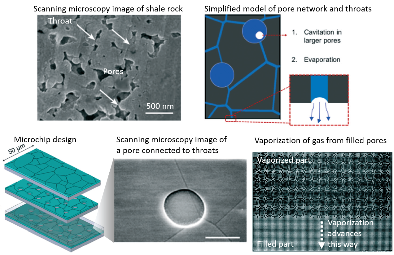
In a recent study published in Lab on Chip, David Sinton and co-authors review the current state of the technology and demonstrate a nano-scale physical model of shale with pores. The authors also study the dynamics of gas production in nanopores via imaging the system optically and developing an analytical model for gas vaporization. They first created a microchip model matching shale nanoporous matrix properties (e.g., dominant pore sizes and permeability) (Figure 2). The microchip model contained approximately 5800 pores connected via 23000 throats, where a hydrocarbon mixture was injected. In the model, the number of the small pores (≤10 nm) is designed to be greater than the number of the larger pores (∼100 nm) to store most of the accessible hydrocarbons. This pore size distribution captures the influence of nanoscale throats connecting the larger pores and is relevant to shale production. High, medium, and low superheat was applied to the filled microchip to investigate the spatiotemporal dynamics of vaporization via optical imaging. An analytical model and experimental results showed that phase change (liquid to vapor) in a pore is largely independent of phase change in neighboring pores.

This work supports the hypothesis that the rapid decline in production rates is due to a shift from the large connected features to the nanoporous matrix, as over time the smallest pores become enriched with heavier fractions. The authors reveal that vaporization rate slows down 3000 times thanks to the nanoscale throat bottlenecks at high temperatures, while the rates reduce further with vaporization of light components in large pores at low temperatures. Even the pores with 10 nm and fewer diameters can significantly influence the production from larger pores by severely gating transport. The authors found that this problem can be solved by applying very low pressures, although currently not available in the field, during the later stages of hydraulic fracturing. This finding seems to open a new avenue in the field of shale rock processing for energy.

Figure 2. Close up view of shale rock, the description of how the evaporation works, and the description of the microchip operation.

To download the full article for free* click the link below:

Natural gas vaporization in a nanoscale throat connected model of shale: multi-scale, multicomponent and multi-phase

Arnav Jatukaran, Junjie Zhong, Ali Abedini, Atena Sherbatian, Yinuo Zhao, Zhehui Jin, Farshid Mostowfi and David Sinton

Lab Chip, 2018, Lab on a Chip Articles

DOI: 10.1039/c8lc01053f

*Article free to read until 7th May 2019

About the Webwriter

Burcu Gumuscu is a researcher in Mesoscale Chemical Systems Group at University of Twente in the Netherlands. Her research interests include the development of microfluidic devices for quantitative analysis of proteins from single-cells, next generation sequencing, compartmentalized organ-on-chip studies, and desalination of water on the microscale.


Digg This
Reddit This
Stumble Now!
Share on Facebook
Bookmark this on Delicious
Share on LinkedIn
Bookmark this on Technorati
Post on Twitter
Google Buzz (aka. Google Reader)

Emerging Investigator Series – Jessie S. Jeon

Dr. Jessie S. Jeon received her SB, SM, and PhD in Mechanical Engineering from Massachusetts Institute of Technology (2008, 2010, 2014), and worked as a research fellow at Beth Israel Deaconess Medical Center, (2014-2015). She has joined the KAIST faculty in the fall of 2015 as an assistant professor in the Department of Mechanical Engineering. Her research focuses on the development of microfluidic platform with applications in investigating biological systems. She plans to further develop the microfluidic system with the emphasis in fluidic aspects and also to extend its applications in mimicking various organ disease systems as well as other biological microenvironments. By doing so, she hopes to bridge the needs of biomedical research with the knowledge of mechanical engineering principles.

Read Jessie S. Jeon’s Emerging Investigator article “On-chip phenotypic investigation of combinatory antibiotic effects by generating orthogonal concentration gradients and find out more about her in the interview below: 

Your recent Emerging Investigator Series paper focuses on on-chip phenotypic investigation of combinatory antibiotic effects. How has your research evolved from your first article to this most recent article?

My group first worked on microfluidic-based single antibiotic testing platform where we could reduce the time it takes for antibiotic susceptibility testing (AST). As we learn more about AST, we realized that recently most studies on antibiotics focus on investigation of combinatory antibiotic effects. Since microfluidic platform enables combination of multiple channels, it was quite natural to try a combination of antibiotics in one chip.

What aspect of your work are you most excited about at the moment?

Broadly speaking, I am excited that we could potentially utilize our platform to screen for personalized medicine. That is to screen for patient specific therapy using microfluidic platform. The thought that our technology would contribute to enhance our lives definitely motivates me working on this topic.

In your opinion, what is the future of chip-based screening for clinical therapies?

I believe that with the development of lab-on-chips, we would be able to screen for the most optimal therapeutic strategy using a patient’s own cells, and this technology would bring the biggest impact to the society. This includes selection of strategy in terms of therapeutic methods as well as possibility in combinatory therapy either for antibiotics or anti-cancer drugs. That is also in line with my answer for the question above that I am very excited for the opportunities in personalized medicine with lab-on-a-chip technology.

What do you find most challenging about your research?

As a researcher in an interdisciplinary field, it is always challenging for me to identify meaningful biological and biomedical questions that I can address with my expertise. I realize that it is very important to keep keen relationships with clinicians and biologists.

In which upcoming conferences or events may our readers meet you?

I plan to attend the 2019 Annual Meeting of the Biomedical Engineering Society in coming October.

How do you spend your spare time?

I enjoy playing a variety of sports, mostly tennis these days, and I also try to spend more time with family on short trips whenever possible.

Which profession would you choose if you were not a scientist?

Perhaps I would be serving in military as I briefly took a part in the ROTC program when I was in college.

Can you share one piece of career-related advice or wisdom with other early career scientists?

While I’m still in a position needing much advice from others, I would like to share my thought that if you don’t give up, there will be opportunities to come.

Digg This
Reddit This
Stumble Now!
Share on Facebook
Bookmark this on Delicious
Share on LinkedIn
Bookmark this on Technorati
Post on Twitter
Google Buzz (aka. Google Reader)

Outstanding Reviewers for Lab on a Chip in 2018

We would like to highlight the Outstanding Reviewers for Lab on a Chip in 2018, as selected by the editorial team, for their significant contribution to the journal. The reviewers have been chosen based on the number, timeliness and quality of the reports completed over the last 12 months.

We would like to say a big thank you to those individuals listed here as well as to all of the reviewers that have supported the journal. Each Outstanding Reviewer will receive a certificate to give recognition for their significant contribution.

Dr Chia Hung Chen, National University of Singapore, Singapore
Professor Daniel Citterio, Keio University, Japan
Dr David Collins, MIT, United States
Professor Dino Di Carlo, University of California, Los Angeles, United States
Dr Mei He, Kansas State University, United States
Dr Daniel Irimia, Harvard Medical School, United States
Dr Séverine Le Gac, University of Twente, Netherlands
Dr Robert Meagher, Sandia National Laboratories, United States
Professor Michael Roper, Florida State University, United States
Dr Edmond Young, University of Toronto, Canada

We would also like to thank the Lab on a Chip board and the Lab on a Chip community for their continued support of the journal, as authors, reviewers and readers.

If you would like to become a reviewer for our journal, just email us at LOC-RSC@rsc.org with details of your research interests and an up-to-date CV or résumé. You can find more details in our author and reviewer resource centre

Digg This
Reddit This
Stumble Now!
Share on Facebook
Bookmark this on Delicious
Share on LinkedIn
Bookmark this on Technorati
Post on Twitter
Google Buzz (aka. Google Reader)